Abstract
Purpose
To evaluate the tolerability of escalating doses of stereotactic body radiation therapy in the treatment of localized prostate cancer.
Patients and Methods
Eligible patients included those with Gleason score 2 to 6 with prostate-specific antigen (PSA) ≤ 20, Gleason score 7 with PSA ≤ 15, ≤ T2b, prostate size ≤ 60 cm3, and American Urological Association (AUA) score ≤ 15. Pretreatment preparation required an enema and placement of a rectal balloon. Dose-limiting toxicity (DLT) was defined as grade 3 or worse GI/genitourinary (GU) toxicity by Common Terminology Criteria of Adverse Events (version 3). Patients completed quality-of-life questionnaires at defined intervals.
Results
Groups of 15 patients received 45 Gy, 47.5 Gy, and 50 Gy in five fractions (45 total patients). The median follow-up is 30 months (range, 3 to 36 months), 18 months (range, 0 to 30 months), and 12 months (range, 3 to 18 months) for the 45 Gy, 47.5 Gy, and 50 Gy groups, respectively. For all patients, GI grade ≥ 2 and grade ≥ 3 toxicity occurred in 18% and 2%, respectively, and GU grade ≥ 2 and grade ≥ 3 toxicity occurred in 31% and 4%, respectively. Mean AUA scores increased significantly from baseline in the 47.5-Gy dose level (P = .002) as compared with the other dose levels, where mean values returned to baseline. Rectal quality-of-life scores (Expanded Prostate Cancer Index Composite) fell from baseline up to 12 months but trended back at 18 months. In all patients, PSA control is 100% by the nadir + 2 ng/mL failure definition.
Conclusion
Dose escalation to 50 Gy has been completed without DLT. A multicenter phase II trial is underway treating patients to 50 Gy in five fractions to further evaluate this experimental therapy.
INTRODUCTION
Stereotactic body radiation therapy (SBRT) is an even newer form of external-beam radiotherapy capable of accurately and precisely directed irradiation of localized tumors outside of the CNS. It involves delivering high daily doses using unique beam arrangements, stable patient immobilization, motion assessment/control, and daily image guidance. This technique has been successfully applied to early-stage lung cancer and liver metastasis.1–5 Prostate cancer may be uniquely appropriate for treatment with hypo-fractionation (large dose per fraction) because of a lower α/β ratio (∼1.5) for prostate cancer that is similar to normal tissue late effects.6–9
Trials using modestly larger radiation dose per fraction treatments have been published using external-beam radiation therapy, or brachytherapy.10–13 The group at the William Beaumont Hospital used high-dose rate (HDR) brachytherapy with a four-fraction regimen of 9.5 Gy to a total dose of 38 Gy and published an acceptable toxicity profile,14,15 whereas Yashioka et al16,17 from Japan used higher total doses up to 48 to 50 Gy in 6-Gy fractions without untoward toxicity. It has been shown that SBRT can mimic these highly conformal brachytherapy dose distributions.18
Clinical trials with conventionally fractionated radiation have shown that increased total radiation doses result in improved outcomes.19,20 Our preclinical data showed increasing dose response without plateau up to 45 Gy in three fractions with SBRT techniques using nude mouse xenografts with C4-2 human prostate cancer cell lines.21 Radiobiologic modeling of the previous HDR monotherapy experience indicates that the five-fraction equivalent would be 36 to 43 Gy.22 For similar minimum prescription doses, the integral dose for brachytherapy would be considerably higher than for SBRT. As such, it is likely that SBRT treatment would require an even higher dose for equivalent tumor control. Indeed, a pilot trial of SBRT in low-risk prostate cancer from Virginia Mason University using 33.5 Gy in five fractions demonstrated relatively poor prostate-specific antigen (PSA) control by the American Society for Therapeutic Radiology and Oncology definition (70%) compared with the HDR series.23
We initiated a multicenter prospective dose-escalation study to assess toxicity and quality of life (QOL) in patients treated with SBRT for localized prostate cancer. We chose to start at a dose similar to the biologic equivalent margin dose of the HDR brachytherapy experience (ie, 45 Gy in five fractions) and escalate this noninvasive, outpatient therapy to more potent dose levels at which PSA control might be more favorable.
PATIENTS AND METHODS
In October 2006, we began an institutional review board–approved phase I clinical trial of SBRT for low and intermediate risk prostate cancer. This prospective study's goals were to assess acute (< 90 days) urinary and rectal toxicity, QOL measures, and PSA response for SBRT of the prostate.
Patient Eligibility
Patients were included with newly diagnosed and previously untreated prostate cancer. Patients had American Joint Committee on Cancer stage T1 or T2 (a and b) adenocarcinoma of the prostate gland. The patients had no evidence of regional or distant metastases after appropriate staging studies. Zubrod performance status was between 0 and 2. The serum PSA was required to be ≤ 20 ng/mL for patients with Gleason score 2 to 6 and ≤ 15 ng/mL for patients with Gleason score 7. As such, the risk of pelvic lymph node involvement according to the Roach formula would be less than 20%.24 Patients were excluded if the pre-SBRT prostate volume on ultrasound was greater than 60 cm3. Hormonal therapy was allowed for up to 9 months before SBRT to downsize the prostate gland as confirmed by ultrasound. Patients were also excluded if they had prior transurethral resection of the prostate, American Urological Association (AUA) score more than 15 (α blockers allowed), history of inflammatory colitis, or other active severe comorbidities.
Planning
Fiducial markers consisting of gold seeds or Calypso beacons were placed within the prostate approximately 1 week before radiation simulation. A bowel regimen consisting of 30 mL of milk of magnesia the evening before and a Fleet enema 30 to 60 minutes before simulation and each treatment was used, along with the insertion of a 60- to 100-cm3 rectal balloon. Patients were instructed to have a full bladder for simulation and treatment. A flexible catheter was used to delineate the urethra at simulation only. Magnetic resonance imaging (MRI) or computed tomography (CT) was used to define the prostate and organs at risk. The prostate was expanded uniformly by 3 mm to create the planning target volume (PTV).
SBRT was delivered via ring gantry helical accelerator (Tomotherapy; TomoTherapy Inc, Madison, WI) or step and shoot on a linear accelerator (Trilogy; Varian Medical Systems, Palo Alto, CA, and Synergy; Elekta AB, Stockholm, Sweden) with energies of 6 to 15 MV. The dose was prescribed to cover ≥ 95% of the PTV. Rapid dose falloff outside the PTV was prioritized over PTV dose uniformity, resulting in considerable dose heterogeneity within the PTV. Tissue heterogeneity correction was used in all cases.
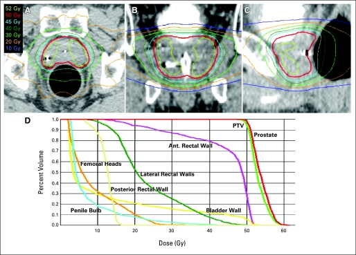
A rectal balloon was used to expand the rectum and push the lateral and posterior walls away from the high-dose region. The rectal wall was divided and separately contoured into anterior, lateral, and posterior walls in the region of the PTV. The anterior wall was allowed to receive no more than 105% of the prescription dose. No more than 3 cm3 of the lateral walls were allowed to receive 90% of the prescription dose. The posterior rectal wall maximum dose was limited to ≤ 45% of the prescription dose. The bladder wall (outer 5 mm of the entire bladder contour) was limited to 105% of the prescription dose with no more than 10 cm3 receiving 18.3 Gy or greater. The maximum prostatic urethra dose was limited to ≤ 105% of the prescription dose. A sample isodose plan is shown in Figure 1for a patient on the 50-Gy dose level.
Fig 1.
Isodose distribution of a patient receiving 50 Gy of radiation in the axial plane (A), coronal plane (B), and sagittal plane (C). (D) is a dose-volume histogram showing coverage of the planning target volume (PTV)/prostate and low dose to surrounding organs. Ant., anterior.
Treatment
Daily image guidance was used to localize the prostate via megavoltage or kilovoltage computed tomography before each fraction. Proper position of the fiducial markers, rectal balloon, and filling of the bladder was confirmed before each treatment. SBRT was delivered in five fractions separated by a minimum of 36 hours. Radiation dose started at 9 Gy per fraction to a total dose of 45 Gy for first 15 patients and was escalated in subsequent cohorts to total doses of 47.5 Gy and then 50 Gy. Patients were premedicated with 4 mg of dexamethasone before each SBRT treatment.
Study End Points and Statistics
This study was designed as a prospective dose-escalation study. The goal was to escalate the dose of five-fraction SBRT to the maximum-tolerated dose or 50 Gy. Dose-limiting toxicity (DLT) was defined as grade 3 to 5 GI, genitourinary, sexual, or neurologic toxicity attributed to therapy occurring within 90 days of registration using Common Terminology Criteria of Adverse Events (version 3). Escalation was allowed to occur if four or fewer patients of 15 experienced DLT within 90 days of follow-up at a given dose level. This escalation rule is the same as the traditional 3 + 3 design in which less than 33% DLT rate at the current dose level leads to further dose escalation. Using a larger number of patients per dose level is justified for this trial on the basis of the previously referenced SBRT and HDR experiences that predict efficacy even with the starting dose. As with the traditional 3 + 3 design, sequential enrollment serves as a protection to limit potential overdosing. Furthermore, 15 patients at each level would allow us to more accurately estimate the DLT rate and to study other end points related to the enrolled patients. The maximum-tolerated dose was defined as the dose level immediately below the intolerable dose. Secondary end points were late toxicity (occurring > 90 days from treatment), patient-reported toxicity/QOL, and PSA response. The Expanded Prostate Cancer Index Composite (EPIC) questionnaire and AUA scores were collected at baseline and 1.5, 3, 12, and 18 months after treatment. Patients were observed with PSA, history, and physical examination every 3 months for the first year, every 6 months for years 2 to 3, and yearly starting 4 years after treatment. The nadir + 2 ng/mL failure definition was used for biochemical control.
In the statistical analyses, mixed models and generalized estimating equations with empirical SE estimates were used to compare the EPIC scores for urinary, bowel, and sexual and the normalized AUA scores among the radiation doses at different times. The models consist of terms of dose group and time. For EPIC sexual scores, the model also contains a term of hormone therapy. The term of interaction between dose group and time was added to the models and was found not to be significant and hence was dropped from the models. Fisher's exact test was performed to the number of patients among the study sites and dose groups. One-way analysis of variance (ANOVA) was conducted to test the AUA scores and prostate sizes among the dose groups. All reported P values are two-sided. The statistical analyses were done in SAS 9.2 for Windows (SAS Institute, Cary, NC).
RESULTS
A total of 48 patients were enrolled from November 2006 to May 2009. Two people withdrew consent before treatment and one person became ineligible after attempted downsizing with hormones when his AUA score increased to above 15 before any study treatment. Therefore, 45 patients were treated and are evaluable for study end points. Of these 45 patients, one patient was treated but was later found to have been ineligible when re-review of his pathology showed Gleason score 9. He was observed for toxicity but not PSA control. The median follow-up was 30 months (range, 3 to 36 months) for the 45-Gy cohort, 18 months (range, 0 to 30 months) for the 47.5-Gy cohort, and 12 months (range, 3 to 18 months) for the 50-Gy group. Follow-up as a function of time since treatment is displayed in Appendix Table A1 (online only). Four patients died of unrelated causes during follow-up (two patients as a result of myocardial infarction, one patient as a result of suicide, and one patient as a result of unknown cause without autopsy). The dose groups were balanced with no statistical differences in pretreatment characteristics, as shown in Table 1.
Table 1.
Baseline Patient Characteristics
| Characteristic | 45 Gy |
47.5 Gy |
50 Gy |
Total |
||||
|---|---|---|---|---|---|---|---|---|
| No. | % | No. | % | No. | % | No. | % | |
| No. of patients | 15 | 15 | 15 | 45 | ||||
| Age, years | ||||||||
| Median | 67 | 67 | 67 | 67 | ||||
| Range | 55-82 | 58-76 | 53-78 | 53-82 | ||||
| Prostate size, cm3 | ||||||||
| Median | 31 | 38 | 30 | 31 | ||||
| Range | 19-60 | 17-52 | 17-55 | 17-60 | ||||
| AUA score | ||||||||
| Median | 4.5 | 4 | 7 | 5 | ||||
| Range | 0-15 | 0-13 | 2-12 | 0-15 | ||||
| Hormones | ||||||||
| Yes | 4 | 27 | 2 | 13 | 4 | 27 | 10 | 22 |
| No | 11 | 73 | 13 | 87 | 11 | 73 | 35 | 78 |
| PSA | ||||||||
| Median | 6.40 | 5.68 | 4.49 | 5.60 | ||||
| Range | 3.28-12.36 | 1.30-11.54 | 0.19-7.94 | 0.19-12.36 | ||||
| T stage | ||||||||
| T1c | 11 | 73 | 13 | 87 | 8 | 53 | 32 | 71 |
| T2a | 1 | 7 | 1 | 7 | 5 | 33 | 7 | 16 |
| T2b | 3 | 20 | 1 | 7 | 2 | 13 | 6 | 13 |
| GS | ||||||||
| 6 (3 + 3) | 4 | 27 | 8 | 53 | 9 | 60 | 21 | 47 |
| 7 (3 + 4) | 8 | 53 | 5 | 33 | 3 | 20 | 16 | 36 |
| 7 (4 + 3) | 3 | 20 | 2 | 13 | 3 | 20 | 8 | 18 |
| Treatment site | ||||||||
| A | 14 | 8 | 10 | 32 | ||||
| B | 1 | 4 | 4 | 9 | ||||
| C | 0 | 3 | 1 | 4 | ||||
| Low risk (GS ≤ 6, PSA < 10, ≤ T2a) | 3 | 20 | 8 | 53 | 7 | 47 | 18 | 40 |
| Intermediate risk (GS = 7 or PSA > 10 or T2b) | 12 | 80 | 7 | 47 | 8 | 53 | 27 | 60 |
Abbreviations: AUA, American Urological Association; PSA, prostate-specific antigen; GS, Gleason score.
No DLT was seen within 90 days from the start of treatment, and thus dose escalation proceeded through all planned dose levels. Only GI and genitourinary (GU) toxicity was observed. The number of patients experiencing GI and GU toxicity by grade for each dose level is shown in Tables 2 and 3. The most common early GU toxicity was urinary frequency and urgency. The initial patients treated on trial experienced urinary frequency after SBRT. Therefore, the protocol was amended early to include Tamsulosin for 6 weeks from the start of treatment as is commonly done after brachytherapy procedures. There was no grade 3 toxicity over the complete course of follow-up within the first dose level. Only a single patient in the 45-Gy dose level experienced a grade 2 GI toxicity, and one third of the patients had grade 2 GU toxicity during follow-up. One patient treated with 47.5 Gy experienced grade 3 or worse toxicity. Two separate patients treated on the 50-Gy dose level accounted for a grade 3 GU toxicity and a grade 4 GI toxicity. Grade 3 GU toxicities were dysuria and cystitis, and the time to grade 3 GU toxicity was 12 months for both patients. The patient with grade 4 GI toxicity had a rectal ulcer that appeared shortly after SBRT and slowly enlarged. This patient was being treated with and continued immunosuppressive medication (including sirolimus and tacrolimus) as antirejection prophylaxis for a previous kidney transplant. The transplanted kidney became dysfunctional before the protocol therapy, but he was continued on the antirejection medicine to avoid host-versus-graft problems. Hyperbaric oxygen and a diverting colostomy were used to aid in healing after SBRT. The patient's toxicity was scored as a grade 4 when he was admitted to the hospital for rectal bleeding with a hemoglobin of 6 g/dL. His antirejection medicines were ultimately withdrawn, resulting in improvements of symptoms and healing of radiation damage at last follow-up. No other patients on trial were taking these classes of immunosuppressive drugs. In response to these events, the protocol was amended to make patients on such immunosuppressive medicines ineligible for protocol treatment.
Table 2.
Worst GI Toxicity per Patient According to Time and Total Radiation Dose Level
| Highest Grade Toxicity | 45 Gy |
47.5 Gy |
50 Gy |
|||||||||||||||
|---|---|---|---|---|---|---|---|---|---|---|---|---|---|---|---|---|---|---|
| < 90 Days |
> 90 Days |
Worst |
< 90 Days |
> 90 Days |
Worst |
< 90 Days |
> 90 Days |
Worst |
||||||||||
| No. | % | No. | % | No. | % | No. | % | No. | % | No. | % | No. | % | No. | % | No. | % | |
| 0 | 9 | 60 | 13 | 87 | 8 | 53 | 9 | 60 | 10 | 67 | 7 | 47 | 7 | 47 | 9 | 60 | 4 | 27 |
| 1 | 6 | 40 | 1 | 7 | 6 | 40 | 2 | 13 | 4 | 27 | 3 | 20 | 7 | 47 | 5 | 33 | 9 | 60 |
| 2 | 0 | 0 | 1 | 7 | 1 | 7 | 4 | 27 | 1 | 7 | 5 | 33 | 1 | 7 | 0 | 0 | 1 | 7 |
| 3 | 0 | 0 | 0 | 0 | 0 | 0 | 0 | 0 | 0 | 0 | 0 | 0 | 0 | 0 | 0 | 0 | 0 | |
| 4 | 0 | 0 | 0 | 0 | 0 | 0 | 0 | 0 | 0 | 0 | 0 | 0 | 0 | 0 | 1 | 7 | 1 | 7 |
NOTE. Toxicity graded according to Common Terminology Criteria of Adverse Events, version 3.
Table 3.
Worst Genitourinary Toxicity per Patient According to Time and Total Radiation Dose Level
| Highest Grade Toxicity |
45 Gy |
47.5 Gy |
50 Gy |
|||||||||||||||
|---|---|---|---|---|---|---|---|---|---|---|---|---|---|---|---|---|---|---|
| < 90 Days |
> 90 Days |
Worst |
< 90 Days |
> 90 Days |
Worst |
< 90 Days |
> 90 Days |
Worst |
||||||||||
| No. | % | No. | % | No. | % | No. | % | No. | % | No. | % | No. | % | No. | % | No. | % | |
| 0 | 8 | 53 | 10 | 67 | 6 | 40 | 9 | 60 | 9 | 60 | 6 | 40 | 5 | 33 | 14 | 93 | 4 | 27 |
| 1 | 3 | 20 | 3 | 20 | 4 | 27 | 5 | 33 | 3 | 20 | 6 | 40 | 5 | 33 | 0 | 0 | 5 | 33 |
| 2 | 4 | 27 | 2 | 13 | 5 | 33 | 1 | 7 | 2 | 13 | 2 | 13 | 5 | 33 | 0 | 0 | 5 | 33 |
| 3 | 0 | 0 | 0 | 0 | 0 | 0 | 0 | 0 | 1 | 7 | 1 | 7 | 0 | 0 | 1 | 7 | 1 | 7 |
| 4 | 0 | 0 | 0 | 0 | 0 | 0 | 0 | 0 | 0 | 0 | 0 | 0 | 0 | 0 | 0 | 0 | 0 | 0 |
NOTE. Toxicity graded according to Common Terminology Criteria of Adverse Events, version 3.
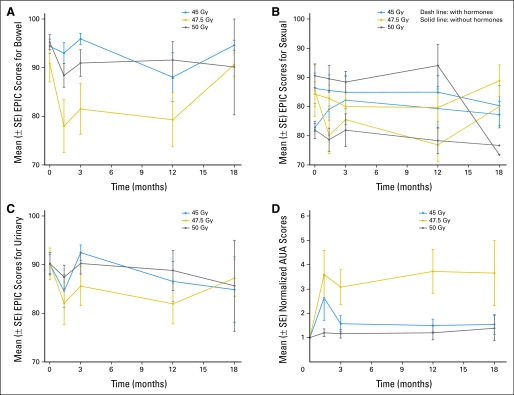
Compliance with obtaining questionnaires was 92% for the 45-Gy dose level, 93% for the 47.5-Gy dose level, and 93% for the 50-Gy dose level. Figure 2 shows trends in EPIC scores separated by dose level. Patients' bowel related QOL decreased initially at 6 weeks of follow-up but trended back to baseline at 18 months. There was no difference between the 45-Gy and 50-Gy dose levels (P = .5), but a worse bowel-related QOL was seen for the 47.5-Gy dose level (P = .01). Patients who received hormonal therapy for gland downsizing before treatment had significantly worse sexual QOL (P = .01).
Fig 2.
Means and SEs for Expanded Prostate Cancer Index Composite (EPIC) scores for (A) bowel, (B) sexual, and (C) urinary quality of life. (D) American Urological Association (AUA) mean scores with SEs that were normalized using a given patient's baseline score as the denominator. Data points with a single measurement are not shown.
Patient-reported urinary function was assessed with the EPIC QOL instrument and also by their AUA scores. There was a drop and subsequent recovery of urinary QOL within the first 3 months after treatment. Overall there is no significant difference among the dose levels, and there is a nonsignificant decrease in urinary QOL at 12 and 18 months compared with baseline. This is mirrored in the normalized AUA scores trend (Fig 2). AUA score increases returned to baseline in the 45-Gy and 50-Gy group but persisted in the 47.5-Gy dose level patients. There was no difference in pretreatment AUA scores across all dose levels (P = .22) and no difference between 45-Gy and 50-Gy dose levels during follow-up (P = .5). The 47.5-Gy dose level patients had significantly elevated AUA scores after treatment (P = .002) compared with those in the other groups.
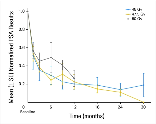
One patient experienced a persistently increasing PSA after treatment, and on re-review of his pathology, he was found to have Gleason score 9 disease. He was then classified ineligible and ultimately experienced disease recurrence distantly. To date, all of the other 44 men have declining or stable PSA measurements. Figure 3 shows the decline in PSA as a function of patients' initial PSA. Bounces have been seen in multiple patients. No patient has experienced a biochemical failure.
Fig 3.
Mean prostate-specific antigen (PSA) with SEs. PSA was normalized using a given patient's baseline as the denominator.
DISCUSSION
This prospective multicenter study that was intended to define a prudent SBRT treatment dose represents a critical step in implementing this technology. For practical reasons, a 90-day observation window was used to guide dose escalation. Subsequent to the 90-day window, only three patients have experienced high-grade toxicity. Although so-called late effects from radiation can technically occur decades after the therapy,25 the paucity of observed high-grade toxic events within our median follow-up range of 12 to 30 months is encouraging. Although the assessment of acute toxicity (< 90 days) is mature for each dose level studied in this report, the follow-up on our trial is inadequate to reliably assess late toxicity.
Subsequent to the initiation of our study, there have been several single-institution reports regarding the use of SBRT to total radiation doses ranging from 33.5 to 36.25 in five fractions (6.7 to 7.25 Gy).23,26,27 These studies reported acceptable rates of grade 1 and 2 toxicities, with rare grade 3 toxicities. Although these studies used SBRT techniques, they did not use doses as high as those in our current study. The acute toxicity in our trial compares favorable with the acute toxicity that was seen in the dose-escalated arm of the MD Anderson and Proton Radiation Oncology Group trials in which GU/GI grade 2 or worse toxicity was seen in 49% to 62%/57% to 64% and grade 3 or worse toxicity was seen in 2%/0% to 2%, respectively.19,20,28
We did not identify the maximum-tolerated dose for a five-fraction SBRT regimen despite escalating to potent dose levels not tested in previous reports. We partially attribute this to inherent tissue tolerance as well as the strict conduct of the treatment. We used a rectal balloon to distance the lateral and posterior rectal walls to avoid circumferential damage to the rectum. We preemptively gave patients tamsulosin for 6 weeks to reduce the risk of urinary complications. Patients were treated every other day to give time for tissue recovery. King et al27 found that every-other-day treatment reduced the rate of rectal toxicity.
We assessed the impact of SBRT on urinary, rectal, and sexual QOL using the EPIC questionnaire. Results using this questionnaire were reported by Sanda et al29 for patients treated with prostatectomy, conventional radiation, or brachytherapy. SBRT seems to be similar to brachytherapy with an early bowel-related decrease in QOL related to irritation that eventually returns to baseline. There is also an early urinary irritation-related dip in QOL that does not recover completely back to baseline. The 47.5-Gy dose level had significantly worse QOL scores for bowel and increase in AUA scores at early time points. There were no differences in baseline characteristics that may contribute to this such as prostate size, baseline AUA score, or age. This is not likely just a phenomenon of follow-up in the later dose level as median follow-up for the 50-Gy group is greater than where the changes were observed in the 47.5-Gy group.
This trial included more patients per dose level than previous SBRT dose-escalation trials.2,5,30 Drug discovery phase I trials commonly use only three patients per dose level to treat as few patients as possible at likely subtherapeutic dose levels. In our trial, however, even the starting dose is predicted to have efficacy on the basis of the previously published SBRT and HDR experiences.14,16 With increased follow-up, an optimal dose level may be identified that has few treatment failures and the lowest toxicity or change in QOL.
In conclusion, dose escalation on this multi-institutional phase I trial focusing on acute toxicity (< 90 days from completion of therapy) in patients with localized prostate cancer was possible to 50 Gy. Early PSA response is promising. We are currently enrolling patients to a phase II trial (70 patients) using the 50-Gy level in which the primary end point is 18-month late toxicity.
Appendix
Table A1.
Patients Seen in Follow-Up at Various Time Points Since Therapy
| Dose Level | No. of Patients |
|||||||
|---|---|---|---|---|---|---|---|---|
| 3 Months | 6 Months | 9 Months | 12 Months | 18 Months | 24 Months | 30 Months | 36 Months | |
| 45 Gy | 15 | 14 | 14 | 14 | 14 | 14 | 8 | 1 |
| 47.5 Gy | 14 | 14 | 14 | 14 | 12 | 4 | 0 | 0 |
| 50 Gy | 15 | 14 | 11 | 8 | 4 | 0 | 0 | 0 |
Footnotes
See accompanying editorial on page 1940
Supported by a Clinical Trial Award from the US Department of Defense (Grant No. PC061629; R.T., principal investigator).
Presented in part at the 51st Annual Meeting of the American Society for Therapeutic Radiology and Oncology, October 31-November 5, 2009, Chicago, IL; and the American Society of Clinical Oncology Genitourinary Cancers Symposium, March 5-7, 2010, San Francisco, CA.
Authors' disclosures of potential conflicts of interest and author contributions are found at the end of this article.
Clinical trial information can be found for the following: NCT00547339.
AUTHORS' DISCLOSURES OF POTENTIAL CONFLICTS OF INTEREST
Although all authors completed the disclosure declaration, the following author(s) indicated a financial or other interest that is relevant to the subject matter under consideration in this article. Certain relationships marked with a “U” are those for which no compensation was received; those relationships marked with a “C” were compensated. For a detailed description of the disclosure categories, or for more information about ASCO's conflict of interest policy, please refer to the Author Disclosure Declaration and the Disclosures of Potential Conflicts of Interest section in Information for Contributors.
Employment or Leadership Position: None Consultant or Advisory Role: None Stock Ownership: None Honoraria: None Research Funding: Robert Timmerman, Varian Medical Systems, Accuray Expert Testimony: None Other Remuneration: None
AUTHOR CONTRIBUTIONS
Conception and design: Yair Lotan, L. Chinsoo Cho, Xian-Jin Xie, David Pistenmaa, Robert Timmerman
Financial support: Robert Timmerman
Administrative support: David Pistenmaa, Alida Perkins, Susan Cooley, Robert Timmerman
Provision of study materials or patients: Thomas P. Boike, Yair Lotan, L. Chinsoo Cho, Jeffrey Brindle, Paul DeRose, David Pistenmaa, Robert Timmerman
Collection and assembly of data: Thomas P. Boike, L. Chinsoo Cho, Jeffrey Brindle, Paul DeRose, Ryan Foster, David Pistenmaa, Alida Perkins, Susan Cooley, Robert Timmerman
Data analysis and interpretation: Thomas P. Boike, Yair Lotan, L. Chinsoo Cho, Paul DeRose, Xian-Jin Xie, Jingsheng Yan, David Pistenmaa, Alida Perkins, Robert Timmerman
Manuscript writing: Thomas P. Boike, Robert Timmerman
Final approval of manuscript: Thomas P. Boike, Yair Lotan, L. Chinsoo Cho, Jeffrey Brindle, Paul DeRose, Xian-Jin Xie, Jingsheng Yan, Ryan Foster, David Pistenmaa, Alida Perkins, Susan Cooley, Robert Timmerman
REFERENCES
- 1.Timmerman R, Paulus R, Galvin J, et al. Stereotactic body radiation therapy for inoperable early stage lung cancer. JAMA. 2010;303:1070–1076. doi: 10.1001/jama.2010.261. [DOI] [PMC free article] [PubMed] [Google Scholar]
- 2.Timmerman R, Papiez L, McGarry R, et al. Extracranial stereotactic radioablation: Results of a phase I study in medically inoperable stage I non-small cell lung cancer. Chest. 2003;124:1946–1955. doi: 10.1378/chest.124.5.1946. [DOI] [PubMed] [Google Scholar]
- 3.Timmerman R, McGarry R, Yiannoutsos C, et al. Excessive toxicity when treating central tumors in a phase II study of stereotactic body radiation therapy for medically inoperable early-stage lung cancer. J Clin Oncol. 2006;24:4833–4839. doi: 10.1200/JCO.2006.07.5937. [DOI] [PubMed] [Google Scholar]
- 4.Schefter TE, Kavanagh BD, Timmerman RD, et al. A phase I trial of stereotactic body radiation therapy (SBRT) for liver metastases. Int J Radiat Oncol Biol Phys. 2005;62:1371–1378. doi: 10.1016/j.ijrobp.2005.01.002. [DOI] [PubMed] [Google Scholar]
- 5.Rusthoven KE, Kavanagh BD, Cardenes H, et al. Multi-institutional phase I/II trial of stereotactic body radiation therapy for liver metastases. J Clin Oncol. 2009;27:1572–1578. doi: 10.1200/JCO.2008.19.6329. [DOI] [PubMed] [Google Scholar]
- 6.Fowler J, Chappell R, Ritter M. Is alpha/beta for prostate tumors really low? Int J Radiat Oncol Biol Phys. 2001;50:1021–1031. doi: 10.1016/s0360-3016(01)01607-8. [DOI] [PubMed] [Google Scholar]
- 7.Brenner DJ, Hall EJ. Fractionation and protraction for radiotherapy of prostate carcinoma. Int J Radiat Oncol Biol Phys. 1999;43:1095–1101. doi: 10.1016/s0360-3016(98)00438-6. [DOI] [PubMed] [Google Scholar]
- 8.D'Souza WD, Thames HD. Is the alpha/beta ratio for prostate cancer low? Int J Radiat Oncol Biol Phys. 2001;51:1–3. doi: 10.1016/s0360-3016(01)01650-9. [DOI] [PubMed] [Google Scholar]
- 9.Dasu A. Is the alpha/beta value for prostate tumours low enough to be safely used in clinical trials? Clin Oncol (R Coll Radiol) 2007;19:289–301. doi: 10.1016/j.clon.2007.02.007. [DOI] [PubMed] [Google Scholar]
- 10.Martin T, Baltas D, Kurek R, et al. 3-D conformal HDR brachytherapy as monotherapy for localized prostate cancer. A pilot study. Strahlenther Onkol. 2004;180:225–232. doi: 10.1007/s00066-004-1215-4. [DOI] [PubMed] [Google Scholar]
- 11.Vargas CE, Martinez AA, Boike TP, et al. High-dose irradiation for prostate cancer via a high-dose-rate brachytherapy boost: Results of a phase I to II study. Int J Radiat Oncol Biol Phys. 2006;66:416–423. doi: 10.1016/j.ijrobp.2006.04.045. [DOI] [PubMed] [Google Scholar]
- 12.Yeoh EE, Holloway RH, Fraser RJ, et al. Hypofractionated versus conventionally fractionated radiation therapy for prostate carcinoma: Updated results of a phase III randomized trial. Int J Radiat Oncol Biol Phys. 2006;66:1072–1083. doi: 10.1016/j.ijrobp.2006.06.005. [DOI] [PubMed] [Google Scholar]
- 13.Kupelian PA, Willoughby TR, Reddy CA, et al. Hypofractionated intensity-modulated radiotherapy (70 Gy at 2.5 Gy per fraction) for localized prostate cancer: Cleveland Clinic experience. Int J Radiat Oncol Biol Phys. 2007;68:1424–1430. doi: 10.1016/j.ijrobp.2007.01.067. [DOI] [PubMed] [Google Scholar]
- 14.Grills IS, Martinez AA, Hollander M, et al. High dose rate brachytherapy as prostate cancer monotherapy reduces toxicity compared to low dose rate palladium seeds. J Urol. 2004;171:1098–1104. doi: 10.1097/01.ju.0000113299.34404.22. [DOI] [PubMed] [Google Scholar]
- 15.Martinez AA, Pataki I, Edmundson G, et al. Phase II prospective study of the use of conformal high-dose-rate brachytherapy as monotherapy for the treatment of favorable stage prostate cancer: A feasibility report. Int J Radiat Oncol Biol Phys. 2001;49:61–69. doi: 10.1016/s0360-3016(00)01463-2. [DOI] [PubMed] [Google Scholar]
- 16.Yoshioka Y, Nose T, Yoshida K, et al. High-dose-rate brachytherapy as monotherapy for localized prostate cancer: A retrospective analysis with special focus on tolerance and chronic toxicity. Int J Radiat Oncol Biol Phys. 2003;56:213–220. doi: 10.1016/s0360-3016(03)00081-6. [DOI] [PubMed] [Google Scholar]
- 17.Yoshioka Y, Nose T, Yoshida K, et al. High-dose-rate interstitial brachytherapy as a monotherapy for localized prostate cancer: Treatment description and preliminary results of a phase I/II clinical trial. Int J Radiat Oncol Biol Phys. 2000;48:675–681. doi: 10.1016/s0360-3016(00)00687-8. [DOI] [PubMed] [Google Scholar]
- 18.Fuller DB, Naitoh J, Lee C, et al. Virtual HDR CyberKnife treatment for localized prostatic carcinoma: Dosimetry comparison with HDR brachytherapy and preliminary clinical observations. Int J Radiat Oncol Biol Phys. 2008;70:1588–1597. doi: 10.1016/j.ijrobp.2007.11.067. [DOI] [PubMed] [Google Scholar]
- 19.Zietman AL, Bae K, Slater JD, et al. Randomized trial comparing conventional-dose with high-dose conformal radiation therapy in early-stage adenocarcinoma of the prostate: Long-term results from proton radiation oncology group/american college of radiology 95-09. J Clin Oncol. 2010;28:1106–1111. doi: 10.1200/JCO.2009.25.8475. [DOI] [PMC free article] [PubMed] [Google Scholar]
- 20.Kuban DA, Tucker SL, Dong L, et al. Long-term results of the M. D. Anderson randomized dose-escalation trial for prostate cancer. Int J Radiat Oncol Biol Phys. 2008;70:67–74. doi: 10.1016/j.ijrobp.2007.06.054. [DOI] [PubMed] [Google Scholar]
- 21.Lotan Y, Stanfield J, Cho LC, et al. Efficacy of high dose per fraction radiation for implanted human prostate cancer in a nude mouse model. J Urol. 2006;175:1932–1936. doi: 10.1016/S0022-5347(05)00893-1. [DOI] [PubMed] [Google Scholar]
- 22.Park C, Papiez L, Zhang S, et al. Universal survival curve and single fraction equivalent dose: Useful tools in understanding potency of ablative radiotherapy. Int J Radiat Oncol Biol Phys. 2008;70:847–852. doi: 10.1016/j.ijrobp.2007.10.059. [DOI] [PubMed] [Google Scholar]
- 23.Madsen BL, Hsi RA, Pham HT, et al. Stereotactic hypofractionated accurate radiotherapy of the prostate (SHARP), 33.5 Gy in five fractions for localized disease: First clinical trial results. Int J Radiat Oncol Biol Phys. 2007;67:1099–1105. doi: 10.1016/j.ijrobp.2006.10.050. [DOI] [PubMed] [Google Scholar]
- 24.Roach M, 3rd, Marquez C, Yuo HS, et al. Predicting the risk of lymph node involvement using the pre-treatment prostate specific antigen and Gleason score in men with clinically localized prostate cancer. Int J Radiat Oncol Biol Phys. 1994;28:33–37. doi: 10.1016/0360-3016(94)90138-4. [DOI] [PubMed] [Google Scholar]
- 25.Friberg S, Ruden BI. Hypofractionation in radiotherapy: An investigation of injured Swedish women, treated for cancer of the breast. Acta Oncol. 2009;48:822–831. doi: 10.1080/02841860902824917. [DOI] [PubMed] [Google Scholar]
- 26.Katz AJ, Santoro M, Ashley R, et al. Stereotactic body radiotherapy for organ-confined prostate cancer. BMC Urol. 2010;10:1. doi: 10.1186/1471-2490-10-1. [DOI] [PMC free article] [PubMed] [Google Scholar]
- 27.King CR, Brooks JD, Gill H, et al. Stereotactic body radiotherapy for localized prostate cancer: Interim results of a prospective phase II clinical trial. Int J Radiat Oncol Biol Phys. 2009;73:1043–1048. doi: 10.1016/j.ijrobp.2008.05.059. [DOI] [PubMed] [Google Scholar]
- 28.Zietman AL, DeSilvio ML, Slater JD, et al. Comparison of conventional-dose vs high-dose conformal radiation therapy in clinically localized adenocarcinoma of the prostate: A randomized controlled trial. JAMA. 2005;294:1233–1239. doi: 10.1001/jama.294.10.1233. [DOI] [PubMed] [Google Scholar]
- 29.Sanda MG, Dunn RL, Michalski J, et al. Quality of life and satisfaction with outcome among prostate-cancer survivors. N Engl J Med. 2008;358:1250–1261. doi: 10.1056/NEJMoa074311. [DOI] [PubMed] [Google Scholar]
- 30.Rusthoven KE, Kavanagh BD, Burri SH, et al. Multi-institutional phase I/II trial of stereotactic body radiation therapy for lung metastases. J Clin Oncol. 2009;27:1579–1584. doi: 10.1200/JCO.2008.19.6386. [DOI] [PubMed] [Google Scholar]


