Abstract
In diffusion tensor imaging (DTI), spatial and temporal variations of the static magnetic field (B0) caused by susceptibility effects and time-varying eddy currents result in severe distortions, blurring, and misregistration artifacts, which in turn lead to errors in DTI metrics and in fiber tractography. Various correction methods have been proposed, but typically assume that the eddy current-induced magnetic field can be modeled as a constant or a single exponential decay within the DTI readout window. Here, we show that its temporal dependence is more complex because of the interaction of multiple eddy currents with different time constants, but that it remains very consistent over time. As such, we propose a novel dynamic B0 mapping and off-resonance correction method that measures the exact spatial, temporal, and diffusion-weighting direction dependence of the susceptibility- and eddy current-induced magnetic fields to effectively and efficiently correct for artifacts caused by both susceptibility effects and time-varying eddy currents, thereby resulting in a high spatial fidelity and accuracy.
Keywords: dynamic, artifact correction, susceptibility, eddy currents, diffusion tensor imaging, magnetic field mapping
1. Introduction
Diffusion tensor imaging (DTI) (Basser et al., 1994) has become a valuable neuroimaging technique for assessing white matter connectivity and integrity noninvasively in both basic neuroscience research (Mori and Zhang, 2006) and clinical applications, such as the investigation of stroke, multiple sclerosis, epilepsy, and neurodegenerative diseases (Ciccarelli et al., 2008). It is, however, typically performed with fast imaging sequences, such as echo-planar imaging (EPI), which are vulnerable to spatial and temporal variations of the static magnetic field (B0) due to susceptibility differences at air/tissue interfaces, B0susc(x), and eddy currents induced by the strong diffusion-weighting (DW) gradients, B0eddy(x, t, d). These off-resonance effects vary with space (x), time (t), and/or DW direction (d), resulting in severe geometric distortions, image blurring, and misregistration among different diffusion-weighted images, respectively. All of these artifacts in turn lead to errors in the derivation of the diffusion tensor and consequently in DTI metrics, such as mean diffusivity or fractional anisotropy (FA) maps, as well as in fiber tractography.
A number of correction methods have been developed to address these issues, involving the acquisition of B0 maps with the same DW gradients as the DTI scan (Chen et al., 2006; Truong et al., 2008), the acquisition of two DTI datasets with reversed DW directions (Bodammer et al., 2004) or reversed phase-encoding directions (Embleton et al., 2010; Gallichan et al., 2010), and the coregistration of DTI images with various post-processing algorithms (Rohde et al., 2004; Ardekani and Sinha, 2005; Zhuang et al., 2006). However, reversed gradient methods typically require a twice longer scan time, which is particularly impractical in pediatric or patient populations, whereas post-processing methods require the coregistration of images with highly variable contrasts. Furthermore, all of these methods assume that B0eddy remains constant within the DTI readout window and can therefore not correct for artifacts (e.g., blurring) caused by time-varying eddy currents. Such methods include the affine registration algorithms available in major post-processing packages such as FSL (http://www.fmrib.ox.ac.uk/fsl) or AIR (http://bishopw.loni.ucla.edu/AIR5), which are used by most DTI end users to correct for eddy currents.
In reality, B0eddy varies over time and is generally modeled as a sum of exponential decays with different time constants, ranging from a few microseconds to hundreds of seconds (Bernstein et al., 2004). The twice refocused spin-echo method (Reese et al., 2003) further assumes that it can be characterized by a single exponential decay, which is known not to be the case (Frank et al., 2010). As such, this method can only compensate for the dominant eddy currents, moreover at the cost of a longer echo time (TE) and hence lower signal-to-noise ratio (SNR), which is particularly undesirable in DTI. A modified version that can compensate for eddy currents with two different time constants has been proposed (Finsterbusch, 2010), but requires an even longer TE. Finally, one reversed gradient method (Shen et al., 2004) may be able to correct for the higher-order distortions caused by the time-varying component of B0eddy, but only provided that they are small in comparison to the linear distortions caused by the static component of B0eddy, which is not necessarily true, as will be shown in the Results section.
In this study, we show that B0eddy varies substantially within the DTI readout window and cannot be accurately modeled as a constant or a single exponential decay. We thus propose a novel dynamic B0 mapping and off-resonance correction method that measures the exact spatial, temporal, and DW direction dependence of B0susc and B0eddy to effectively and efficiently correct for artifacts caused by both susceptibility effects and time-varying eddy currents.
2. Methods
2.1 Dynamic B0eddy mapping
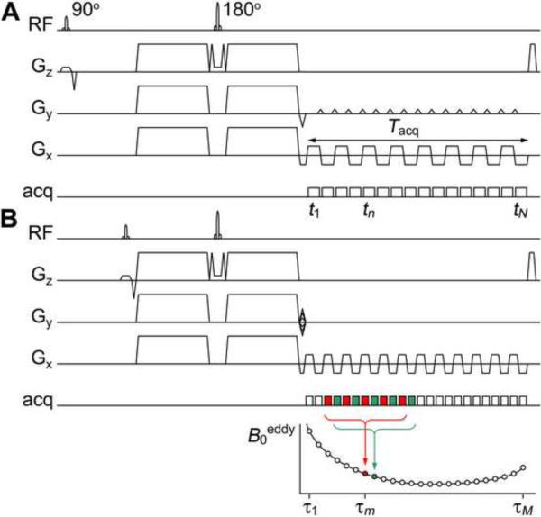
The first step consists in acquiring a series of dynamic B0eddy maps for a range of time points spanning the DTI readout window and for each DW direction. To this end, a train of diffusion-weighted asymmetric spin-echo images are acquired with a multiecho spin-echo pulse sequence at different time points τm = τ1, …, τM spanning the readout duration Tacq of the DTI scan (Fig. 1). The resulting phase images are then unwrapped along the time dimension and, for each time point τm and DW direction d, fitted with:
| [1] |
where ϕ0 is a constant, γ is the gyromagnetic ratio, and t is a 5-point moving window {τm−4, τm−2, τm, τm+2, τm+4}, to derive a time series of B0eddy maps. The phase unwrapping and fitting are performed separately on odd and even echoes, which are acquired with readout gradients of opposite polarity, to avoid errors due to off-resonance effects (as shown in different colors in Fig. 1B). Although dynamic B0eddy maps could also be generated from successive pairs of phase images, we found that a 5-point moving window provides a higher SNR with no significant loss in temporal resolution, because B0eddy varies relatively slowly with respect to the echo spacing.
Fig. 1.
DTI (A) and dynamic B0eddy mapping (B) pulse sequence diagrams (RF: radiofrequency excitation; Gz, Gy, Gx: slice-select, phase-encoding, and readout gradients; acq: data acquisition). For clarity, only a limited number of echoes are shown in both (A) and (B). In reality, the width of the moving window is much smaller in comparison to the DTI readout duration Tacq. Odd and even echoes are shown in different colors in (B).
Since eddy currents depend on the DW gradients, but not on the subject being imaged, this dynamic B0eddy mapping is performed with the same DW gradients as the DTI scan, but only once on a phantom. The resulting B0eddy maps can then be repeatedly used to correct for artifacts caused by time-varying eddy currents in all previous and subsequent DTI scans acquired with the same protocol, without requiring any additional scan time. Furthermore, since B0eddy varies slowly in space, the B0eddy mapping can be performed at a lower spatial resolution than that of the DTI scan, which can be traded for a higher SNR and a higher temporal resolution (because of the reduced echo spacing) to accurately measure the temporal dependence of B0eddy within Tacq (Fig. 1).
To remove any common B0 inhomogeneities not caused by the eddy currents, such as those due to imperfect shimming, non-diffusion-weighted B0 maps of the phantom are also acquired and are subtracted from the diffusion-weighted B0 maps for all time points and DW directions. To improve the SNR, the resulting B0eddy maps are then fitted with a third-order polynomial function in space within the phantom, which was previously shown to be an adequate model (Truong et al., 2008), and extrapolated outside the phantom. However, in contrast to existing eddy current correction methods, the proposed method does not make any assumptions regarding the temporal dependence of B0eddy, so that it can effectively correct for artifacts caused by any time-varying eddy currents.
2.2 Static B0susc mapping
The second step consists in acquiring a static B0susc map by using the same pulse sequence and post-processing method as for the dynamic B0eddy mapping, but by simultaneously fitting all odd or even echoes with:
| [2] |
(instead of using a moving window) and by averaging the two resulting B0susc maps. In contrast to eddy currents, since susceptibility effects depend on the subject being imaged, but not on the DW gradients, this B0susc mapping is performed in vivo, but without diffusion-weighting. In addition, since B0susc can vary rapidly in space, but does not vary in time, the B0susc mapping is performed at the same spatial resolution as that of the DTI scan, but with fewer echoes than the dynamic B0eddy mapping.
2.3 Dynamic off-resonance correction
The third step consists in using the static B0susc map and dynamic B0eddy maps to correct for distortions and blurring artifacts in the DTI images caused by both susceptibility effects and time-varying eddy currents (Fig. 2). Specifically, for each time point tn = t1, …, tN corresponding to the acquisition of a ky line in k-space (where y denotes the phase-encoding direction) and for each DW direction d, the uncorrected DTI image is multiplied by exp[−iϕ(x, tn, d)], where
| [3] |
Each of these N images is Fourier transformed to k-space and the nth ky line (acquired at time tn) is extracted from the nth k-space to form a new k-space, which is inverse Fourier transformed to yield the corrected image. If partial Fourier imaging is used, the partial Fourier reconstruction is performed at the last step.
Fig. 2.
Schematic diagram of the dynamic off-resonance correction method (FT: Fourier transform, PF: partial Fourier reconstruction (optional), FT−1: inverse Fourier transform).
Note that the acquired B0susc and B0eddy maps require further processing before they can be used in Eq. [3]. First, the B0eddy maps are interpolated in space and downsampled in time (from {τm = τ1, …, τM} to {tn = t1, …, tN}) to match the spatial resolution and echo spacing of the DTI scan. Second, the B0susc and B0eddy maps, which are initially in undistorted coordinates, need to be transformed into distorted coordinates, so that they are coregistered with the uncorrected DTI images. In practice, we found it sufficient to perform this transformation only on the B0susc map, because the B0eddy maps vary slowly in space so that the difference is negligible. To this end, each undistorted asymmetric spin-echo image acquired for the B0susc mapping is distorted by using the same procedure as above, except that Eq. [3] is replaced by
| [4] |
where B0susc(x) is the undistorted B0susc map. The resulting distorted asymmetric spin-echo images are then used to generate a distorted B0susc map as described in section 2.2.
2.4 Experiments
We studied three healthy volunteers, who provided written informed consent as approved by our Institutional Review Board, on a 3 T Excite MRI scanner (GE Healthcare, Milwaukee, WI) equipped with an eight-channel phased-array head coil and a gradient system with 40 mT/m maximum amplitude and 150 T/m/s slew rate. High-order shimming was applied to minimize the global B0 inhomogeneity. Axial DTI images of the brain were acquired with a single-shot spin-echo EPI pulse sequence and the following parameters: repetition time (TR) = 5 s, TE = 73 ms (minimum), field-of-view = 24×24 cm, matrix size = 96×96 (interpolated to 128×128 by zero padding), slice thickness = 2.5 mm, number of slices = 20, frequency-encoding direction = right/left, partial Fourier encoding = 5/8, echo spacing = 944 μs, and Tacq = 56 ms.
Static B0susc mapping was performed with TR = 2 s, TE = 32 ms, and number of echoes = 24, whereas dynamic B0eddy mapping was performed with TR = 2.5 s, TE = 55 ms, matrix size = 48×48, slice thickness = 5 mm, number of slices = 10, number of echoes = 96, and echo spacing = 624 μs on a 20-cm diameter spherical gel phantom. For the DTI scan and B0eddy mapping, DW gradients were applied with the following parameters: amplitude = 39.4 mT/m, duration (δ) = 22 ms, separation (Δ) = 26.7 ms, b-factor = 1000 s/mm2, and 15 DW directions. To assess the temporal stability of the eddy currents, the B0eddy mapping was repeated four times over a period of six months. For anatomical reference, high-resolution T2-weighted images of the brain were also acquired with a fast spin-echo pulse sequence and TR = 3 s, TE = 86 ms, matrix size = 256×256, and slice thickness = 2.5 mm.
For comparison with the proposed dynamic off-resonance correction, a static off-resonance correction was also performed by using the same B0susc(x) map, but time-averaged B0eddy(x, d) maps instead of the dynamic B0eddy(x, t, d) maps. For each of the three DTI datasets obtained with no correction, static correction, and dynamic correction, FA maps were computed and color-coded according to the direction of the first eigenvector. In addition, maps of the total FA error were computed as (FAuncorrected – FAdynamic)/FAdynamic and maps of the residual FA error after static correction were computed as (FAstatic – FAdynamic)/FAdynamic, where FAuncorrected, FAstatic, and FAdynamic are the FA values with no correction, static correction, and dynamic correction, respectively. All image reconstruction and analysis were performed in Matlab (The MathWorks, Natick, MA).
3. Results and Discussion
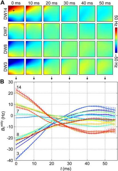
Representative B0eddy maps at different time points within the DTI readout window (Fig. 3A) as well as dynamic B0eddy time courses in a given voxel (Fig. 3B) for different DW directions show that B0eddy varies substantially with space, time, and DW direction. In particular, these results demonstrate that B0eddy cannot simply be modeled as a constant within the DTI readout window, as assumed in the vast majority of existing eddy current correction methods. Furthermore, the zero-crossing clearly shows that it cannot be accurately characterized by a single exponential decay either, as assumed in the twice-refocused spin-echo method, because of the interaction of multiple eddy currents with different amplitudes and time constants. The range of variation of B0eddy within the DTI readout window is 2 to 3 orders of magnitude larger than its temporal average for all DW directions.
Fig. 3.
(A) Representative B0eddy maps at different time points within the DTI readout window for 4 out of 15 DW directions. (B) Dynamic B0eddy time courses in the voxel marked as + in (A) for all DW directions (mean ± standard error of the mean for four repeated measurements performed over six months).
A representative uncorrected diffusion-weighted image shows severe distortions and blurring artifacts caused by susceptibility effects and time-varying eddy currents, particularly near the anterior horns of the lateral ventricles and in the frontal lobes (Fig. 4A, arrows). These artifacts are only partially reduced with the static off-resonance correction (Fig. 4B), whereas they are completely eliminated with the proposed dynamic off-resonance correction (Fig. 4C).
Fig. 4.
Representative diffusion-weighted images with no correction (A), static correction (B), and dynamic correction (C). Contour lines generated from an undistorted fast spin-echo image are overlaid on one hemisphere. The arrows show severe distortions and blurring artifacts caused by susceptibility effects and time-varying eddy currents.
Similarly, uncorrected FA maps in three representative slices show severe susceptibility-induced distortions, particularly near the genu of the corpus callosum (Fig. 5A, dashed lines), as well as eddy current-induced FA errors, most prominently at the anterior and posterior edges of the brain (arrows). The static off-resonance correction can only correct for the susceptibility-induced distortions as well as the eddy current-induced FA errors at the posterior edge of the brain, but not at the anterior edge (Fig. 5B, arrows). In contrast, the proposed dynamic off-resonance correction can effectively correct for all artifacts (Fig. 5C).
Fig. 5.
Color-coded FA maps in three representative slices with no correction (A), static correction (B), and dynamic correction (C) (red: right–left, green: anterior–posterior, blue: superior–inferior). The dashed lines and arrows highlight regions with severe susceptibility-induced distortions and eddy current-induced FA errors, respectively. The middle slice is the same as the one shown in Fig. 4.
Although the susceptibility- and eddy current-induced artifacts may initially appear fairly localized, a representative map of the total FA error reveals that they are actually extensive throughout the whole brain (Fig. 6A), with (75.9 ± 1.4)% of the voxels in the brain having an absolute FA error larger than 10% (Fig. 6C, solid line). Similarly, a map of the residual FA error shows that even after static off-resonance correction, residual eddy current-induced artifacts are still widespread (Fig. 6B), with (26.2 ± 1.9)% of the voxels in the brain having an absolute FA error larger than 10% (Fig. 6C, dashed line).
Fig. 6.
Maps of the total FA error (A) and residual FA error after static off-resonance correction (B) in the same slice as the middle slice of Fig. 5. (C) Percentage of voxels in the brain having an absolute FA error exceeding a given value (mean ± standard error of the mean across subjects).
Dynamic B0eddy mapping repeated four times over a period of six months yielded virtually identical B0eddy maps (Fig. 3B, error bars), whereas dynamic off-resonance correction of the same DTI data with these four sets of B0eddy maps resulted in virtually identical FA maps (e.g., FA = 0.744 ± 0.004 in the genu of the corpus callosum). These results demonstrate that the eddy currents induced by the DW gradients remain very stable, even over a long time period, so that the B0eddy mapping can be performed very infrequently (i.e., no more than once or twice a year).
Furthermore, although in this study the B0eddy maps were acquired with the same DW gradients as the DTI scan, it is in principle possible to use linear combinations of these B0eddy maps for the correction of DTI data acquired with other DW gradient amplitudes and directions as well, provided that the eddy currents induced by gradients applied along the x, y, and z axes can be modeled as a linear system (Zhuang et al., 2006), which can be verified in future studies.
Since the focus of this study was the correction of artifacts caused by time-varying eddy currents, the static B0susc mapping was performed with the same multiecho spin-echo sequence as the dynamic B0eddy mapping (but without diffusion-weighting) for convenience. However, more efficient B0susc mapping methods, such as the acquisition of two asymmetric spin-echo EPI images at different TEs, can also be used to further reduce the scan time.
In contrast to existing eddy current correction methods, the proposed method can effectively correct for artifacts caused by any time-varying eddy currents. Furthermore, it does not require any additional scan time as compared to static B0 mapping methods, is much more efficient than reversed gradient methods, and does not suffer from the SNR penalty of twice-refocused spin-echo methods. In this study, we used a single-shot EPI pulse sequence because it is the most commonly used for DTI. However, the proposed method is also compatible with multishot acquisitions, other imaging sequences (e.g., spiral or radial imaging), as well as parallel imaging techniques.
This method will be beneficial in longitudinal studies of the same subjects, because different head positions with respect to the B0 field or the gradient axes will lead to different susceptibility- and eddy current-induced artifacts, resulting in different DTI metrics, if not corrected for. It will also be particularly beneficial in multicenter studies, which may use different scanner models and/or manufacturers with potentially very different eddy currents, even though the scan parameters are the same. Finally, it will benefit both basic neuroscience research and clinical applications such as presurgical planning, for which a high spatial fidelity is especially critical.
4. Conclusions
The results of this study demonstrate that B0eddy remains very consistent over time, but varies substantially within the DTI readout window and cannot be accurately modeled as a constant or a single exponential decay, as assumed in nearly all existing eddy current correction methods. The proposed dynamic B0 mapping and off-resonance correction method can measure the exact spatial, temporal, and DW direction dependence of B0susc and B0eddy to effectively and efficiently correct for the severe distortions, blurring, and misregistration artifacts caused by susceptibility effects and time-varying eddy currents, thereby leading to a high spatial fidelity and accuracy in the resulting DTI metrics.
4. Highlights
Susceptibility effects and time-varying eddy currents cause severe artifacts in DTI
Eddy current-induced magnetic fields vary substantially within the readout window
They cannot be modeled as a constant or a monoexponential decay, as usually assumed
A novel dynamic off-resonance correction method is proposed to address these issues
This method can effectively and efficiently correct for both types of artifacts
Acknowledgments
We thank Susan Music for her assistance with MRI scanning. This work was, in part, supported by grants NS41328, NS65344, EB09483, and EB12586 from the National Institutes of Health.
Footnotes
Publisher's Disclaimer: This is a PDF file of an unedited manuscript that has been accepted for publication. As a service to our customers we are providing this early version of the manuscript. The manuscript will undergo copyediting, typesetting, and review of the resulting proof before it is published in its final citable form. Please note that during the production process errors may be discovered which could affect the content, and all legal disclaimers that apply to the journal pertain.
References
- Ardekani S, Sinha U. Geometric distortion correction of high-resolution 3 T diffusion tensor brain images. Magn. Reson. Med. 2005;54:1163–1171. doi: 10.1002/mrm.20651. [DOI] [PubMed] [Google Scholar]
- Basser PJ, Mattiello J, LeBihan D. MR diffusion tensor spectroscopy and imaging. Biophys. J. 1994;66:259–267. doi: 10.1016/S0006-3495(94)80775-1. [DOI] [PMC free article] [PubMed] [Google Scholar]
- Bernstein MA, King KF, Zhou XJ. Handbook of MRI pulse sequences. Elsevier Academic Press; Burlington, MA: 2004. [Google Scholar]
- Bodammer N, Kaufmann J, Kanowski M, Tempelmann C. Eddy current correction in diffusion-weighted imaging using pairs of images acquired with opposite diffusion gradient polarity. Magn. Reson. Med. 2004;51:188–193. doi: 10.1002/mrm.10690. [DOI] [PubMed] [Google Scholar]
- Chen B, Guo H, Song AW. Correction for direction-dependent distortions in diffusion tensor imaging using matched magnetic field maps. NeuroImage. 2006;30:121–129. doi: 10.1016/j.neuroimage.2005.09.008. [DOI] [PubMed] [Google Scholar]
- Ciccarelli O, Catani M, Johansen-Berg H, Clark C, Thompson A. Diffusion-based tractography in neurological disorders: concepts, applications, and future developments. Lancet Neurol. 2008;7:715–727. doi: 10.1016/S1474-4422(08)70163-7. [DOI] [PubMed] [Google Scholar]
- Embleton KV, Haroon HA, Morris DM, Lambon Ralph MA, Parker GJM. Distortion correction for diffusion-weighted MRI tractography and fMRI in the temporal lobes. Human Brain Mapp. 2010;31:1570–1587. doi: 10.1002/hbm.20959. [DOI] [PMC free article] [PubMed] [Google Scholar]
- Finsterbusch J. Double-spin-echo diffusion weighting with a modified eddy current adjustment. Magn. Reson. Imaging. 2010;28:434–440. doi: 10.1016/j.mri.2009.12.004. [DOI] [PubMed] [Google Scholar]
- Frank LR, Jung Y, Inati S, Tyszka JM, Wong EC. High efficiency, low distortion 3D diffusion tensor imaging with variable density spiral fast spin echoes (3D DW VDS RARE) NeuroImage. 2010;49:1510–1523. doi: 10.1016/j.neuroimage.2009.09.010. [DOI] [PMC free article] [PubMed] [Google Scholar]
- Gallichan D, Andersson JLR, Jenkinson M, Robson MD, Miller KL. Reducing distortions in diffusion-weighted echo planar imaging with a dual-echo blip-reversed sequence. Magn. Reson. Med. 2010;64:382–390. doi: 10.1002/mrm.22318. [DOI] [PubMed] [Google Scholar]
- Mori S, Zhang J. Principles of diffusion tensor imaging and its applications to basic neuroscience research. Neuron. 2006;51:527–539. doi: 10.1016/j.neuron.2006.08.012. [DOI] [PubMed] [Google Scholar]
- Reese TG, Heid O, Weisskoff RM, Wedeen VJ. Reduction of eddy-current-induced distortion in diffusion MRI using a twice-refocused spin echo. Magn. Reson. Med. 2003;49:177–182. doi: 10.1002/mrm.10308. [DOI] [PubMed] [Google Scholar]
- Rohde GK, Barnett AS, Basser PJ, Marenco S, Pierpaoli C. Comprehensive approach for correction of motion and distortion in diffusion-weighted MRI. Magn. Reson. Med. 2004;51:103–114. doi: 10.1002/mrm.10677. [DOI] [PubMed] [Google Scholar]
- Shen Y, Larkman DJ, Counsell S, Pu IM, Edwards D, Hajnal JV. Correction of high-order eddy current induced geometric distortion in diffusion-weighted echo-planar images. Magn. Reson. Med. 2004;52:1184–1189. doi: 10.1002/mrm.20267. [DOI] [PubMed] [Google Scholar]
- Truong T-K, Chen B, Song AW. Integrated SENSE DTI with correction of susceptibility- and eddy current-induced geometric distortions. NeuroImage. 2008;40:53–58. doi: 10.1016/j.neuroimage.2007.12.001. [DOI] [PMC free article] [PubMed] [Google Scholar]
- Zhuang J, Hrabe J, Kangarlu A, Xu D, Bansal R, Branch CA, Peterson BS. Correction of eddy-current distortions in diffusion tensor images using the known directions and strengths of diffusion gradients. J. Magn. Reson. Imaging. 2006;24:1188–1193. doi: 10.1002/jmri.20727. [DOI] [PMC free article] [PubMed] [Google Scholar]


