Abstract
The aim of this study was to design a tele-radiology imaging system for rapid emergency care via mobile networks and to assess the diagnostic feasibility of the Joint Photographic Experts Group 2000 (JPEG2000) radiological imaging using portable devices. Rapid patient information and image exchange is helpful to make clinical decisions. We assessed the usefulness of the mobile tele-radiology system by measuring both a quantitative method, PNSR calculation, for image qualities, and its transmission time via mobile networks in different mobile networks, respectively; code division multiple access evolution-data optimized, wireless broadband, and high-speed downlink packet access; and the feasibility of the JPEG2000 computed tomography (CT) images by qualitatively assessing with the Alberta stroke program early CT score method with 12 CT image cases (seven normal and five abnormal cases). We found that the quality of the JPEG2000 radiological images was satisfied quantitatively and was judged as acceptable qualitatively at 5:1 and 10:1 compression levels for the mobile tele-radiology imaging system. The JPEG2000-format radiological images achieved a fast transmission while maintaining a diagnosis quality on a portable device via mobile networks. Unfortunately, a PDA device, having a limited screen resolution, posed difficulties in reviewing the JPEG2000 images regardless of the compression levels. An ultra mobile PC was preferable to study the medical image. The mobile tele-radiology imaging systems supporting JPEG2000 image transmission can be applied to actual emergency care services under mobile computing environments.
Key words: Mobile tele-radiology, JPEG2000, radiological CT image, emergency care
Introduction
Emergency situations can unexpectedly occur anytime and anyplace. Making a rapid clinical decision is a crucial factor in emergency medical care. A mobile emergency tele-radiology system can be helpful to support rapid clinical decisions in emergency situations by specialists having difficulty accessing a stationary system outside of hospitals.1–3 Radiological images need to be interpreted by trained radiologists to achieve an accurate diagnosis. Immediate communications must be accomplished by rapid image transfer so that specialists can understand thoroughly the radiological results. Unfortunately, if the special radiologist is outside the hospitals or a resident is not able to interpret the images accurately, the emergency dispatch could be delayed. Such delays may result in emergency patient fatality because of deferred image interpretations. Therefore, we designed the mobile emergency patient information and imaging communication system that provide rapid access to the patient information and images either inside or outside of hospitals via a wireless mobile communication link such as code division multiple access evolution-data optimized (CDMA 1x-EVDO), wireless broadband (WIBRO), and high-speed downlink packet access (HSDPA) networks.
Many studies about radiological image quality evaluation for tele-radiology systems were assessed according to the Joint Photographic Experts Group (JPEG) compression ratios because preserving acceptable qualities of medical images is crucial. However, in order to decrease misinterpretation upon a diagnostically relevant loss of image quality with an efficient compression method and increase transmission performances varying wireless mobile networks to support the rapid reviewing and the demand of referring physicians to have quick access to the image data of their patients even from outside the hospital using a mobile device, evaluating influence of compression on radiological images is needed to be studied. Therefore, in order to overcome the data bandwidth limitation at wireless communication links and improve the efficiency of mobile tele-radiology systems for transferring massive Digital Imaging and Communications in Medicine (DICOM) medical images, we adopted the JPEG2000 coding method.4,5
In this study, we designed an integrated mobile tele-radiology imaging system. In emergency departments, emergency physicians can acquire radiological images and patient information and transmit those images via Web service operation, and remote clinicians can access those compressed images and patient information using portable devices through various mobile links. We assessed the feasibility of using this system for the rapid transmission of emergency patient information and images. Also, we investigated its usefulness for making an effective medical decision in an emergency with JPEG2000 images displayed on a portable device.
Mobile Tele-Radiology System
System Configuration
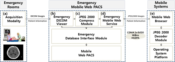
The overall system configuration was described as shown in Figure 1. The designed system was composed of five components, representatively; (a) acquisition modality, (b) emergency DICOM viewer, (c) JPEG2000 compress module, (d) emergency mobile Web service, and (e) mobile browser as end-user mobile devices. The acquisition modality (a) is the CT modality (LightSpeed Plus; GE Healthcare, Milwaukee, WI, USA). The emergency DICOM viewer (b) was designed to allow review of patient information and images from the acquisition modality. The JPEG2000 compression module (c) was composed to convert DICOM images to JPEG 2000 format images and to record them on the mobile Web PACS system including patient information. The LEADTOOLS® (LEAD Technologies, Inc., NC, USA) JPEG2000 encoding library and Korean-DICOM (KDICOM) library were customized for this system. The mobile Web PACS system (d) was composed of the mobile Web service using a mobile internet toolkit with a Microsoft.NET platform in order to support the service in the mobile web browser regardless of the type of portable embedded system, mobile phone, and embedded operating system. We also included security using a log-in feature to provide an effective mobile Web service. The Web application software (e) in the end-user devices was developed using C# programming language in conjunction with the Windows Mobile 5 software development kit (SDK).
Fig. 1.
The configuration of a mobile tele-radiology imaging system composed of an emergency mobile Web PACS and mobile system with five components; a acquisition modality, b emergency DICOM viewer, c JPEG2000 compress module, d emergency mobile Web service, and e mobile browser as end-user mobile devices.
System Operation
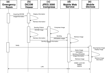
The designed mobile tele-radiology imaging system can support transmitting images and patient information via Web services operation and displayed on mobile devices in real time. This system was designed to transmit radiological images from a physician at an emergency room to a specialist located outside a hospital for emergency consultations. The radiological images were compressed with the JPEG2000 format in real time, and transmitted via various mobile networks such as CDMA 1x EV-DO, WIBRO, and HSDPA. The system operation and workflow were illustrated as a sequence diagram as shown in Figure 2.
Fig. 2.
The possible service scenario for mobile tele-radiology imaging system between emergency room and remote physicians via mobile networks.
The possible scenarios of emergency image transfer services are described as follows:
When an emergency patient arrived at an emergency department, the urgency of treatment and crucial symptom were determined by residents.
In spite of initial first aid and radiography, the residents could not achieve an accurate diagnosis on images through our designed Web PACS viewer because of an inability of the radiological image to convey the needed clinical information.
The acquired image from emergency patients were stored to the Web PACS system and simultaneously transmitted to a remote radiologist as JPEG2000 compressed format via mobile networks.
The remote radiologist studied the transmitted images along with detailed patient information and relevant images later by accessing the Web PACS system with a portable device.
Materials and Methods
System Design
JPEG2000
The two-dimensional, wavelet-based, lossy image compression algorithm, JPEG2000, has been implemented in the DICOM standard since the year 2000.6
JPEG2000 provides high compression efficiency valuable in a medical imaging PACS environment. JPEG2000 can provide significantly higher compression ratios than the JPEG technique with less degradation and distortion. JPEG2000 adopts embedded block coding with optimal truncation as a source coder. In particular, a two-dimensional discrete wavelet transform is at the heart of JPEG2000. For image compression, a JPEG2000 codec (Aware JPEG; Aware, Bedford, MA) was used. The original images from the CT modality were of 16-bit depth and 512 × 512 pixels. The controlled compression levels of JPEG2000 codec were 5:1, 10:1, 20:1, 30:1, 40:1, 50:1, and 100:1, respectively.
ASPECTS
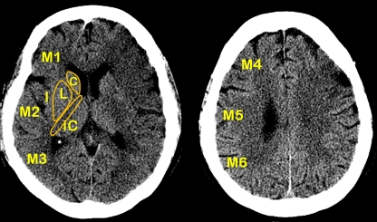
The Alberta stroke program early CT score (ASPECTS) was developed to offer the reliability and utility of a standard CT examination with a reproducible grading system to assess early ischemic changes on pretreatment CT studies in patients with acute ischemic stroke of anterior circulation.7 The score divides the middle cerebral artery (MCA) territory into ten regions of interest as shown in Figure 3.8 The ASPECTS is a topographic scoring system applying a quantitative approach that does not ask physicians to estimate volumes from two-dimensional images. The amount of clinical discrepancy can be evaluated using scoring of a ten-point scale. A score of zero indicated diffuse ischemic involvement throughout the territory of middle cerebral artery in brain CT images, and contrary to ten evidenced normal status in brain CT images. In this study, for the assessment of image quality, we used APSECTS as a scoring system to compare between the original image and compressed image.
Fig. 3.
The ten regions for APSECTS on a brain CT image; M1 anterior MCA cortex; M2 MCA cortex lateral to insular ribbon; M3 posterior MCA cortex; M4, M5, and M6 are anterior, lateral, and posterior MCA territories immediately superior to M1, M2, and M3, rostral to basal ganglia; I insular ribbon; L lentiform; C caudate, and IC internal capsule.
System Evaluation
We investigated the usefulness of the designed mobile tele-radiology imaging system with two kinds of objective experiments.
Calculating the peak signal-to-noise ratio (PNSR) values in radiological images with respect to different compression ratios in JPEG 2000 format
Measuring the image transmission times at mobile networks, such as CDMA 1x EVDO, WIBRO, and HSDPA in terms of different compression ratios in JPEG2000 format
The PSNR method is the most widely used image quality metrics and was calculated from the expression;
 |
where I(x, y) is the original image, J(x, y) is the decompressed image, and M and N are the dimensions of the image.
For evaluating the clinical diagnosis ability on portable devices subjectively, for the feasibility of medical images, considering an emergency involving an ischemia brain, we reviewed brain DICOM images of radiological lesions in 12 (seven normal and five abnormal) cases produced by the computerized tomography to compare between the original image and compressed image. The original image was displayed on a single monitor (CV812R, 18.1-in. CRT panel, TOTOKU Co., Japan). Compressed images were displayed on an ultra mobile PC (UMPC, VGN-UX58LN, 4.5-in. LCD panel, Sony Co., Japan). The monitor resolutions of both the single monitor and UMPC were 1,280 × 1,024 pixels, and 1,024 × 600 pixels, respectively. The image reviewing time was 10 s at each study.
Results
Usefulness of Mobile Tele-Radiology System
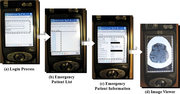
The mobile system could query patient information and images on the mobile Web PACS instantly. The application user interface of the designed mobile system was handy to operate based on a Web browser application, as shown in Figure 4. The image transmission of mobile tele-radiology system was composed of four supported functional steps; (1) In a log-in process, only related or available doctors could review and query the emergency medical information through the user authentication process. When an incorrect identification or password was entered, guidelines for the correct process were suggested on the Web browser of mobile devices, (2) after the user authentication, the related patient’s name were listed up on a mobile device, (3) the patient information from a database in the hospital was displayed, (4) clicking the “image” button on a web browser, and the JPEG2000 radiological image was displayed.
Fig. 4.
The application user interface of the designed mobile system; a log-in process, b patients’ name list, c patient’s information, and d patient’s image display.
The test results for a PSNR calculation of JPEG 2000 images in terms of compression ratios were tabulated in Table 1. Higher compression levels provided quantitatively lower image quality. Comparatively, the PSNR values of 5:1 and 10:1 compression images having 52.64 dB and 40.54 dB, respectively, were higher than others. High PSNR values (≥40 dB) for compression ratios up to 10:1 were clinically reasonable compression ratios according to the preliminary studies in this study.
Table 1.
The result values of the PSNR test for various compression ratios
| Compression ratios | PSNR |
|---|---|
| 5:1 | 52.64 ± 0.22 |
| 10:1 | 40.54 ± 0.17 |
| 20:1 | 31.12 ± 0.25 |
| 30:1 | 26.47 ± 0.24 |
| 40:1 | 24.18 ± 0.53 |
| 50:1 | 22.70 ± 0.32 |
| 100:1 | 19.08 ± 0.55 |
Moreover, Table 2 shows the test results for transmission time of JPEG2000 images in terms of compression ratios at CDMA 1x EVDO, WIBRO, and HSDPA networks, respectively. The average transmission time was measured by 30 time repetitive transmission trials and calculated with five images randomly selected among 12 brain CT images. Higher compression ratios provided faster transmission speeds. A significant difference of transmission time (over 1 s) was realized at the 5:1 compression ratio, as compared to the original. Transmission time of JPEG2000 images at a 5:1 compression ratio was 1.23 s in CDMA 1x EVDO, 1.18 s in WIBRO, and 1.17 s in HSDPA networks, respectively. The transmission time of the HSDPA network was the fastest between both CDMA 1x EVDO and WIBRO networks, slightly. While considering the satisfaction of the PSNR test and the measured transmission time, the radiological CT images with the compression level of 5:1 was acceptable in this study.
Table 2.
Average transmission time using various compression ratios from hospital to portable mobile device—brain CT image (transmission trial number was 30)
| CDMA 1x EVDO | WIBRO | HSDPA | |||||
|---|---|---|---|---|---|---|---|
| Compression ratios | File size (KB) | Theoretical transmission time (seconds) | Measured transmission time (seconds) | Theoretical transmission time (seconds) | Measured transmission time (seconds) | Theoretical transmission time (seconds) | Measured transmission time (seconds) |
| Original | 524 | 1.74 (at 2.4 Mbps) | 6.18 | 1 (at 4 Mbps) | 5.91 | 0.3 (at 14 Mbps) | 5.81 |
| 5:1 | 105 | 0.35 (at 2.4 Mbps) | 1.23 | 0.2 (at 4 Mbps) | 1.18 | 0.06 (at 14 Mbps) | 1.17 |
| 10:1 | 53 | 0.17 (at 2.4 Mbps) | 0.62 | 0.1 (at 4 Mbps) | 0.60 | 0.03 (at 14 Mbps) | 0.59 |
| 20:1 | 26.2 | 0.08 (at 2.4 Mbps) | 0.30 | 0.05 (at 4 Mbps) | 0.29 | 0.01 (at 14 Mbps) | 0.29 |
| 30:1 | 17.5 | 0.05 (at 2.4 Mbps) | 0.20 | 0.03 (at 4 Mbps) | 0.19 | <0.01 (at 14 Mbps) | 0.19 |
| 40:1 | 13.2 | 0.04 (at 2.4 Mbps) | 0.16 | 0.02 (at 4 Mbps) | 0.15 | <0.01 (at 14 Mbps) | 0.15 |
| 50:1 | 10.6 | 0.03 (at 2.4 Mbps) | 0.13 | 0.02 (at 4 Mbps) | 0.12 | <0.01 (at 14 Mbps) | 0.12 |
| 100:1 | 5.4 | 0.01 (at 2.4 Mbps) | 0.06 | 0.01 (at 4 Mbps) | 0.06 | <0.01 (at 14 Mbps) | 0.06 |
Feasibility of Image Quality
Allowing for the results of the measured transmission times and PSNR values, related compressed images with 5:1 and 10:1 ratios were included in the ASPECTS test. Tables 3, 4, and 5 show the test results of ASPECTS scoring experiment for the 12 original brain CT images with original images, 5:1 compression image, and 10:1 compression image, respectively. When the total score was 10 it was considered as a normal status, otherwise nearing to 0 as an abnormal status. The measured scores of ASPECTS in original image accorded with 5:1 and 10:1 compression image in terms of 12 brain CT images. As shown in Table 6, correspondence about the ASPECTS test was satisfied in terms of different compression ratios with brain CT images. There was no significant clinical discrepancy on according to the ASPECT scores of the brain CT images as shown in Table 6. Therefore, subjective of image quality did not differ significantly between original and both 5:1 and 10:1 compressed images in the APSECTS test.
Table 3.
The ASPECTS test results of the original image
| File no. | ASPECT scores according to ten different regions at brain CT images | Normality | ||||||||||
|---|---|---|---|---|---|---|---|---|---|---|---|---|
| M1 | M2 | M3 | M4 | M5 | M6 | C | L | IC | I | Total | ||
| 1 | 1 | 0 | 0 | 0 | 0 | 0 | 0 | 0 | 1 | 0 | 2 | N |
| 2 | 1 | 1 | 1 | 1 | 1 | 1 | 1 | 0 | 1 | 1 | 9 | Y |
| 4 | 1 | 1 | 1 | 1 | 1 | 1 | 1 | 1 | 1 | 0 | 9 | Y |
| 5 | 1 | 1 | 0 | 1 | 1 | 0 | 1 | 0 | 1 | 0 | 6 | N |
| 6 | 0 | 0 | 0 | 0 | 0 | 0 | 0 | 0 | 1 | 0 | 1 | N |
| 7 | 1 | 1 | 1 | 1 | 1 | 1 | 1 | 1 | 1 | 0 | 9 | Y |
| 8 | 0 | 0 | 0 | 0 | 0 | 0 | 0 | 0 | 1 | 0 | 1 | N |
| 9 | 1 | 1 | 1 | 0 | 0 | 0 | 1 | 1 | 1 | 0 | 6 | N |
| 10 | 1 | 1 | 1 | 1 | 1 | 1 | 1 | 1 | 1 | 1 | 10 | Y |
| 11 | 0 | 1 | 1 | 1 | 1 | 1 | 1 | 1 | 1 | 0 | 8 | N |
| 12 | 1 | 1 | 1 | 1 | 1 | 1 | 1 | 0 | 1 | 0 | 8 | Y |
Table 4.
The ASPECTS test results of the 5:1 compression image
| File no. | ASPECT scores according to ten different regions at brain CT images | Normality | ||||||||||
|---|---|---|---|---|---|---|---|---|---|---|---|---|
| M1 | M2 | M3 | M4 | M5 | M6 | C | L | IC | I | Total | ||
| 1 | 1 | 0 | 0 | 0 | 0 | 0 | 0 | 0 | 1 | 0 | 2 | N |
| 2 | 1 | 1 | 1 | 1 | 1 | 1 | 1 | 0 | 1 | 0 | 8 | Y |
| 3 | 1 | 1 | 1 | 1 | 1 | 1 | 1 | 0 | 1 | 1 | 9 | Y |
| 4 | 1 | 1 | 1 | 1 | 1 | 1 | 1 | 1 | 1 | 0 | 9 | Y |
| 5 | 1 | 1 | 0 | 1 | 1 | 0 | 1 | 0 | 1 | 0 | 6 | N |
| 6 | 0 | 0 | 0 | 0 | 0 | 0 | 0 | 0 | 1 | 0 | 1 | N |
| 7 | 1 | 1 | 1 | 1 | 1 | 1 | 1 | 1 | 1 | 0 | 9 | Y |
| 8 | 0 | 0 | 0 | 0 | 0 | 0 | 0 | 0 | 1 | 0 | 1 | N |
| 9 | 1 | 1 | 1 | 0 | 0 | 0 | 1 | 1 | 1 | 0 | 6 | N |
| 10 | 1 | 1 | 1 | 1 | 1 | 1 | 1 | 1 | 1 | 1 | 10 | Y |
| 11 | 0 | 0 | 1 | 1 | 1 | 1 | 1 | 1 | 1 | 0 | 7 | N |
| 12 | 1 | 1 | 1 | 1 | 1 | 1 | 1 | 0 | 1 | 0 | 8 | Y |
Table 5.
The ASPECTS test results of the 10:1 compression image
| File no. | ASPECT scores according to ten different regions at brain CT images | Normality | ||||||||||
|---|---|---|---|---|---|---|---|---|---|---|---|---|
| M1 | M2 | M3 | M4 | M5 | M6 | C | L | IC | I | Total | ||
| 1 | 1 | 0 | 0 | 0 | 0 | 0 | 0 | 0 | 1 | 0 | 2 | N |
| 2 | 1 | 1 | 1 | 1 | 1 | 1 | 1 | 0 | 1 | 0 | 8 | Y |
| 3 | 1 | 1 | 1 | 1 | 1 | 1 | 1 | 0 | 1 | 1 | 9 | Y |
| 4 | 1 | 1 | 1 | 1 | 1 | 1 | 1 | 1 | 1 | 0 | 9 | Y |
| 5 | 1 | 1 | 0 | 1 | 1 | 0 | 1 | 0 | 1 | 0 | 6 | N |
| 6 | 0 | 0 | 0 | 0 | 0 | 0 | 0 | 0 | 1 | 0 | 1 | N |
| 7 | 1 | 1 | 1 | 1 | 1 | 1 | 1 | 1 | 1 | 0 | 9 | Y |
| 8 | 0 | 0 | 0 | 0 | 0 | 0 | 0 | 0 | 1 | 0 | 1 | N |
| 9 | 1 | 1 | 1 | 0 | 0 | 0 | 1 | 1 | 1 | 0 | 6 | N |
| 10 | 1 | 1 | 1 | 1 | 1 | 1 | 1 | 1 | 1 | 1 | 10 | Y |
| 11 | 0 | 0 | 1 | 1 | 1 | 1 | 1 | 1 | 1 | 0 | 7 | N |
| 12 | 1 | 1 | 1 | 1 | 1 | 1 | 1 | 0 | 1 | 0 | 8 | Y |
Table 6.
ASPECTS results in terms of different compression ratios with cases (C = correspondence observed in different brain CT images)
| File no. | Compression ratios | |||||
|---|---|---|---|---|---|---|
| Original | 5:1 | 10:1 | ||||
| ASPECTS total scores | Correspondence in ASPECTS | ASPECTS total scores | Agreement (%) | ASPECTS total scores | Agreement (%) | |
| 1 | 2 | C | 2 | 100 | 2 | 100 |
| 2 | 8 | C | 8 | 100 | 8 | 100 |
| 3 | 9 | C | 8 | 100 | 8 | 100 |
| 4 | 9 | C | 9 | 100 | 9 | 100 |
| 5 | 6 | C | 5 | 100 | 5 | 100 |
| 6 | 1 | C | 1 | 100 | 1 | 100 |
| 7 | 9 | C | 9 | 100 | 9 | 100 |
| 8 | 1 | C | 1 | 100 | 1 | 100 |
| 9 | 6 | C | 6 | 100 | 6 | 100 |
| 10 | 10 | C | 10 | 100 | 10 | 100 |
| 11 | 8 | C | 7 | 100 | 7 | 100 |
| 12 | 8 | C | 8 | 100 | 8 | 100 |
Discussion
When an emergency department cannot be staffed by specialized physicians, residents can only rely on direction provided to them by specialists. In these emergency cases, where immediate clinical treatment is the important issue, mobile tele-radiology systems reduce the possibility of serious injuries. We conducted the present study to assess the diagnosis feasibility of a JPEG2000 radiological image viewed in portable devices with wireless transmission for rapid emergency care. Consequently, there was no significant difference between the JPEG 2000 compressed images at 5:1 and 10:1 compression ratios, quantitatively or qualitatively; diagnosis on portable mobile devices using those images was possible.
The designed mobile tele-radiology system was useful to access patient images and related patient information on the mobile devices. The use of mobile device systems with medical images has been reported.1,9,10 Some systems are very user-friendly and fast to communicate with remote physicians because of an instant image transmission as multimedia messaging services or a direct image capturing transmission; however, sending patient information corresponded to the hospital information system and preserving image quality was necessary to be considered for the point-of-expertise.
In this study, inquiry of both patient images and relevant information helped to make clinical decisions through the mobile Web PACS. Manipulation for identification access, image inquiry, and transmission was useful and not annoying. Functionalities for image manipulation, such as zoom-in, zoom-out, rotation, and magnification, were useful on a portable device.1–3
Regarding the transmission of medical images, there are essentially no theoretical bandwidth requirements, but the transmission time is crucial to the applicability of the mobile tele-radiology system in the emergency situations. It is associated with the bandwidth performance of mobile network. In this study, although the CDMA 1x EVDO data transmission speed was slightly slower than of other networks; in WIBRO and HSDPA, the transmission speed was acceptable for transmitting JPEG2000 compressed images in emergency situations. If the mobile tele-radiology system has connected with a CDMA 1x EVDO network, then the brain CT images with lossless JPEG2000 compression (5:1) can be transmitted in approximately 1.5 s. Transmitting images using mobile devices requires not only data transmission time, but also associated with some static network setup time or log-in process time within 10 s. An actual image transmission service time with mobile tele-radiology systems can be considered more than its transmission time. If we consider the average number of images per examination to be assumed between 50 and 60 (one volume) for a CT brain case,11 total transmission time using JPEG2000 compressed images at a compression level of 5:1 will be less than 1.5 min via CDMA 1x EVDO, WIBRO, and HSDPA networks. Besides timely rapid transmission of radiological images, preserving the fine quality of the images is also important for emergency care. It was reported that the JPEG2000 image referred to in the Web browser of the mobile devices was compressed into 15:1 in order to be diagnosed effectively by the radiologist using a personal digital assistant (PDA) device.12 This study suggested a compression ratio of 10:1, as long as the original images were subsequently reviewed and a decrease in sensitivity at a ratio of 10:1 and above, but focused on the cerebral artery in the brain CT images. That study showed that compression ratios of up to 10:1 still provide diagnostically satisfactory image quality in the cerebral artery in brain CT images. Since the JPEG2000 format had efficient compression ratios, both fast transmission and acceptable quality was achievable.
As aforementioned, the use of an image viewer application in a mobile tele-radiology imaging system for multiple imaging processing would be quite useful. The user interface of a PDA application was handy to operate, and image studying and transmission time were not considerable, but reviewing image details, such as region of interest, on a PDA was difficult. Furthermore, it was reported that inspecting CT images can be medically sensible to review in detail using pocket-sized tele-radiology PDA terminals (2.8-in. LCD screen size and 320 × 240 pixels) for consultation purposes.10 Normally, a CT image has a 512 × 512-pixel resolution; however, a PDA device has a limited screen resolution (240 × 320 pixels). The resolution of the CT images (512 × 512 pixels) did not correspond with the maximum display resolution of the PDA device (240 × 320 pixels). The disparity pixels (272 × 192 pixels) to the each side of the CT images from 512 to 512 were discarded to match the maximum resolution when displaying the CT image images on the PDA screen. Physicians may require a more operational step to manipulate images such as zooming or rotating to supplement the limited resolution of PDA devices. In addition, performing various manipulations, such as scrolling, zooming, and tilting, with multiple CT images on a PDA device was aggravating. Clinically, minor discrepancies could occur because of low-resolution and ambient conditions such as reflected lights. On the other hand, the resolution of 1,024 × 600 pixels that was used with UMPCs may display original CT image resolutions advantageously so that the UMPC was appropriate to compensate for the insufficient screen resolutions of the PDA devices. Therefore, radiological image review was preferable on a UMPC device containing higher screen resolutions than PDAs. Finally, the major concern over a wireless network is its security, especially when personal information is involved. Although Web-based access control method was applied in this study, role-based access control methods for concrete user access controls and mobile virtual private network techniques for secure transmission can be involved in the designed system for proposing further practical systems.
Conclusions
In conclusion, wireless transmission of JPEG2000 radiological images of emergency patients via mobile networks to remote specialists can help achieve proper first aid of emergency patients. We developed the mobile tele-radiology imaging system with the JPEG2000 for emergency care. This system is provided to remote physicians that require immediate access to patient’s medical image and information from random locations. The results of the quantitative and qualitative evaluation about the designed mobile tele-radiology system showed that the application was useful for remote physicians due to fast image transmissions, and brain CT images with JPEG 2000 compression level 10:1 did not differ significantly associated with the original image. The performance of the system has been technically demonstrated as application of mobile tele-radiology system will help both physicians and remote with sufficient image quality and rapid transmission rates for emergency cases.
Acknowledgement
This research was financially supported by the Ministry of Knowledge Economy and Korea Industrial Technology Foundation through the Human Resource Training Project for Strategic Technology, and by the Basic Science Research Program through the National Research Foundation of Korea funded by the Ministry of Education, Science and Technology (2009-0074717)
References
- 1.Kim DK, Yoo SK, Kim SH. Instant wireless transmission of radiological images using a personal digital assistant phone for emergency teleconsultation. J Telemed Telecare. 2005;11(2):58–61. doi: 10.1258/135763305775124786. [DOI] [PubMed] [Google Scholar]
- 2.Kim DK, Yoo SK, Park JJ, Kim SH. PDA-phone-based instant transmission of radiological images over a CDMA network by combining the PACS screen with a Bluetooth-interfaced local wireless link. J Digit Imaging. 2007;20:131–139. doi: 10.1007/s10278-007-9002-2. [DOI] [PMC free article] [PubMed] [Google Scholar]
- 3.Jung SM, Yoo SK, Kim BS, Yun HY, Kim SR. Design of mobile emergency telemedicine system based on CDMA2000 1X-EVDO. J Korean Soc Med Inform. 2007;9:401–406. [Google Scholar]
- 4.Sung MM, Kim HJ, Kim EK, Kwak JY, Yoo JK, Yoo HS. Clinical evaluation of JPEG2000 compression algorithm for digital mammography. IEEE Trans Nucl Sci. 2002;49:827–832. doi: 10.1109/TNS.2002.1039571. [DOI] [Google Scholar]
- 5.Sung MM, Kim HJ, Yoo SK, Choi BW, Nam JE, Kim HS, Lee JH, Yoo HS. Clinical evaluation of compression ratios using JPEG2000 on computed radiography chest images. J Digit Image. 2002;15:78–83. doi: 10.1007/s10278-002-0007-6. [DOI] [PMC free article] [PubMed] [Google Scholar]
- 6.Ringl H, Schernthaner RE, Bankier AA, Weber M, Herold PM, CJ PCS. JPEG2000 compression of thin-section CT images of the lung: effect of compression ratio on image quality. Radiology. 2006;240:869–877. doi: 10.1148/radiol.2403050519. [DOI] [PubMed] [Google Scholar]
- 7.Pexman JHW, Barber PA, Hill MD, Servick RJ, Demchuk AM, Hudon ME, Hu WY, Buchan AM. Use of the Alberta Stroke Program Early CT Score (ASPECTS) for assessing CT scans in patient with acute stroke. Am J Neuroradiol. 2001;22:1534–1542. [PMC free article] [PubMed] [Google Scholar]
- 8.Barber PA, Demchuk AM, Zhang J, Buchan AM. Validity and reliability of a quantitative computed tomography score in predicting outcome of hyperacute stroke before thrombolytic therapy. Lancet. 2000;355:1670–1674. doi: 10.1016/S0140-6736(00)02237-6. [DOI] [PubMed] [Google Scholar]
- 9.Ng WH, Wang E, Ng G, Ng I. Multimedia Messaging Service teleradiology in the provision of emergency neurosurgery services. Surg Neurol. 2007;67:338–341. doi: 10.1016/j.surneu.2006.10.056. [DOI] [PubMed] [Google Scholar]
- 10.Reponen J, Niinimäki J, Kumpulainen T, Ilkko E, Karttunen A, Jartti P. Mobile teleradiology with smartphone terminals as a part of a multimedia electronic patient record. Comput Assist Radiol Surg. 2005;1281:916–921. [Google Scholar]
- 11.Bankman In: Handbook of medical imaging processing and analysis. Academic Press, 2000
- 12.Yang KH, Cho HM, Jung HJ, Jang BM, Han DH, Lee CL, Kim HJ. Design of emergency medical imaging and informatics system for mobile computing environment. World Congr Med Phys Biomed Eng. 2006;14:4072–4076. doi: 10.1007/978-3-540-36841-0_1031. [DOI] [Google Scholar]


