Abstract
Acute kidney injury (AKI) leads to increased lung microvascular permeability, leukocyte infiltration and upregulation of soluble inflammatory proteins in rodents. Most work investigating connections between AKI and pulmonary dysfunction, however, has focused on characterizing whole lung tissue changes associated with AKI. Studies at the cellular level are essential to understanding the molecular basis of lung changes during AKI. Given that the pulmonary microvascular barrier is functionally abnormal during AKI, we hypothesized that AKI induces a specific pro-inflammatory and pro-apoptotic lung endothelial cell (EC) response. Four and 24 hours after kidney ischemia/reperfusion injury (IRI) or bilateral nephrectomy (BNx), murine pulmonary endothelial cells were isolated via tissue digestion followed by magnetic bead sorting. Purified lung ECs were analyzed for changes in mRNA expression using real time “Superarray” PCR analysis of genes related to endothelial cell function. In parallel experiments, confluent rat pulmonary microvascular ECs were treated with AKI or control serum to evaluate functional cellular alterations. Immunocytochemistry and FACS analysis of Annexin V/PI staining were employed to evaluate cytoskeletal changes and promotion of apoptosis. Isolated murine pulmonary endothelial cells exhibited significant changes in expression of gene products related to inflammation, vascular reactivity and programmed cell death. Further experiments using an in vitro rat pulmonary microvascular EC system revealed that AKI serum induced functional cellular changes related to apoptosis, including structural actin alterations and phosphatidylserine translocation. Analysis and segregation of both upregulated and downregulated genes into functional roles suggests that these transcriptional events likely participate in the transition to an activated pro-inflammatory and pro-apoptotic endothelial cell phenotype during AKI. Further mechanistic analysis of EC-specific events in the lung during AKI might reveal potential novel therapeutic targets for the deleterious kidney-lung crosstalk in the critically ill patient.
Keywords: AKI, Ischemia-reperfusion, inflammation, apoptosis, Superarray, MODS
INTRODUCTION
Acute kidney injury (AKI, as defined in (24)) occurs frequently in critically ill patients and is a crucial contributor to morbidity and mortality in both adults and children (10), (3), (2). Severe kidney injury is associated with increased ICU mortality and length of stay (1), and often occurs in the setting of multiple organ dysfunction. In particular, there appears to be a clinical relationship between kidney injury and acute lung injury (ALI) and/or the Acute Respiratory Distress Syndrome (ARDS). In the intensive care setting, AKI is associated with a significantly higher need for mechanical ventilation (25). In addition, oliguria and increases in serum creatinine greater than 85% above baseline have been associated with more difficult weaning from mechanical ventilation in adults (32). Studies have demonstrated that acute lung injury is often associated with AKI and that the presence of both conditions predicts mortality (26), (20).
Despite this frequent association, little is known about the mechanistic relationships between AKI and ALI. Historically, the pathogenesis of ALI was attributed to alterations in mechanical and physiologic factors such as decreased urine output leading to pulmonary edema. More recent studies, however, have explored the cellular and molecular mechanisms of pulmonary injury after an insult remote to the lungs. For example, AKI promotes activation of pro-inflammatory and pro-apoptotic molecules, and the development of lung injury following AKI can be attenuated with anti-inflammatory compounds (9), (4), (34). Additionally, experimental AKI leads to alterations in pulmonary vascular permeability and inflammation as well as sodium and water transporter expression (18), (29). Finally, extensive genomic changes occur in both lung and kidney tissue following AKI, particularly in inflammatory and apoptotic processes (7).
All previous studies, however, have only evaluated changes in whole lung tissue after kidney injury (7, 8). We hypothesized that the complex interactions between the lung and kidney might be better delineated by evaluating cell-specific changes after AKI. We identified the pulmonary vascular endothelium, in particular, as a potential mediator between kidney and lung injury. During AKI, circulating soluble factors or inflammatory cells might lead to changes in lung endothelial cell structure and function, leading to microvascular permeability, alveolar inflammation and ultimately acute lung injury.
In the current study, AKI resulted in significant changes in pulmonary endothelial cell gene expression of multiple factors involved in inflammation, vascular reactivity and apoptosis. In addition, in vitro studies revealed that pulmonary microvascular cells treated with AKI serum underwent functional changes consistent with initiation of apoptosis, including actin rearrangement and phosphatidylserine translocation. This is the first study detailing the pulmonary endothelial cell response to AKI; future studies will continue to investigate the underlying mechanisms responsible for deleterious kidney-lung crosstalk in the critically ill.
MATERIALS AND METHODS
Animal Care
All procedures were approved by the Johns Hopkins Animal Care and Use Committee, and were consistent with the National Institutes of Health (NIH) Guide for the Care and Use of Laboratory Animals. Male 6–8 week-old mice (C57/BL6), 25–30 grams, were obtained from Jackson Laboratory (Bar Harbor, ME) and male 6–8 week-old Sprague-Dawley rats (200–250g) were obtained from Charles River (Wilmington, MA). All animals were housed under pathogen-free conditions at least five days prior to operative procedures. All procedures were performed using sterile techniques under general anesthesia with pentobarbital (50–75 mg/kg i.p.). Assessment of adequate anesthesia was obtained by paw and tail pinch.
Surgical Procedures
After anesthesia, animals were placed on a heating table to maintain a rectal temperature of 37°C and underwent midline laparotomy with isolation of bilateral renal pedicles. For rodents assigned to experimental ischemia-reperfusion injury (IRI), non-traumatic microvascular clamps were applied across both renal pedicles for 60 minutes. After placement of clamps, 1cc warm sterile saline was administered intraperitoneally. We used 60 minutes of ischemia because previous experiments in our lab demonstrated that 30 minutes of kidney ischemia did not lead to significant changes in the lung transcriptome compared to sham surgery alone (7). After the allotted ischemia time, clamps were gently removed, a second intraperitoneal 1cc saline bolus was given, and the incision closed in one layer with 4-0 silk suture. The animals were then allowed to recover with free access to food and water. The rodents assigned to bilateral nephrectomy (BNx) underwent similar procedures except both renal pedicles were ligated with 5-0 silk suture and the kidneys removed. Sham animals underwent the identical procedure without placement of the vascular clamps or ligature. At 4 or 24 hours following the experimental procedure, the animals were euthanized by exsanguination under pentobarbital anesthesia and tissue and blood were collected for analysis.
Renal Function
Blood samples (0.2 ml) were obtained from each animal prior to surgery and at sacrifice, centrifuged for 10 minutes at 8000 rpm to obtain serum and stored at −20°C until analysis. Serum creatinine (SCr) levels were measured as a marker of renal function, using a 557A Creatinine kit (Sigma Diagnostics, St. Louis, MO) and analyzed on a Cobas Mira S Plus automated analyzer (Roche Diagnostics Corp. Indianapolis, IN).
Pulmonary Vascular Endothelial Cell Isolation
Pulmonary vascular endothelial cell isolation was adapted from previously published protocols (19), (22). Unless otherwise noted, all steps were performed on ice/at 4 °C and under sterile conditions. Pulmonary tissue was dissected from animals after exsanguination. After gross mechanical dissociation with razor blades, tissue was incubated for 45 minutes at 37°C in a type-1 collagenase solution. Tissue was then homogenized into a single cell suspension by disruption via a Seward Stomacher 80 Biomaster for 240 seconds on high, and then passage through a 70-μm cell strainer. Endothelial cells [CD45−/CD31+] were then sorted using a magnetic microbead sorting system (MACS, Miltenyi Biotec). Nonspecific antibody binding was prevented by pre-incubation with 5% normal rabbit serum for 10 minutes prior to sorting.
RNA isolation
After isolation, endothelial cells were suspended in 1ml of TriZol reagent and then snap frozen in liquid nitrogen. Total RNA was later extracted using the Trizol Reagent method (Invitrogen, catalog number 15596-026). The quality of total RNA samples was assessed using an Agilent 2100 Bioanalyzer (Agilent Technologies, Palo Alto, CA).
Superarray quantitative RT-PCR (QRT-PCR) Analysis
Reverse transcription was performed on total RNA isolated from mouse pulmonary microvascular endothelial cells and processed with Applied Biosystems (Foster City, CA) High-Capacity cDNA Archive kit first-strand synthesis system for RT-PCR according to the manufacturer’s protocol. QRT-PCR was performed using the RT2 Profiler™ PCR Array from SuperArray (Gaithersburg MD). RT2 Profiler PCR Arrays are designed for relative quantitative QRT-PCR based on SybrGreen detection and performed on a one sample/one plate 96-well format using primers for a preset list of genes corresponding to a particular biological pathway. The specific array type included here was the Endothelial Cell Biology PCR Arrays (PAMM-015). In brief, cDNA volumes were adjusted to ~2.5ml with SuperArray RT2 Real-Time SYBR Green/ROX PCR 2X Master Mix (PA-012). 25 μl of cDNA mix was added to all wells. The PCR plate was sealed, spun at 1500 rpm X 4min and real time PCR was performed on an Applied Biosystem (Foster City, CA) 7300 Real Time PCR System. ABI instrument settings include setting reporter dye as “SYBR”, passive reference is “ROX”. Delete UNG Activation, and add Dissociation Stage.
Relative gene expressions were calculated by using the 2−ΔΔCt method, in which Ct indicates cycle threshold, the fractional cycle number where the fluorescent signal reaches detection threshold (21). The normalized ΔCt value of each sample is calculated using up to a total of 5 endogenous control genes (18S rRNA, HPRT1, RPL13A, GAPDH, and ACTB). Fold change values are presented as average fold change = 2−(averageΔΔCt) for genes in treated relative to control samples.
Cluster and heat map analysis was performed using the Cluster and Treeview software from the Eisen Lab (http://rana.lbl.gov/EisenSoftware.htm).
Cell Culture
Rat lung microvascular endothelial cells (RLMVEC) were purchased from Vec Technologies (Rensselaer, NY; http://www.vectechnologies.com/). Cells were cultured at 37°C, 5% CO2 on fibronectin-coated flasks in MCDB-131 Complete media according to the recommendations of Vec Technologies. All experiments were performed on early passage cells (p4–p9) in order to preserve true endothelial cell phenotype.
Actin Staining
RLMVEC (passage 4) were plated on fibronectin-coated sides and allowed to reach confluence. They were treated with rat serum from animals that underwent either sham surgery or IRI at 5% and 10% for 24 hours and fixed in 4% PFA for 15 minutes. Cells were permeabilized with 3% Triton X-100 and blocked with 10% normal goat serum for 1 hour before staining with both AlexaFluor 488 phalloidin and DAPI staining. All images were taken at 20X magnification.
Annexin V/PI analysis
RLMVECs (at passage 4) were plated in twelve-well plates on fibronectin and allowed to reach confluence. They were treated with rat serum from animals that underwent either sham surgery or IRI at varying concentrations (5%, 10, or 25%). At 24 hours, apoptosis was assessed by Annexin V/PI staining. Briefly, both floating and adherent cells were collected from each well and stained with Annexin V/PI for 15 min at RT. Cells were fixed in 1% PFA and FACS analysis was performed the next day. Results are expressed as % cells that were Annexin V (+)/PI (−) as a marker of apoptosis. N = 3 wells per group, and experiments were repeated 3 times.
ICAM-1 Western blot
RLMVECs (at passage 4) were plated in twelve-well plates on fibronectin and allowed to reach confluence. They were treated with 10% rat serum from animals that underwent either sham surgery or IRI. At 24 hours, the cells were homogenized immediately after isolation in buffer containing 20 mM HEPES, 51.5 mM MgCl2, 150 mM NaCl, 10% glycerol, 1% Triton X-100, 2 mM EDTA, 2 mM Na3VO4 (Sodium Orthovandate), 50 mM NaF (Sodium fluoride), 1 mM PMSF (Phenylmethylsulfonyl fluoride), 1X Protease Inhibitor Cocktail Set I (Calbiochem #539131), 500 uM AEBSF, 150nM Aprotinin, 1 uM E-64, 500 uM EDTA, and 1 uM Leupeptin. Homogenates were centrifuged at 10,000g for 15 minutes, and the supernatants stored at −80°C until used for Western immunoblot.
Aliquots from cellular homogenates prepared as described above were assayed for protein measurement using the Bradford protein assay. Equal amounts of protein (50μg) were then loaded in each well of 12% Tris-glycine gels (BioRad, Hercules, CA). After electrophoresis for 90min at 125 V of constant voltage, the gel was blotted onto a nitrocellulose membrane by electrophoretic transfer at 70 V of constant voltage for 1–2 hours. The membrane was then washed, blocked with 5% blocking solution, and probed with ICAM-1 antibody (Cell Signaling, Danvers, MA). The immunoreactive bands were visualized using a secondary antibody conjugated to horseradish peroxidase and a chemiluminescent detection system (Pierce, Rockford IL). Nitrocellulose blots were visualized using a BioRad Gel doc 1000 (BioRad, Hercules, CA).
Statistical Analysis
Data are expressed as mean +/− standard deviation and were analyzed via one-way ANOVA, with pairwise multiple comparison procedures performed via the Holm-Sidak method. P-values less than 0.05 were considered statistically significant.
RESULTS
Renal Function During Experimental AKI
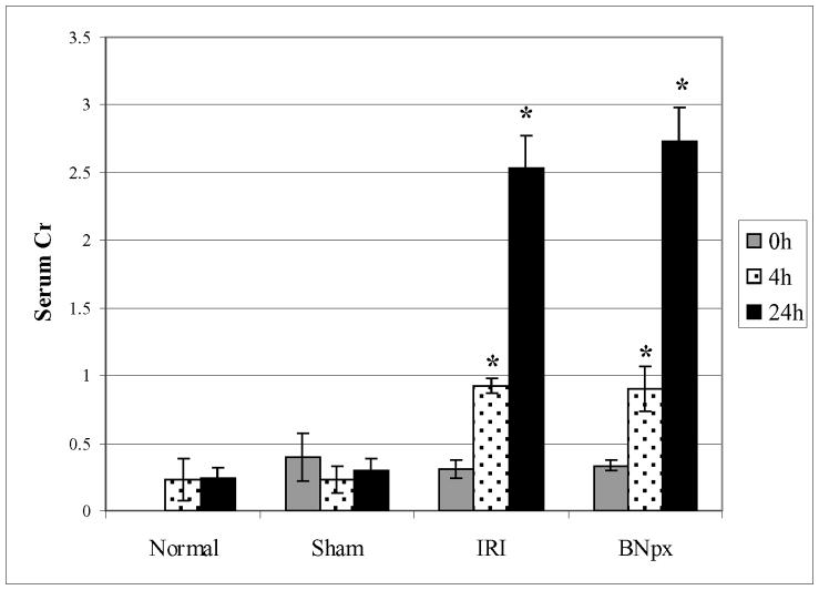
Mice (~25 gm) underwent 60 minutes of bilateral kidney IRI, bilateral nephrectomy, or sham surgery, and were then sacrificed at either 4 or 24 hours post-procedure. Serum creatinine was measured prior to surgery and at time of sacrifice. Both bilateral IRI and nephrectomy resulted in significant increase in serum creatinine at both time points, while sham surgery did not (Figure 1).
Figure 1. Experimental AKI Leads to Renal Injury.
Prior to endothelial cell isolation, mice underwent 60 minutes of bilateral IRI, bilateral nephrectomy or sham surgery, and were then sacrificed at either 4 or 24 hours post-procedure. Normal mice (without any surgical intervention) were also sacrificed at the final time point as internal controls. Serum creatinines were measured prior to surgery (0h) and at time of sacrifice (4h or 24h). Both bilateral IRI and nephrectomy result in significant renal injury (*) while sham surgery does not increase creatinine levels above normal baseline. There was no significant difference between the rise in serum creatinine induced by IRI sersus bilateral nephrectomy. Results shown are average serum creatinine values for a representative experiment with error bar signifying standard deviation (n=5 mice per group).
Microbead Sorting Leads to Isolation of a Pure Endothelial Cell Population
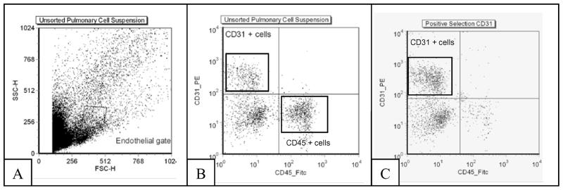
Whole lung tissue was digested and homogenized into a single cell suspension and pulmonary endothelial cells were isolated using a microbead sorting system (Miltenyi Biotec). Cells were first incubated with antibodies against CD31 and CD45 (Figure 2B). Cells were then negatively sorted to remove CD45+ cells, and then a second, positive sort enriched the CD31+ pool. This resulting in a homogenous population (approximately 90% pure) of CD31+ endothelial cells (Figure 2C). For each experiment, lung tissue from 5 mice was pooled in order to isolate enough cells/RNA for analysis.
Figure 2. Magnetic Isolation of Pulmonary Endothelial Cells.
Whole lung tissue from mice (n = 5 mice per surgical group) was pooled and homogenized into a single cell suspension and pulmonary endothelial cells isolated using a microbead sorting system. (A) Total cells prior to sorting, without fluorescent antibody label. (B) Cells stained with CD45-FITC and CD31-PE (C) Pure population of CD31(+), CD45(−) cells after a negative sort for CD 45 and a second, positive selection for CD31.
AKI-induced Changes in Pulmonary Vascular Endothelial Gene Expression
In order to investigate the effects of AKI on pulmonary endothelial cells, we performed either kidney IRI, BNx, or sham surgery on mice as previously described. At either 4 or 24 hours after injury, mice were sacrificed and lung tissue was pooled and harvested. After isolation of pulmonary endothelial cells, total mRNA was extracted and analyzed for endothelial-specific gene expression by a novel quantitative RT-PCR array (SABiosciences).
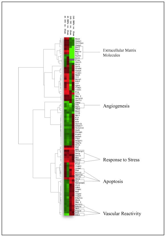
Figure 3 is a heatmap representation of mRNA expression in pulmonary endothelial cells after acute kidney injury. Fold changes compared to sham surgery are represented by color and intensity, with red indicating increase in gene expression and green signifying a decrease. Additionally, hierarchal cluster analysis reveals functional groupings for several genes, including those associated with inflammation, apoptosis and stress response, vascular reactivity and angiogenesis. Notably, there was no significant difference in gene expression levels between normal mice that did not undergo surgery and those that underwent sham surgeries (data not shown). Specific genes exhibiting fold changes greater than 1.5x (increased or decreased) are presented in Table 1.
Figure 3. Renal Injury Leads to Alterations in Pulmonary Vascular Endothelial Gene Expression.
Vascular endothelial cell gene expression was assessed via RT-PCR array 4 and 24 hours after injury. Fold changes in gene expression were clustered by average linkage analysis using the Cluster and Treeview software and illustrated in heatmap format. Red indicates increased gene expression while green indicates decreased gene expression. All fold changes are relative to appropriate sham surgeries. Clustering reveals similar gene expression profiles for several functional groupings, including apoptosis and stress response, vascular reactivity as well as angiogenesis.
TABLE 1.
Functional Grouping of Pulmonary Endothelial Cell Gene Expression During Acute Kidney Injury
| Gene Name | Gene Symbol | Fold Change | Fold Change | ||
|---|---|---|---|---|---|
| IRI
|
BNx
|
||||
| 4hr | 24hr | 4hr | 24hr | ||
| Inflammation | |||||
| E-selectin | Sele | 2.07 | 26.46 | 1.98 | 13.76 |
| Intercellular adhesion molecule 1 (CD54) | Icam1 | -- | 5.28 | -- | 10.89 |
| Tissue plasminogen activator (tPA) | Plat | -- | 4.77 | −1.57 | 4.01 |
| P-selectin | Selp | 1.83 | 2.19 | 1.70 | 2.21 |
| Integrin, beta 3 (CD61) | Itgb3 | -- | 2.0 | -- | 2.51 |
| Integrin, alpha 5 | Itga5 | -- | 1.58 | -- | 1.52 |
| (Fibronectin receptor, alpha polypeptide) | |||||
| Ras homolog gene family, member B | Rhob | -- | 1.55 | -- | 2.38 |
| Von Willebrand Factor | Vwf | -- | -- | −1.52 | 2.27 |
| L-selectin (CD62L) | Sell | 1.54 | -- | 1.66 | −3.16 |
| Selectin P ligand (CD162) | Selplg | -- | -- | 1.45 | −2.21 |
| Angiogenesis | |||||
| Plasminogen activator inhibitor-1 | Serpine1 | -- | 3.50 | -- | 2.28 |
| Apoptosis/Injury Response | |||||
| Baculovirus IAP repeat-containing 2 | Birc2 | -- | 1.89 | -- | 3.06 |
| Bcl2-like 1 | Bcl2l1 | -- | -- | -- | 2.49 |
| CASP8 and FADD-like apoptosis regulator | Cflar | -- | -- | -- | 1.73 |
| RANTES | Ccl5 | −1.71 | −13.12 | -- | −10.59 |
| Fas ligand | Fasl | -- | −7.84 | 1.78 | −2.84 |
| Interleukin-6 | Il6 | 1.73 | −4.21 | 2.25 | −3.03 |
| Chemokine receptor (BLR-1) | Cxcr5 | -- | −2.23 | -- | −3.02 |
| Vascular Reactivity | |||||
| Endothelin-1 | Edn1 | −2.49 | 1.62 | −2.76 | 1.58 |
| Nitric Oxide Synthase 2 (inducible nitric oxide synthase/iNOS) | Nos2 | 2.07 | -- | 2.56 | −1.97 |
| Endothelin-2 | Edn2 | 5.26 | −1.81 | 12.97 | 4.85 |
| Angiotensin-Renin System | |||||
| Angiotensinogen | Agt | 3.21 | 4.91 | 4.15 | 9.14 |
| Angiotensin II receptor, type Ia | Agtr1a | -- | 1.7 | -- | 3.73 |
All fold changes are compared to sham surgery
Murine pulmonary endothelial cells were isolated at 4 and 24 hours after ischemia-reperfusion injury (IRI), bilateral nephrectomy (BNx) or sham surgery. After mRNA isolation, an RT-PCR superarray was used to evaluate changes in endothelial cell biology-specific gene expression. Genes with changes in expression greater than 1.5-fold when compared with sham surgery were organized according to broad functional categories. Significant changes occurred primarily in genes associated with inflammation, apoptosis and angiogenesis.
We identified prominent early and sustained activation of genes related to leukocyte activation and trafficking including E-selectin, P-selectin, and ICAM-1. We also noted activation of early injury response genes interleukin-6 (IL-6) and inducible nitric oxide synthase (NOS-2), and later activation of genes related to cell motility and angiogenesis such as plasminogen activator inhibitor-1 (Serpine-1). At 24 hours, there was significant down-regulation of the prominent pro-apoptotic gene Fas ligand. Interestingly, we have previously demonstrated a prominent role for the tumor necrosis factor receptor-1 (TNFR1) in AKI-induced lung apoptosis and this data may lend further support that TNFR1 is the primary death signaling receptor involved in this model [17].
AKI Induces Actin Reorganization and Apoptosis in Pulmonary Microvascular Endothelial Cells
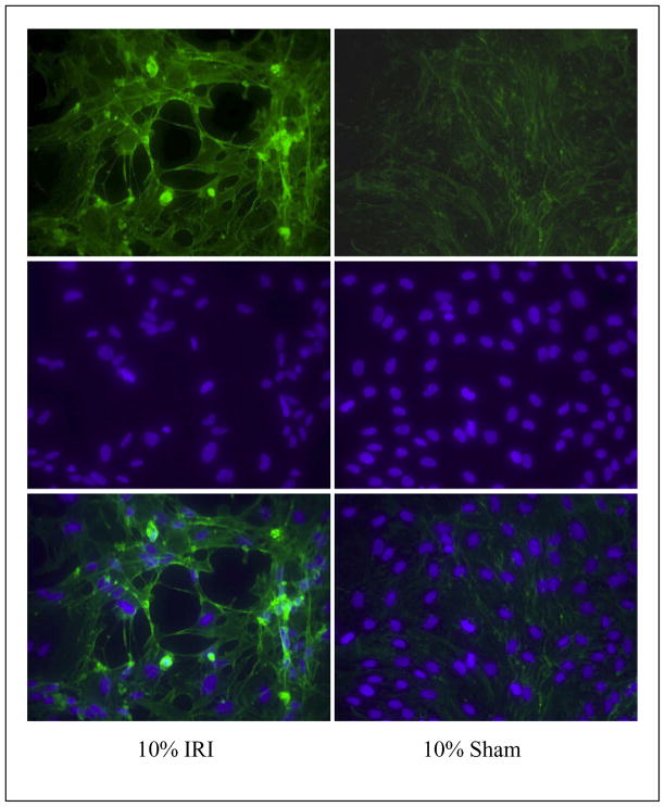
In order to further investigate the apoptosis-specific effects of AKI on pulmonary endothelial cells, an in vitro system was developed. Rat Lung Microvascular Endothelial Cells (RLMVECs) were obtained and cultured in recommended conditions. At early passage, cells were allowed to reach confluence and then grown for 24 hours in serum from rats that had undergone either IRI or sham surgery. Cells were then fixed and double stained with fluorescent-conjugated phalloiden and DAPI to evaluate actin cytoskeletal organization as well as nuclear morphology. Exposure to IRI serum for 24 hours led to noticeable changes in actin and nuclear morphology (Figure 4). Compared to sham serum, actin stress fiber formation was evident in cells grown in IRI serum. In addition, there were notable peri-nuclear actin fibers in the IRI group. Finally, by DAPI staining, the IRI-exposed cells exhibit nuclei that are large and abnormally shaped compared with sham cells (4B).
Figure 4. AKI induces Actin Reorganization.
Rat Lung Microvascular Endothelial Cells (RLMVECs) were treated for 24 hours with 10% serum from animals that underwent either sham surgery or bilateral IRI. Cells were stained with phalloiden and DAPI to evaluate actin organization and nuclear structure. In cells exposed to IRI serum, actin stress fiber formation and nuclear disorganization are apparent compared with cells grown in sham serum. All pictures are at 20X magnification
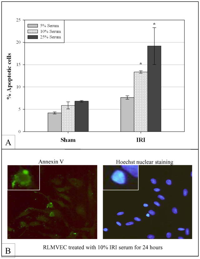
Apoptosis was then assessed and quantified by Annexin V/PI analysis (Figure 5). 24 hours after cells were exposed to either IRI or sham serum at varying concentrations (5%, 10% or 25%), they were stained with fluorescein-tagged Annexin V and PI and then analyzed by FACS. Cells exposed to 10% and 25% IRI serum had significantly more apoptosis (Annexin V positive, PI negative) than those exposed to sham serum in a dose-dependent fashion.
Figure 5. AKI Induces Apoptosis in Vascular Endothelial Cells.
Rat Lung Microvascular Endothelial Cells (RLMVECs) were cultured in the presence of serum from animals that underwent either bilateral IRI or sham surgery. Serum was isolated 24 hours after injury; cells were then cultured in either 5%, 10% or 25% serum for 24 hours. After serum exposure, cells were stained with a FITC-conjugated antibody against Annexin V as well as PI and then evaluated by FACS. Cells exposed to IRI serum showed increased evidence of apoptosis in a dose-dependent manner (A) compared to cells exposed to sham serum. There was a statistically significant difference in apoptosis between IRI and Sham in the 10% and 25% serum groups. Notably there was no significant difference in apoptosis amongst the sham groups. (B) An RLMVEC stained with Annexin V and Hoechst for nuclear visualization reveals membrane localization of Annexin V as well as nuclear changes consistent with apoptosis.
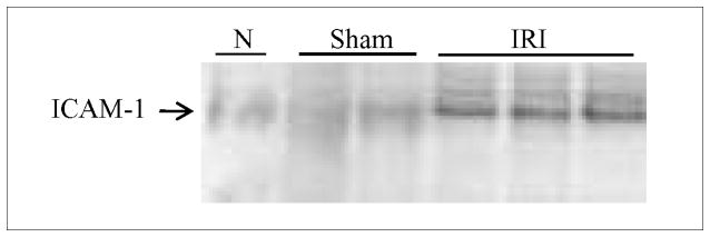
Utilizing this novel in vitro system, we further validated our findings of specific EC transcriptional changes by measuring ICAM-1 protein expression by Western blot in RLMVECs following 24 hour exposure to rat serum from untreated (ie. normal), sham, and IRI treated rats. Cells exposed to IRI serum demonstrated increased protein expression compared to normal or sham treated rats (Figure 6).
Figure 6. AKI Induces ICAM-1 protein expression in Vascular Endothelial Cells.
Representative Western blot of homogenates obtained from Rat Lung Microvascular Endothelial Cells (RLMVECs) treated for 24 hours with 10% serum from animals that underwent either sham surgery or bilateral IRI. ICAM-1 protein expression was increased in RLMVECs treated with serum from IRI-treated mice when compared to those treated with sham serum.
DISCUSSION
Clinical data suggests that AKI contributes to the development and exacerbation of multi-organ failure, although the physiologic and molecular mechanisms responsible are largely unknown. The link between AKI and pulmonary dysfunction is particularly important given the significant clinical morbidity and mortality associated with the presence of both pathologies. Understanding the ways in which AKI influences pulmonary pathophysiology is a crucial step in improving outcomes during multiple organ dysfunction syndrome (MODS).
Previous animal studies have shown that there are derangements in the innate immune response, inflammatory cascade, and the response to oxidative stress during AKI (reviewed in (5)). For example, AKI in several models (60 minute bilateral IRI, bilateral nephrectomy or bilateral pedicle ligation) induced pulmonary injury and neutrophil infiltration that was similar in nature to that induced by sepsis (16). Additional studies have revealed that pro- and anti-inflammatory cytokines appear in some fashion to mediate AKI-associated lung damage (9), (17).
Given that interactions between the kidney and the lung in the setting of renal injury have so far been shown to be mediated by either serum factors such as interleukins or serum cellular components such as neutrophils or macrophages, we hypothesized that one likely target for such factors would be the pulmonary endothelium. The pulmonary microvascular endothelium is central to the development of both acute lung injury (ALI) and acute respiratory distress syndrome (ARDS) (reviewed in (23)). Cytoskeletal rearrangement, changes in intracellular signaling and interactions with circulating inflammatory cells promote the breakdown of the endothelial barrier and development of pulmonary edema. There is also evidence that early endothelial cell activation is both necessary and sufficient for the further development of MODS (31).
Many alterations in the pathophysiology of pulmonary vascular endothelial cells should be reflected by changes in mRNA expression, particularly in those genes responsible for cellular adhesion, intracellular signaling and initiation of apoptosis. We therefore developed a strategy to isolate murine pulmonary endothelial cells after AKI and to analyze mRNA expression of various genes using novel RT-PCR arrays. Unlike cDNA arrays, quantitative RT-PCR arrays allow for reliable analysis of expression of a focused panel of genes, with amplification of various gene-specific products simultaneously under uniform cycling conditions. This provides the PCR array with high specificity and high amplification efficiencies. Housekeeping genes are included in the experiment to control for variability in PCR performance.
Pulmonary endothelial cells were isolated 4 or 24 hours after injury by a magnetic sorting protocol using a negative/positive sort strategy (CD45−/CD31+). One limitation of this study is that even after magnetic bead sorting, our population of pulmonary endothelial cells was, at best, approximately 90% pure by flow cytometry (Figure 2). We found pulmonary endothelial cells to be fragile, and attempted to maintain an appropriate balance between purity of population and loss of cells during the isolation process. Even though our populations were not 100% pure, however, we felt that this still represented a major improvement over previous experiments using whole lung tissue and was warranted given the opportunity to examine cell response in an in vivo model.
We performed differential RT-PCR on total RNA from these pulmonary endothelial cells at both early and late time points. Key genes involved in inflammation, such as E-selectin, revealed small fold increases (approximately 2x) 4 hours after injury, and much larger increases in expression 24 hours after injury when compared with sham. ICAM-1 expression also increased 24 hours after AKI, approximately 5-fold after IRI and 10-fold after bilateral nephrectomy. E-selectin and ICAM-1 expression are known to correlate with inflammatory damage in pulmonary endothelium and surrounding tissues by promoting inflammatory cell recruitment (30, 33); recent studies have also revealed increased levels of membrane-bound and circulating E-selectin and ICAM-1 in patients with sickle cell disease, with elevated protein levels associated with increased morbidity and mortality (15). Upregulation of leukocyte adhesion factors is one of the hallmarks of endothelial cell activation (35).
There were also several changes in gene expression related to angiogenesis, vascular reactivity and thrombosis. Plasminogen activator inhibitor-1 (PAI-1, or Serpine 1) was noted to have increased expression 24 hours after acute kidney injury. Notably, elevated levels of PAI-1 in pulmonary edema fluid have been associated with increased mortality (28). PAI-1 has also recently been shown to inhibit phagocytosis of neutrophils and apoptotic cells (27); increased expression of PAI-1 in the lung could therefore perpetuate and promote the inflammatory cascade and subsequent tissue injury. RT-PCR array analysis also revealed differential expression of endothelin-1 and -2 as well as inducible nitric oxide synthase (iNOS). Endothelin-2 (ET-2) changed most appreciably at both early and late time points, while changes in expression of endothelin-1 and iNOS were most notable 4 hours after injury. Endothelin-2 is a potent chemoattractant for macrophages (6); its upregulation may allow for macrophage infiltration into lung parenchyma, contributing to inflammation and subsequent pulmonary injury. These changes in gene expression suggest a transition to a prothrombotic state, another feature of endothelial cell activation (12).
Clearly, AKI induces distant organ changes in apoptosis-related gene expression; in order to investigate whether or not functional apoptosis might be occurring in pulmonary endothelium after IRI, we turned to an in vitro cell system. Rat pulmonary microvascular endothelial cells (RLMVECs) were cultured in the presence of serum from animals that had undergone IRI or sham surgery. Lung microvascular endothelial cells are very difficult to culture, and at the time of this study the only cells available were rat instead of mouse. We first evaluated actin architecture in order to look for qualitative changes in cytoskeletal arrangement indicative of early apoptosis. Classically, actin reorganization and nuclear disorganization/break-up are hallmarks of apoptosis (13). Compared to cells treated with sham serum, cells exposed to IRI-serum exhibited actin rearrangement including thick stress fiber formation as well as intercellular gap formation. Apoptosis was confirmed by Annexin V/PI staining and analysis via FACS. Annexin V is used to detect phosphatidylserine translocation from the inner to the outer plasma membrane, a hallmark of early apoptosis. We found that cells exposed to IRI serum showed significant increases in apoptosis in a dose-dependent fashion compared to those exposed to sham serum. Endothelial cell apoptosis has been postulated to be another pathway by which lung injury evolves in both sepsis and emphysema (11, 14); it may be possible that it also promotes ALI in the setting of AKI given these findings.
This is the first study to demonstrate detailed pulmonary endothelial-specific downstream consequences of AKI, and it builds upon our prior work which identified whole lung pro-inflammatory and pro-apoptotic changes during ischemic AKI. We used a newly developed method to isolate pulmonary endothelial cells ex vivo after AKI, and analyzed differential mRNA expression to help elucidate interactions between AKI and pulmonary injury. AKI leads to specific changes in the endothelial cell phenotype consistent with activation. These include upregulation of leukocyte adhesion factors as well as changes that may indicate a transition to a more prothrombotic state. In addition, we used an in vitro model to further reveal the functional pulmonary endothelial apoptotic events that occur after AKI. Future studies can focus on the specific AKI regulated genes and pathways we have identified in pulmonary endothelial cells to investigate the mechanisms by which AKI predisposes to pulmonary dysfunction and ARDS.
Acknowledgments
Supported by NHLBI P50 HL073944, K08 HL089181, T32 GM075774 and NIDDK R01 DK54770
References
- 1.Akcan-Arikan A, Zappitelli M, Loftis LL, Washburn KK, Jefferson LS, Goldstein SL. Modified RIFLE criteria in critically ill children with acute kidney injury. Kidney Int. 2007;71:1028–1035. doi: 10.1038/sj.ki.5002231. [DOI] [PubMed] [Google Scholar]
- 2.Bailey D, Phan V, Litalien C, Ducruet T, Merouani A, Lacroix J, Gauvin F. Risk factors of acute renal failure in critically ill children: A prospective descriptive epidemiological study. Pediatr Crit Care Med. 2007;8:29–35. doi: 10.1097/01.pcc.0000256612.40265.67. [DOI] [PubMed] [Google Scholar]
- 3.Chertow GM, Burdick E, Honour M, Bonventre JV, Bates DW. Acute kidney injury, mortality, length of stay, and costs in hospitalized patients. J Am Soc Nephrol. 2005;16:3365–3370. doi: 10.1681/ASN.2004090740. [DOI] [PubMed] [Google Scholar]
- 4.Deng J, Hu X, Yuen PS, Star RA. Alpha-melanocyte-stimulating hormone inhibits lung injury after renal ischemia/reperfusion. Am J Respir Crit Care Med. 2004;169:749–756. doi: 10.1164/rccm.200303-372OC. [DOI] [PubMed] [Google Scholar]
- 5.Feltes CM, Van Eyk J, Rabb H. Distant-organ changes after acute kidney injury. Nephron Physiol. 2008;109:p80–84. doi: 10.1159/000142940. [DOI] [PubMed] [Google Scholar]
- 6.Grimshaw MJ, Wilson JL, Balkwill FR. Endothelin-2 is a macrophage chemoattractant: implications for macrophage distribution in tumors. Eur J Immunol. 2002;32:2393–2400. doi: 10.1002/1521-4141(200209)32:9<2393::AID-IMMU2393>3.0.CO;2-4. [DOI] [PubMed] [Google Scholar]
- 7.Hassoun HT, Grigoryev DN, Lie ML, Liu M, Cheadle C, Tuder RM, Rabb H. Ischemic acute kidney injury induces a distant organ functional and genomic response distinguishable from bilateral nephrectomy. Am J Physiol Renal Physiol. 2007;293:F30–40. doi: 10.1152/ajprenal.00023.2007. [DOI] [PubMed] [Google Scholar]
- 8.Hassoun HT, Lie ML, Grigoryev DN, Liu M, Tuder RM, Rabb H. Kidney Ischemia-Reperfusion Injury Induces Caspase-Dependent Pulmonary Apoptosis. Am J Physiol Renal Physiol. 2009 doi: 10.1152/ajprenal.90666.2008. [DOI] [PMC free article] [PubMed] [Google Scholar]
- 9.Hoke TS, Douglas IS, Klein CL, He Z, Fang W, Thurman JM, Tao Y, Dursun B, Voelkel NF, Edelstein CL, Faubel S. Acute renal failure after bilateral nephrectomy is associated with cytokine-mediated pulmonary injury. J Am Soc Nephrol. 2007;18:155–164. doi: 10.1681/ASN.2006050494. [DOI] [PubMed] [Google Scholar]
- 10.Hoste EA, Clermont G, Kersten A, Venkataraman R, Angus DC, De Bacquer D, Kellum JA. RIFLE criteria for acute kidney injury are associated with hospital mortality in critically ill patients: a cohort analysis. Crit Care. 2006;10:R73. doi: 10.1186/cc4915. [DOI] [PMC free article] [PubMed] [Google Scholar]
- 11.Hu H, Li X, Li Y, Wang L, Mehta S, Feng Q, Chen R, Peng T. Calpain-1 induces apoptosis in pulmonary microvascular endothelial cells under septic conditions. Microvasc Res. 2009;78:33–39. doi: 10.1016/j.mvr.2009.04.005. [DOI] [PubMed] [Google Scholar]
- 12.Hunt BJ, Jurd KM. Endothelial cell activation. A central pathophysiological process. BMJ. 1998;316:1328–1329. doi: 10.1136/bmj.316.7141.1328. [DOI] [PMC free article] [PubMed] [Google Scholar]
- 13.Huot J, Houle F, Rousseau S, Deschesnes RG, Shah GM, Landry J. SAPK2/p38-dependent F-actin reorganization regulates early membrane blebbing during stress-induced apoptosis. J Cell Biol. 1998;143:1361–1373. doi: 10.1083/jcb.143.5.1361. [DOI] [PMC free article] [PubMed] [Google Scholar]
- 14.Kasahara Y, Tuder RM, Cool CD, Lynch DA, Flores SC, Voelkel NF. Endothelial cell death and decreased expression of vascular endothelial growth factor and vascular endothelial growth factor receptor 2 in emphysema. Am J Respir Crit Care Med. 2001;163:737–744. doi: 10.1164/ajrccm.163.3.2002117. [DOI] [PubMed] [Google Scholar]
- 15.Kato GJ, Martyr S, Blackwelder WC, Nichols JS, Coles WA, Hunter LA, Brennan ML, Hazen SL, Gladwin MT. Levels of soluble endothelium-derived adhesion molecules in patients with sickle cell disease are associated with pulmonary hypertension, organ dysfunction, and mortality. Br J Haematol. 2005;130:943–953. doi: 10.1111/j.1365-2141.2005.05701.x. [DOI] [PMC free article] [PubMed] [Google Scholar]
- 16.Kim do J, Park SH, Sheen MR, Jeon US, Kim SW, Koh ES, Woo SK. Comparison of experimental lung injury from acute renal failure with injury due to sepsis. Respiration. 2006;73:815–824. doi: 10.1159/000095588. [DOI] [PubMed] [Google Scholar]
- 17.Klein CL, Hoke TS, Fang WF, Altmann CJ, Douglas IS, Faubel S. Interleukin-6 mediates lung injury following ischemic acute kidney injury or bilateral nephrectomy. Kidney Int. 2008;74:901–909. doi: 10.1038/ki.2008.314. [DOI] [PubMed] [Google Scholar]
- 18.Kramer AA, Postler G, Salhab KF, Mendez C, Carey LC, Rabb H. Renal ischemia/reperfusion leads to macrophage-mediated increase in pulmonary vascular permeability. Kidney Int. 1999;55:2362–2367. doi: 10.1046/j.1523-1755.1999.00460.x. [DOI] [PubMed] [Google Scholar]
- 19.Lim YC, Luscinskas FW. Isolation and culture of murine heart and lung endothelial cells for in vitro model systems. Methods Mol Biol. 2006;341:141–154. doi: 10.1385/1-59745-113-4:141. [DOI] [PubMed] [Google Scholar]
- 20.Lin CY, Kao KC, Tian YC, Jenq CC, Chang MY, Chen YC, Fang JT, Huang CC, Tsai YH, Yang CW. The RIFLE score increases the accuracy of outcome prediction in patients with acute respiratory distress syndrome undergoing open lung biopsy. Respiration. 2009;77:398–406. doi: 10.1159/000183756. [DOI] [PubMed] [Google Scholar]
- 21.Livak KJ, Schmittgen TD. Analysis of relative gene expression data using real-time quantitative PCR and the 2(−Delta Delta C(T)) Method. Methods. 2001;25:402–408. doi: 10.1006/meth.2001.1262. [DOI] [PubMed] [Google Scholar]
- 22.Mahabeleshwar GH, Somanath PR, Byzova TV. Methods for isolation of endothelial and smooth muscle cells and in vitro proliferation assays. Methods Mol Med. 2006;129:197–208. doi: 10.1385/1-59745-213-0:197. [DOI] [PubMed] [Google Scholar]
- 23.Maniatis NA, Orfanos SE. The endothelium in acute lung injury/acute respiratory distress syndrome. Curr Opin Crit Care. 2008;14:22–30. doi: 10.1097/MCC.0b013e3282f269b9. [DOI] [PubMed] [Google Scholar]
- 24.Mehta RL, Kellum JA, Shah SV, Molitoris BA, Ronco C, Warnock DG, Levin A. Acute Kidney Injury Network: report of an initiative to improve outcomes in acute kidney injury. Crit Care. 2007;11:R31. doi: 10.1186/cc5713. [DOI] [PMC free article] [PubMed] [Google Scholar]
- 25.Metnitz PG, Krenn CG, Steltzer H, Lang T, Ploder J, Lenz K, Le Gall JR, Druml W. Effect of acute renal failure requiring renal replacement therapy on outcome in critically ill patients. Crit Care Med. 2002;30:2051–2058. doi: 10.1097/00003246-200209000-00016. [DOI] [PubMed] [Google Scholar]
- 26.Parikh CR, Abraham E, Ancukiewicz M, Edelstein CL. Urine IL-18 is an early diagnostic marker for acute kidney injury and predicts mortality in the intensive care unit. J Am Soc Nephrol. 2005;16:3046–3052. doi: 10.1681/ASN.2005030236. [DOI] [PubMed] [Google Scholar]
- 27.Park YJ, Liu G, Lorne EF, Zhao X, Wang J, Tsuruta Y, Zmijewski J, Abraham E. PAI-1 inhibits neutrophil efferocytosis. Proc Natl Acad Sci U S A. 2008;105:11784–11789. doi: 10.1073/pnas.0801394105. [DOI] [PMC free article] [PubMed] [Google Scholar]
- 28.Prabhakaran P, Ware LB, White KE, Cross MT, Matthay MA, Olman MA. Elevated levels of plasminogen activator inhibitor-1 in pulmonary edema fluid are associated with mortality in acute lung injury. Am J Physiol Lung Cell Mol Physiol. 2003;285:L20–28. doi: 10.1152/ajplung.00312.2002. [DOI] [PubMed] [Google Scholar]
- 29.Rabb H, Wang Z, Nemoto T, Hotchkiss J, Yokota N, Soleimani M. Acute renal failure leads to dysregulation of lung salt and water channels. Kidney Int. 2003;63:600–606. doi: 10.1046/j.1523-1755.2003.00753.x. [DOI] [PubMed] [Google Scholar]
- 30.Tsokos M, Fehlauer F, Puschel K. Immunohistochemical expression of E-selectin in sepsis-induced lung injury. Int J Legal Med. 2000;113:338–342. doi: 10.1007/s004149900105. [DOI] [PubMed] [Google Scholar]
- 31.van Meurs M, Wulfert FM, Knol AJ, De Haes A, Houwertjes M, Aarts LP, Molema G. Early organ-specific endothelial activation during hemorrhagic shock and resuscitation. Shock. 2008;29:291–299. doi: 10.1097/SHK.0b013e318145a7c1. [DOI] [PubMed] [Google Scholar]
- 32.Vieira JM, Jr, Castro I, Curvello-Neto A, Demarzo S, Caruso P, Pastore L, Jr, Imanishe MH, Abdulkader RC, Deheinzelin D. Effect of acute kidney injury on weaning from mechanical ventilation in critically ill patients. Crit Care Med. 2007;35:184–191. doi: 10.1097/01.CCM.0000249828.81705.65. [DOI] [PubMed] [Google Scholar]
- 33.Weiland JE, Davis WB, Holter JF, Mohammed JR, Dorinsky PM, Gadek JE. Lung neutrophils in the adult respiratory distress syndrome. Clinical and pathophysiologic significance. Am Rev Respir Dis. 1986;133:218–225. doi: 10.1164/arrd.1986.133.2.218. [DOI] [PubMed] [Google Scholar]
- 34.Zarbock A, Schmolke M, Spieker T, Jurk K, Van Aken H, Singbartl K. Acute uremia but not renal inflammation attenuates aseptic acute lung injury: a critical role for uremic neutrophils. J Am Soc Nephrol. 2006;17:3124–3131. doi: 10.1681/ASN.2006040358. [DOI] [PubMed] [Google Scholar]
- 35.Zimmerman GA, Albertine KH, Carveth HJ, Gill EA, Grissom CK, Hoidal JR, Imaizumi T, Maloney CG, McIntyre TM, Michael JR, Orme JF, Prescott SM, Topham MS. Endothelial activation in ARDS. Chest. 1999;116:18S–24S. doi: 10.1378/chest.116.suppl_1.18s. [DOI] [PubMed] [Google Scholar]


