Abstract
The high prevalence of drug resistance necessitates the development of novel antifungal agents against infections caused by opportunistic fungal pathogens such as Candida albicans. Elucidation of apoptosis in yeast-like fungi may provide a basis for future therapies. In mammalian cells, photodynamic therapy (PDT) has been demonstrated to generate reactive oxygen species, leading to immediate oxidative modifications of biological molecules and resulting in apoptotic cell death. In this report, we assess the in vitro cytotoxicity and mechanism of PDT, using the photosensitizer Pc 4, in planktonic C. albicans. Confocal image analysis confirmed that Pc 4 localizes to cytosolic organelles, including mitochondria. A colony formation assay showed that 1.0 μM Pc 4 followed by light at 2.0 J/cm2 reduced cell survival by 4 logs. XTT (2,3-bis[2-methoxy-4-nitro-5-sulfophenyl]-2H-tetrazolium-5-carboxyanilide) assay revealed that Pc 4-PDT impaired fungal metabolic activity, which was confirmed using the FUN-1 (2-chloro-4-[2,3-dihydro-3-methyl-(benzo-1,3-thiazol-2-yl)-methylidene]-1-phenylquinolinium iodide) fluorescence probe. Furthermore, we observed changes in nuclear morphology characteristic of apoptosis, which were substantiated by increased externalization of phosphatidylserine and DNA fragmentation following Pc 4-PDT. These data indicate that Pc 4-PDT can induce apoptosis in C. albicans. Therefore, a better understanding of the process will be helpful, since PDT may become a useful treatment option for candidiasis.
INTRODUCTION
Photodynamic therapy (PDT) involves the interaction of light, a photosensitizing agent, and molecular oxygen to induce a biological response (1). It has been used in the treatment of both malignant and non-malignant diseases. The silicon phthalocyanine Pc 4 is a second generation photosensitizer developed at Case Western Reserve University. Upon exposure to 675-nm red light, Pc 4 is activated to an excited singlet state which undergoes intersystem crossing to a triplet state (2). The excited triplet Pc 4 interacts with molecular oxygen forming reactive oxygen species, which cause oxidative damage to proteins and lipids within their immediate vicinity (3, 4). Pharmacokinetic data in mice indicate that Pc 4 is much more rapidly cleared than the FDA-approved Photofrin® when delivered systemically, thereby minimizing the possibility of prolonged generalized photosensitivity (5, 6). Pc 4-PDT has demonstrated an excellent safety profile in a Phase 1 trial on cutaneous neoplasms (7).
Infections due to Candida and other fungi continue to represent a significant health burden. Some cases are highly resistant to traditional antifungals such as azoles and polyenes (e.g., fluconazole and amphotericin B, respectively). Even with antifungal therapy, mortality of patients with invasive candidiasis can still be as high as 40 percent (8). Candidiasis is usually associated with indwelling medical devices (e.g., dental implants, catheters, heart valves, vascular bypass grafts, ocular lenses, artificial joints and central nervous system shunts), which can act as substrates for biofilm growth. Often, removal of the catheters or medical devices is warranted to treat the infection. Denture stomatitis is another form of candidiasis that affects 65% of edentulous individuals (9, 10). Despite the use of antifungal drugs to treat denture stomatitis, infection is often re-established soon after treatment (11).
In vitro studies have demonstrated that Candida species are susceptible to PDT utilized by Photofrin® (12), the porphyrin precursor 5-aminolevulinic acid (ALA) (13, 14), or other photosensitizers that are not yet FDA approved (15, 16). In this study, we demonstrate that Pc 4-PDT is highly effective in killing planktonic C. albicans and induces extensive apoptosis as evidenced by metabolic, morphological, and molecular assays.
MATERIALS AND METHODS
Organism and Culture Conditions
C. albicans (strain SC-5134) was grown in yeast nitrogen base (YNB) medium (Difco Laboratories, Detroit, MI, USA) supplemented with 50 mM glucose and incubated for 24 hr at 37°C in an orbital water bath shaker at 150 rpm. Cells were then harvested and washed twice with 0.15 M phosphate-buffered saline (PBS; pH 7.2, calcium- and magnesium-free). Cells were re-suspended in 10 mL of 1× PBS, further serial diluted, counted using a hemocytometer, standardized and used immediately as previously described (17).
Confocal Microscopy
To investigate the uptake and localization of Pc 4, cells in standard growth medium plus 10% Fetal Bovine Serum (FBS) were loaded with 0.2 μM Pc 4 and incubated for 16 to 18 hr. Thirty min prior to imaging Pc 4, 50 nM MitoTracker Red (CMXRos) (Invitrogen, Carlsbad, CA, USA) or 50 μg/mL AlexaFluor 488-conjugated Concanavalin (Con) A (Invitrogen, Carlsbad, CA, USA) and/or 10 μg/mL Hoechst 33342 (Invitrogen, Carlsbad, CA, USA) was loaded onto cells. Five to 10 μL of stained cells were placed on a slide with a glass coverslip and all images were acquired using an UltraVIEW VoX spinning disk confocal system (PerkinElmer, Waltham, MA, USA), which was mounted on a Leica DMI6000B microscope (Leica Microsystems, Inc., Bannockburn, IL, USA) equipped with a HCX PL APO 100×/1.4 oil immersion objective. Confocal images of Pc 4 fluorescence, MitoTracker Red, ConA (or FITC-labeled Annexin V) and Hoechst 33342 were collected using solid-state diode lasers, with 640-nm, 561-nm, 488-nm and 405-nm excitation light, respectively, and with appropriate emission filters.
Pc 4-Photodynamic Treatment Conditions
The phthalocyanine photosensitizer Pc 4 was kindly provided by Dr. Malcolm E. Kenney (Department of Chemistry, Case Western Reserve University, Cleveland, OH, USA). Stock solutions (0.5 mM) of Pc 4 were prepared in N',N'-dimethylformamide (DMF, ThermoFisher, Waltham, MA, USA) and stored at 4 °C in the dark. C. albicans cultures were incubated with Pc 4 concentrations ranging from 0.1 to 1.0 μM in 1× PBS containing 10% FBS for at least 1 hr at 37 °C in the dark and subsequently irradiated with red light using a light-emitting diode array (EFOS, Mississauga, Ontario, Canada) at a fluence ranging from 1.0 to 2.0 J/cm2 (1.0 mW/cm2, λmax ~670–675 nm) at room temperature.
Colony forming unit (CFU) phototoxicity assay
CFU assay was done as previously described (17) with minor modifications. Briefly, following photoirradiation, C. albicans cell suspensions that received various Pc 4-PDT doses plus Pc 4 alone and DMF +/− light were serially diluted in 1× PBS then plated on Sabouraud dextrose agar (Becton, Dickinson and Company, Sparks, MD, USA) plates and incubated at 37°C to allow colony formation. Aliquots of the cell suspension were plated onto 100-mm Petri dishes in amounts sufficient to yield 50–100 colonies per dish. After 24 hours, colonies were counted by eye, and the percentage of surviving cells from the Pc 4-PDT cultures was normalized against that of cultures exposed to Pc 4 or light alone. Note: The maximum amount used for all negative controls was negligible (i.e. ≤ 0.2% vol:vol/ DMF:medium) and comparable to previous studies using mammalian cells and all Pc 4-PDT experiments were done concurrently with Pc 4 alone and DMF +/− light and none of which had any deleterious effects on candida cells.
In order to qualitatively assess C. albicans survivability post-treatment and show the results in one concise image in some cases, one drop from each of the eight cell suspensions were plated on a single 100-mm Petri dish and incubated for 24 hours at 37°C.
Metabolic assays
(A) XTT (2,3-bis[2-methoxy-4-nitro-5-sulfophenyl]-2H-tetrazolium-5-carboxyanilide) - One hour following photoirradiation of Pc 4-loaded cells, the metabolic activity of the C. albicans cultures was assayed using the colorless sodium salt of XTT (Sigma-Aldrich, St. Louis, MO, USA), which is converted by mitochondrial dehydrogenases of viable cells into a water-soluble orange formazan derivative through the reduction of the tetrazolium ring. The absorbance of the resulting orange solution was measured using a spectrophotometer (Spectronic Genesys 5, Analytical Instrument, Golden Valley, MN, USA) at 492 nm. (B) FUN-1 (2-chloro-4-[2,3-dihydro-3-methyl-(benzo-1,3-thiazol-2-yl)-methylidene]-1-phenylquinolinium iodide) is a fluorescent probe used to assess yeast metabolic activity. Metabolically active yeast will endocytose FUN-1 (Invitrogen, Carlsbad, CA, USA) and process it into an orange-red cylindrical intravacuole structure. One hour following Pc 4-PDT, cells were loaded with 10 μM FUN-1 for 30 minutes at 37°C. Cells were washed before placing 10 μL between a slide with a cover slip, and confocal images were acquired with the same spinning disc confocal system mentioned above. FUN-1 fluorescence was excited using a solid state diode laser (488 nm) and collected with a (580–650 nm) band pass emission filter.
Apoptosis Analyses
(A) FITC-labeled Annexin V staining - To assess the externalization of phosphatidylserine, protoplasts of C. albicans were stained with FITC-labeled Annexin V using the Annexin V-FLUOS staining kit (Roche, Germany) with minor modifications (18, 19). Briefly, control and treated cells were washed three times with 1× PBS before incubating at 37°C for 30 min in a protoplasting solution (50 mM KH2PO4, 5 mM EDTA and 50 mM DTT). Then cells were digested at 37°C for 45 min in an enzyme solution (50 mM K2HPO4, 40 mM 2-mercaptoethanol, 3 μg/mL Chitinase, 1.8 μg/mL Lyticase, 0.15 mg/mL Zymolyase and 2.4 M sorbitol). After addition of 1× PBS, each sample was incubated for 30 min at room temperature. Protoplasts were then incubated with 100 μL of Annexin V-FLUOS labeling solution for 15 min at 25°C. FITC-labeled Annexin V fluorescence was detected by both widefield (Leica DMI6000B microscope; Leica Microsystems, Inc., Bannockburn, IL, USA) and confocal fluorescence microscopy. (B) DNA Fragmentation – Control, Pc 4-PDT and hydrogen peroxide (ThermoFisher Scientific, Waltham, MA, USA) treated C. albicans were pelleted, resuspended in 1× PBS and placed on a 37°C orbital shaker overnight. Subsequently, C. albicans total chromosomal DNA was extracted using the MasterPure™ Yeast DNA Purification Kit (Epicentre Biotechnologies, Madison, WI, USA) per the manufacturer's protocol. The concentration of extracted DNA was determined by spectrophotometry (Nanodrop 3000, ThermoFisher Scientific, Waltham, MA, USA) at a wavelength 260 nm. DNA (3.0 μg) was loaded onto a 1.0% (w/v) agarose gel (Bioexpress, Kaysbille, UT, USA) impregnated with ethidium bromide (ThermoFisher, Waltham, MA, USA) and subjected to electrophoresis. The gel was subsequently visualized using a UV transilluminator (VersaDoc, Bio-Rad, Hercules, CA, USA).
RESULTS
Pc 4 is localized to mitochondria
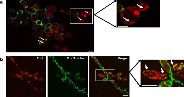
We have previously shown that Pc 4 preferentially, but not exclusively, binds to mitochondria of a variety of mammalian cells (20). To determine whether the binding pattern of Pc 4 is similar in fungi, we loaded C. albicans with Pc 4 in glucose-supplemented YNB and examined its fluorescence by confocal microscopy. As shown in Fig. 1a, the majority of Pc 4 fluorescence was detected in the cytoplasmic compartment but not the cell wall, plasma membrane or nucleus, similar to its distribution in mammalian cells (21). In C. albicans, Pc 4 displayed a punctate pattern of fluorescence primarily localized in a cytoplasmic region distinct from the plasma membrane (Fig. 1a) and resembling mitochondria. To test whether mitochondria are sites of localization of Pc 4, cells were co-loaded with MitoTracker Red, a mitochondrion-specific dye. The bright, punctate fluorescence shown in the Pc 4 (pseudo red) image (Fig. 1b, left panel) usually corresponds to the MitoTracker (pseudo green) fluorescence (Fig. 1b, middle panel), as indicated in the merged image (Fig. 1b, right panel). The absence of a yellow merged image in some cases may reflect an imbalance in the uptake of the two fluorescent dyes. In addition, as previously found in mammalian cells, Pc 4 also binds to other intracellular organelles, presumably ER membranes, as well as other vesicular membranes.
Figure 1. Pc 4 is localized to mitochondrial and other intracellular membranes.
(a) C. albicans were loaded with 0.2 μM Pc 4, 50 μg/mL AlexaFluor 488- conjugated ConA, and 10 μg/mL Hoechst 33342. Pc 4 (pseudo red) was found to penetrate the cell wall and diffuse into the cytoplasmic compartment of the cells. Pc 4 fluorescence did not co-localize with either the cell wall/plasma membrane (pseudo green) or the nucleus (pseudo blue). However, it displayed a punctate fluorescence, some of which is reminiscent of a mitochondrial pattern (see white arrows in main image and in enlarged image from outlined area of main image); Scale bars, 5 μm. (b) C. albicans were loaded with 0.2 μM Pc 4 (pseudo red – left panel) and 50 nM MitoTracker Red (CMXRos) (pseudo green – middle panel) and visualized by confocal microscopy. Pc 4 displayed a punctate pattern of fluorescence that closely resembles that of MitoTracker, as indicated by the yellow/yellow-green fluorescence in a merged image (right panel). The boxed area of the merged image is enlarged at right; white arrows identify regions of clear overlap of Pc 4 and MitoTracker fluorescence; Scale bars, 10 μm.
Photodynamic therapy kills C. albicans
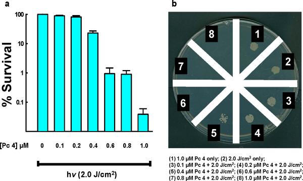
Having established that Pc 4 localizes to mitochondria as well as other intracellular membranes, we next determined a dose of PDT that would yield several logs of cell killing. C. albicans were loaded with 0.1 – 1.0 μM Pc 4 for at least 2 hr, then photoirradiated and plated. With 1.0 μM Pc 4 and 2.0 J/cm2, we consistently detected 3 to 4 logs of cell killing, as determined by a colony forming unit (CFU) assay (Fig. 2). This is shown both by counting colonies (Fig. 2a) and by observation of growth of equal numbers of treated and untreated cells (Fig. 2b). In addition, neither the highest dose of photosensitizer alone nor light alone affected cell proliferation and cell death, when compared to untreated cells (data not shown).
Figure 2. Pc 4-PDT is lethal to C. albicans.
(a) Candida cell cultures at equal concentrations per hemocytometer counts were treated with 0.1–1.0 μM Pc 4 for at least 2 hr before being irradiated with 2.0 J/cm2 of 670–675 nm light and plated on Sabouraud dextrose agar for 24 hr incubation at 37°C. The percentage of surviving cells from the PDT-treated cultures was normalized against that of cultures exposed to Pc 4 or light alone (plating efficiencies of control cultures ≥ 33%). Values represent the mean from at least three independent experiments. Error bars indicate S.E. For all Pc 4-PDT-treated cultures vs. untreated cells, either light alone or Pc 4 alone, p < 0.005. (b) For a qualitative assessment of C. albicans survivability post Pc 4-PDT, all yeast samples from one experiment were plated on a single 100-mm Petri dish. Cell suspensions were prepared as in (a). The Petri dish with Sabouraud dextrose agar was divided into eight equal wedges. After Pc 4-PDT, one drop of each cell suspension as well as light and dark controls were placed on a wedge, and the plate was incubated for 24 hours at 37°C.
Photodynamic therapy inhibits metabolic activity in C. albicans
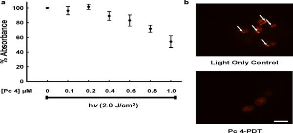
Over the same dose range, we assessed the ability of Pc 4-PDT to affect various short-term measures of metabolic activity of the organisms. Mitochondrial dehydrogenases of viable cells reduce the tetrazolium ring of XTT, a colorless compound, to yield a water-soluble orange formazan derivative. A decrease in absorbance is indicative of a loss of metabolic activity. With the highest Pc 4-PDT dose, metabolic activity was reduced by almost 50% within an hour following photoirradiation of Pc 4-loaded cells (Fig. 3a). As an independent indicator of yeast metabolic activity and viability, we used the fluorescent probe FUN-1. This probe typically enters vacuoles of viable yeast cells and forms cylindrical intravacuole structures that fluoresce orange-green (17, 22). Since this process requires ATP, non-viable or metabolically inactive cells cannot incorporate FUN-1 and will exhibit minimal or no fluorescence. As shown in Fig. 3b, there was much reduced FUN-1 fluorescence in cells exposed to PDT with 1.0 μM Pc 4 as compared to control cells exposed to light but no Pc 4. This provides further evidence of the loss of metabolic activity and, in turn, yeast viability, after Pc 4-PDT.
Figure 3.
(a) Candida cell cultures at equal concentrations per hemocytometer counts were treated with 0.1–1.0 μM Pc 4 for at least 2 hr and then were irradiated with 2.0 J/cm2 of 670–675 nm light. One hour following photoirradiation of Pc 4-loaded cells, cultures were assayed for XTT reduction. Values represent mean ± S.E. from at least three independent experiments. For all Pc 4-PDT-treated cultures, p < 0.005 versus light alone control. (b) Candida cells cultures were treated with (bottom panel) or without (top panel) 1.0 μM Pc 4 for at least 2 hr and then both were irradiated with 2.0 J/cm2 of 670–675 nm light. One hour following photo-irradiation of Pc 4-loaded cells, cultures were loaded with FUN-1 probe (see MATERIALS AND METHODS) which was imaged by confocal microscopy; Scale bar, 10 μm.
Pc 4-PDT induces apoptosis in C. albicans
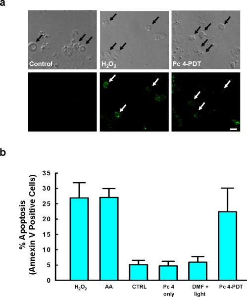
Like mammalian cells, yeasts also have an asymmetric distribution of phosphatidylserine (PS) in the lipid bilayer of the plasma membrane (23). Since the exposure of PS on the outer leaflet of the plasma membrane is a common early marker of apoptosis in mammalian cells, we investigated the extent of this phenomenon in Pc 4-PDT-treated C. albicans, as assessed by the binding of FITC-labeled Annexin V. Plasma membrane-associated FITC-Annexin V was detected in protoplasts of cells that had been treated with 1.0 μM Pc 4-PDT or hydrogen peroxide (Fig. 4a). Externalization of PS was readily detected within 4 to 5 hours of PDT (Fig. 4b), when ~23% of Pc 4-PDT-treated cells were Annexin V positive, as compared to ~27% of cells treated with H2O2 or acetic acid. Fewer than 5% of negative controls were Annexin V positive.
Figure 4. Quantification of apoptosis as characterized by PS exposure in C. albicans.
Candida cell cultures were treated with either 1.0 μM Pc 4 for at least 2 hr followed by 2.0 J/cm2 of 670–675 nm of light, Pc 4 only, vehicle plus light, 20 mM H2O2, 60 mM acetic acid or nothing. Four hr after treatment, protoplasts were prepared, stained with FITC-labeled Annexin V and observed by confocal microscopy. (a) Brightfield and confocal images of FITC-stained protoplasts (white arrows) from cells (black arrows) that were treated with and without Pc 4-PDT, or H2O2; Scale bar, 10 μm. (b) Fifty - 100 protoplasts cells were counted by eye using an epifluorescence microscope. Values represent the mean ± S.E. from at least three independent experiments. For all cultures treated with H2O2, acetic acid or Pc 4-PDT, p < 0.05 vs. the combined controls (untreated cells and those given with Pc 4 alone or vehicle plus light).
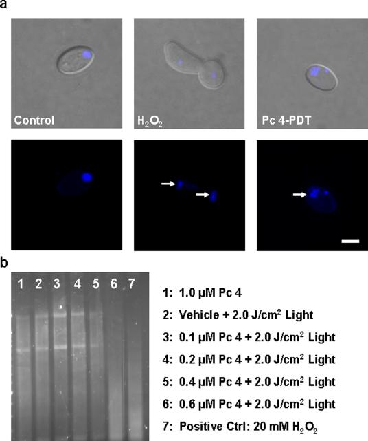
Chromatin condensation, an indicator of a late stage of apoptosis, was detected by confocal microscopy at the highest dose of Pc 4-PDT 24 hr after Pc 4-PDT or treatment with peroxide (Fig. 5a). DNA fragmentation is also a late event in apoptosis. DNA gel electrophoresis revealed increasing DNA fragmentation with increasing dose of Pc 4-PDT. At 0.4 μM Pc 4-PDT, there is clear evidence of DNA degradation with faster migration of the DNA. By 0.6 μM Pc 4-PDT, the DNA fragmentation pattern matched that of the hydrogen peroxide positive control with a complete loss of banding and extensive migration of the small DNA fragments (Fig. 5b).
Figure 5. Pc 4-PDT-induced apoptosis in C. albicans, as characterized by nuclear and DNA fragmentation.
Candida cell cultures were treated with 20 mM H2O2, 1.0 μM Pc 4 for 2 hr followed by 2.0 J/cm2 of 670–675 nm light, or vehicle plus light. (a) Candida cell cultures were incubated overnight followed by labeling with 10 μg/mL Hoechst 33342 and imaging by confocal microscopy. Arrows indicate nuclear condensation; Scale Bar, 5 μm. (b) After overnight incubation of the cells, DNA was extracted and separated on a 1.0% agarose gel.
DISCUSSION
C. albicans has previously been demonstrated to undergo programmed cell death, or apoptosis, following a variety of treatments, including commonly used anti-fungal agents (see review (24)). Although PDT sensitized by Photofrin®, meso-tetra (N-methyl-4-pyridyl) porphine tetra tosylate (TMP-1363) or the porphyrin precursor 5-aminolevulinic acid (12, 14, 15, 25) has been shown to have cytotoxic effects against fungi, the precise mechanism of cell death has not been clearly defined. Our data in this current study suggest that PDT, sensitized by the phthalocyanine Pc 4, induces apoptosis in C. albicans, exhibiting many similarities with mammalian cells undergoing mitochondrion-mediated programmed cell death.
For PDT to be effective against fungi, the photosensitizer must be able to permeate through the cell wall. Photosensitizing drugs, including Photofrin®, TMP-1363, cationic porphyrin 5-phenyl-10,15,20-Tris(N-methyl-4-pyridyl)porphyrin chloride (TriP[4]) and chlorin e6 have been demonstrated to target different cellular domains, such as plasma membrane or mitochondria. Irradiation of these photosensitizers has been shown to generate oxidative damage that leads to cytotoxicity (12, 25–27). In this study, mitochondria are one of the sites of localization of Pc 4 in C. albicans, as evidenced by co-localization experiments shown in Fig. 1 as well as by XTT and FUN-1 assays, which indicated that the mitochondrial pathway may be involved in Pc 4-PDT induced cell death (Fig. 3).
Sensitivity to photodynamic killing has been found to vary considerably among different mammalian cell lines, and it may not necessarily be dependent upon a specific subcellular binding of the photosensitizing agent. Therefore, it was not surprising to find that the effective dose of Pc 4-PDT found in C. albicans differed from doses used to kill tumor cell lines (28). Compared to mammalian cells, the effective Pc 4-PDT dose for C. ablicans was higher (Fig. 2). In order to produce a significant cytotoxic Pc 4-PDT effect, it was necessary to apply as much as 2.0 J/cm2 dose of photoirradiation. The need for higher PDT doses in fungi may be due to the presence of different morphology, i.e., hyphal structure, which could be more recalcitrant to treatment than other yeast forms. It has been recognized that modes of cell death in fungi include necrotic, autophagic as well as apoptotic pathways (29, 30). Our results from the metabolic activity and clonogenic assays demonstrate that Pc 4-PDT was effective in killing planktonic C. albicans in vitro. In addition, similar to its effects seen in mammalian cells, Pc 4-PDT causes C. albicans to undergo apoptosis as a mode of cell death. Our data demonstrate that Pc 4-PDT may have the potential to become an antifungal treatment. Ongoing and future studies involve investigating Pc 4-PDT effects in candidal biofilms.
ACKNOWLEDGMENTS
This study has been supported in part by a NIH grant 5P30AR39750 (NIAMS: Skin Diseases Research Center Grant). We thank Mr. Yuntao Li for his technical assistance and Dr. Scott J. Howell for his assistance regarding image data analyses and his comments on the manuscript.
REFERENCES
- 1.Dougherty TJ. An update on photodynamic therapy applications. J Clin Laser Med Surg. 2002;20:3–7. doi: 10.1089/104454702753474931. [DOI] [PubMed] [Google Scholar]
- 2.He J, Larkin HE, Li YS, Rihter D, Zaidi SI, Rodgers MA, Mukhtar H, Kenney ME, Oleinick NL. The synthesis, photophysical and photobiological properties and in vitro structure-activity relationships of a set of silicon phthalocyanine PDT photosensitizers. Photochem Photobiol. 1997;65:581–586. doi: 10.1111/j.1751-1097.1997.tb08609.x. [DOI] [PubMed] [Google Scholar]
- 3.Dougherty TJ, Gomer CJ, Henderson BW, Jori G, Kessel D, Korbelik M, Moan J, Peng Q. Photodynamic therapy. J Natl Cancer Inst. 1998;90:889–905. doi: 10.1093/jnci/90.12.889. [DOI] [PMC free article] [PubMed] [Google Scholar]
- 4.Oleinick NL, Evans HH. The photobiology of photodynamic therapy: cellular targets and mechanisms. Radiat Res. 1998;150:S146–156. [PubMed] [Google Scholar]
- 5.Oleinick NL, Antunez AR, Clay ME, Rihter BD, Kenney ME. New phthalocyanine photosensitizers for photodynamic therapy. Photochem Photobiol. 1993;57:242–247. doi: 10.1111/j.1751-1097.1993.tb02282.x. [DOI] [PubMed] [Google Scholar]
- 6.Egorin MJ, Zuhowski EG, Sentz DL, Dobson JM, Callery PS, Eiseman JL. Plasma pharmacokinetics and tissue distribution in CD2F1 mice of Pc4 (NSC 676418), a silicone phthalocyanine photodynamic sensitizing agent. Cancer Chemother Pharmacol. 1999;44:283–294. doi: 10.1007/s002800050979. [DOI] [PubMed] [Google Scholar]
- 7.Baron ED, Malbasa CL, Santo-Domingo D, Fu P, Miller JD, Hanneman KK, Hsia AH, Oleinick NL, Colussi VC, Cooper KD. Silicon phthalocyanine (Pc 4) photodynamic therapy is a safe modality for cutaneous neoplasms: results of a phase 1 clinical trial. Lasers Surg Med. 42:728–735. doi: 10.1002/lsm.20984. [DOI] [PMC free article] [PubMed] [Google Scholar]
- 8.Wey SB, Mori M, Pfaller MA, Woolson RF, Wenzel RP. Hospital-acquired candidemia. The attributable mortality and excess length of stay. Arch Intern Med. 1988;148:2642–2645. doi: 10.1001/archinte.148.12.2642. [DOI] [PubMed] [Google Scholar]
- 9.Budtz-Jorgensen E. Histopathology, immunology, and serology of oral yeast infections. Diagnosis of oral candidosis. Acta Odontol Scand. 1990;48:37–43. doi: 10.3109/00016359009012732. [DOI] [PubMed] [Google Scholar]
- 10.Calderone RA, Braun PC. Adherence and receptor relationships of Candida albicans. Microbiol Rev. 1991;55:1–20. doi: 10.1128/mr.55.1.1-20.1991. [DOI] [PMC free article] [PubMed] [Google Scholar]
- 11.MacEntee MI. The prevalence of edentulism and diseases related to dentures--a literature review. J Oral Rehabil. 1985;12:195–207. doi: 10.1111/j.1365-2842.1985.tb00636.x. [DOI] [PubMed] [Google Scholar]
- 12.Bliss JM, Bigelow CE, Foster TH, Haidaris CG. Susceptibility of Candida species to photodynamic effects of photofrin. Antimicrob Agents Chemother. 2004;48:2000–2006. doi: 10.1128/AAC.48.6.2000-2006.2004. [DOI] [PMC free article] [PubMed] [Google Scholar]
- 13.Maisch T. A new strategy to destroy antibiotic resistant microorganisms: antimicrobial photodynamic treatment. Mini Rev Med Chem. 2009;9:974–983. doi: 10.2174/138955709788681582. [DOI] [PubMed] [Google Scholar]
- 14.Monfrecola G, Procaccini EM, Bevilacqua M, Manco A, Calabro G, Santoianni P. In vitro effect of 5-aminolaevulinic acid plus visible light on Candida albicans. Photochem Photobiol Sci. 2004;3:419–422. doi: 10.1039/b315629j. [DOI] [PubMed] [Google Scholar]
- 15.Chabrier-Rosello Y, Foster TH, Mitra S, Haidaris CG. Respiratory deficiency enhances the sensitivity of the pathogenic fungus Candida to photodynamic treatment. Photochem Photobiol. 2008;84:1141–1148. doi: 10.1111/j.1751-1097.2007.00280.x. [DOI] [PubMed] [Google Scholar]
- 16.de Souza SC, Junqueira JC, Balducci I, Koga-Ito CY, Munin E, Jorge AO. Photosensitization of different Candida species by low power laser light. J Photochem Photobiol B. 2006;83:34–38. doi: 10.1016/j.jphotobiol.2005.12.002. [DOI] [PubMed] [Google Scholar]
- 17.Chandra J, Mukherjee PK, Ghannoum MA. In vitro growth and analysis of Candida biofilms. Nat Protoc. 2008;3:1909–1924. doi: 10.1038/nprot.2008.192. [DOI] [PubMed] [Google Scholar]
- 18.Madeo F, Frohlich E, Frohlich KU. A yeast mutant showing diagnostic markers of early and late apoptosis. J Cell Biol. 1997;139:729–734. doi: 10.1083/jcb.139.3.729. [DOI] [PMC free article] [PubMed] [Google Scholar]
- 19.Park C, Lee DG. Melittin induces apoptotic features in Candida albicans. Biochem Biophys Res Commun. 394:170–172. doi: 10.1016/j.bbrc.2010.02.138. [DOI] [PubMed] [Google Scholar]
- 20.Lam M, Oleinick NL, Nieminen AL. Photodynamic therapy-induced apoptosis in epidermoid carcinoma cells. Reactive oxygen species and mitochondrial inner membrane permeabilization. J Biol Chem. 2001;276:47379–47386. doi: 10.1074/jbc.M107678200. [DOI] [PubMed] [Google Scholar]
- 21.Trivedi NS, Wang HW, Nieminen AL, Oleinick NL, Izatt JA. Quantitative analysis of Pc 4 localization in mouse lymphoma (LY-R) cells via double-label confocal fluorescence microscopy. Photochem Photobiol. 2000;71:634–639. doi: 10.1562/0031-8655(2000)071<0634:qaopli>2.0.co;2. [DOI] [PubMed] [Google Scholar]
- 22.Millard PJ, Roth BL, Thi HP, Yue ST, Haugland RP. Development of the FUN-1 family of fluorescent probes for vacuole labeling and viability testing of yeasts. Appl Environ Microbiol. 1997;63:2897–2905. doi: 10.1128/aem.63.7.2897-2905.1997. [DOI] [PMC free article] [PubMed] [Google Scholar]
- 23.Phillips AJ, Sudbery I, Ramsdale M. Apoptosis induced by environmental stresses and amphotericin B in Candida albicans. Proc Natl Acad Sci U S A. 2003;100:14327–14332. doi: 10.1073/pnas.2332326100. [DOI] [PMC free article] [PubMed] [Google Scholar]
- 24.Almeida B, Silva A, Mesquita A, Sampaio-Marques B, Rodrigues F, Ludovico P. Drug-induced apoptosis in yeast. Biochim Biophys Acta. 2008;1783:1436–1448. doi: 10.1016/j.bbamcr.2008.01.005. [DOI] [PubMed] [Google Scholar]
- 25.Chabrier-Rosello Y, Giesselman BR, De Jesus-Andino FJ, Foster TH, Mitra S, Haidaris CG. Inhibition of electron transport chain assembly and function promotes photodynamic killing of Candida. J Photochem Photobiol B. 99:117–125. doi: 10.1016/j.jphotobiol.2010.03.005. [DOI] [PMC free article] [PubMed] [Google Scholar]
- 26.Fuchs BB, Tegos GP, Hamblin MR, Mylonakis E. Susceptibility of Cryptococcus neoformans to photodynamic inactivation is associated with cell wall integrity. Antimicrob Agents Chemother. 2007;51:2929–2936. doi: 10.1128/AAC.00121-07. [DOI] [PMC free article] [PubMed] [Google Scholar]
- 27.Lambrechts SA, Aalders MC, Van Marle J. Mechanistic study of the photodynamic inactivation of Candida albicans by a cationic porphyrin. Antimicrob Agents Chemother. 2005;49:2026–2034. doi: 10.1128/AAC.49.5.2026-2034.2005. [DOI] [PMC free article] [PubMed] [Google Scholar]
- 28.Xue LY, Chiu SM, Oleinick NL. Photodynamic therapy-induced death of MCF-7 human breast cancer cells: a role for caspase-3 in the late steps of apoptosis but not for the critical lethal event. Exp Cell Res. 2001;263:145–155. doi: 10.1006/excr.2000.5108. [DOI] [PubMed] [Google Scholar]
- 29.Abudugupur A, Mitsui K, Yokota S, Tsurugi K. An ARL1 mutation affected autophagic cell death in yeast, causing a defect in central vacuole formation. Cell Death Differ. 2002;9:158–168. doi: 10.1038/sj.cdd.4400942. [DOI] [PubMed] [Google Scholar]
- 30.Eisenberg T, Carmona-Gutierrez D, Buttner S, Tavernarakis N, Madeo F. Necrosis in yeast. Apoptosis. doi: 10.1007/s10495-009-0453-4. [DOI] [PubMed] [Google Scholar]


