Abstract
Artificial mimicry of α-helices offers a basis for development of protein-protein interaction antagonists. Here we report a new type of unnatural peptidic backbone, containing α-, β- and γ-amino acid residues in an αγααβα repeat pattern, for this purpose. This unnatural hexad has the same number of backbone atoms as a heptad of α residues. 2D NMR data clearly establish the formation of an α-helix-like conformation in aqueous solution. The helix formed by our 12-mer α/β/γ-peptide is considerably more stable than the α-helix formed by an analogous 14-mer α-peptide, presumably because of the preorganized β and γ residues employed.
Unnatural oligomers that can reproduce the three-dimensional shapes and side chain projection patterns characteristic of natural polypeptides offer a basis for rational development of protein-protein interaction antagonists.1 Considerable effort has been devoted to mimicry of individual helices, which frequently mediate information transfer in biological systems.2 Many alternatives to the α-helical scaffold have been examined, including oligomers based on unnatural peptidic backbones (e.g., β-peptides3) and entirely non-peptidic oligomers (e.g., oligophenyls4). Evaluation of these new designs has typically focused on α-helices two to four turns in length (e.g., mimicry of the p53 N-terminal domain in binding to hDM2,5 or mimicry of a BH3 domain in binding to Bcl-2-family proteins6); however, epitopes of this size can be effectively mimicked by small molecules.7 We have recently shown that the recognition properties of a 10-turn α-helix, the CHR domain of HIV protein gp41, can be recapitulated with α/β-peptide oligomers in which two of every seven among the original α-amino acid residues are replaced by analogous β-amino acid residues (Figure 1).8 Oligomers with the ααβαααβ heptad repeat can adopt a helical secondary structure in which each turn contains one extra backbone carbon relative to an α-helix. This backbone alteration confers significant resistance to proteolytic degradation while allowing reasonably good mimicry of the side chain projection pattern along one side of the helix. However, this mimicry is not perfect: the resulting α/β-peptides have lower affinity for the target surface than does the original α-peptide.
Figure 1.

(a) Three backbones that correspond to approximately two helical turns: ααααααα, ααβαααβ, αγααβα; (b) helical wheels for the αγααβα hexad and the ααααααα heptad.
Here we introduce a new foldamer design containing α-, β- and γ-amino acid residues in an αγααβα hexad repeat pattern, which is intended to mimic an ααααααα heptad without additional backbone atoms (Figure 1a). Both the α/β/γ hexad and the all-α heptad correspond to two helical turns.9 The helical wheel juxtaposition in Figure 1b shows that the αγααβα repeat potentially allows a direct correspondence between the α residues of this hexad and four of the α residues in a standard peptide heptad. In the parlance of coiled coil-forming sequences, the common α residues could correspond to positions a, d, e and g of an all-α heptad repeat, which define a large and continuous surface that runs along one side of the helix.10
In order to test the α-helix mimicry hypothesis outlined above, we prepared α/β/γ-peptide 12-mer 1 via solid-phase methods. All of the α residues in 1 are derived from proteinogenic amino acids. The helix-forming properties of β-amino acid residues11 and γ-amino acid residues12 have been widely studied, and based on these precedents we employed conformationally preorganized β and γ residues that seemed likely to maximize the propensity of 1 to adopt an α-helix-like conformation. The β residues are derived from (S,S)-trans-2-aminocyclopentanecarboxylic acid (ACPC), which promotes helical α/β-peptide conformations that strongly resemble the α-helix,13 including the gp41 CHR-mimetic oligomers mentioned above.8 The γ residues in 1 are derived from α-ethyl-cis-2-aminocyclohexaneacetic acid (EtACHA; 3), which has recently been shown to participate in helical conformations.14 The α- and β-amino acid residues of 1 were incorporated via microwave-assisted solid-phase synthesis employing common reagents.15 Coupling of Fmoc-EtACHA to the growing polypeptide proved to be challenging, because of intrinsically limited reactivity (perhaps resulting from steric hindrance) and a tendency toward epimerization, presumably at the α-position of the γ-amino acid backbone. The best method for addition of Fmoc-EtACHA proved to be use of 4 equiv. of HOAt and EDCI in DMF for 6 hr at room temperature (no microwave irradiation).

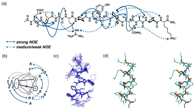
α/β/γ-Peptide 1 is highly water-soluble and displayed sufficient 1H NMR resonance dispersion to enable 2D NMR analysis in aqueous solution (8 mM 1, 9:1 H2O:D2O, 100 mM acetate buffer, pH 3.8, 10°C). Resonances from backbone amides and side chains (6.5–9.0 ppm) were monitored between 0.02 and 14 mM 1; no concentration-dependent variations in chemical shift were observed, which suggests that 1 does not self-associate under these conditions. Numerous NOEs were detected between protons on sequentially non-adjacent residues (i → i+2 or i → i+3; Figure 3a–b), all of which could be accommodated by a single helical conformation. The expected alignment of the β and γ residues along one side of this helix was indicated by characteristic NOE patterns: EtACHA CγH (i) – ACPC NH (i+3) and ACPC CβH (i) – EtACHA CγH (i+3). These NOEs between β and γ residues were detected even at 50°C, which implies that the helical conformation of α/β/γ-peptide 1 is quite robust. Further evidence of α/β/γ-peptide helix stability was obtained from H/D exchange studies. When 1 was dissolved in D2O at room temperature, some NH resonances disappeared completely within 4 min (the backbone NH of Glu 1 and the side chain NH resonances of Lys, Arg and Gln), but most of the backbone NH resonances could be detected even after 1 hour. α-Peptide 14-mer 2 was prepared for direct comparison with 1, but the 1H NMR spectrum of 2 displayed poor dispersion, which precluded resonance assignments and 2D NMR analysis.
Figure 3.
(a) The structure of α/β/γ-peptide 1 with NOEs observed in aqueous buffer between non-adjacent residues indicated by curved arrows (100 mM acetate buffer, pH 3.8); (b) NOEs indicated on the α/β/γ-peptide helix wheel; (c) overlay of the 10 best conformations generated via NOE-restrained dynamics (see text for details); (d) stereoview of the average of the 10 structures from the NOE-strained dynamics simulations (blue backbone) overlaid on a canonical α-helix (black backbone).
NOE-restrained molecular dynamics calculations for α/β/γ-peptide 1 in aqueous buffer were carried out with the CNS program.16 Only i → i+1, i → i+2 and i → i+3 NOEs were used for these calculations. Good overlap among backbone atoms was observed for the 10 best among 1000 calculated structures (rmsd = 0.86 ± 0.24 Å; Figure 3c). Figure 3d shows superimposition of the average of these 10 helical structures for 1 on a canonical α-helix.17 Of particular interest is the overlap between the six central α residues of 1 and the corresponding residues of the α-helix (the terminal α residues of 1 were excluded from this comparison because of expected ‘fraying’ effects). For overlay of the two sets of six α-carbons, rmsd = 0.98 ± 0.50 Å.18 Inspection of the stereoview in Figure 3d shows that the Cα-to-sidechain vectors for these two sets of α residues are largely coincident.
We turned to circular dichroism (CD) to compare α/β/γ-peptide 1 and α-peptide 2, because the latter could not be studied via 2D NMR. Figure 4 shows far-UV CD data for 1 and 2 in aqueous buffer and in 50 vol % aqueous methanol. In aqueous solution, the CD signature of α-peptide 2 suggests a largely unfolded state. In the presence of 50 vol % methanol 2 manifests a typical α-helical CD signature, with minima at ~208 and ~222 nm; the pronounced helix-promoting impact of the organic co-solvent is typical for short α-peptides. The behavior of α/β/γ-peptide 1 is quite different in that the CD signature is not strongly affected by changing the solvent. In both cases a single strong minimum is observed (~204 nm in aqueous buffer, ~205 nm in aqueous methanol), with only a minor intensity difference between solvents. Since NMR analysis indicates substantial population of an α-helix-like conformation by 1 in aqueous buffer, we assign the CD signature observed for 1 to this conformation. A similar far-UV CD signature has been established for α-helix-like conformations adopted by α/β-peptides;13 it is unclear why these heterogeneous peptidic backbones fail to manifest a second minimum in the helical state. The fact that similar CD signatures are observed for 1 in aqueous buffer and in 50% aqueous methanol suggests that the α-helix-like conformation of this α/β/γ-peptide is highly populated even in a fully aqueous environment, an extent of folding that would be unusual for a linear α-peptide of comparable length. Further evidence of the high stability of the α/β/γ-peptide 1 helix is provided by variable-temperature CD: the minimum at ~204 nm becomes less intense on heating from 10 to 90°C, as expected from thermally induced unfolding, but at 90°C this minimum retains ~70% of the intensity observed at 10°C.19
Figure 4.

Circular dichroism data for α/β/γ-peptide 1 and α-peptide 2 in 10 mM aqueous acetate buffer, pH 3.8, or 50 vol % aqueous methanol (one volume of methanol added to the aqueous buffer). The concentration of peptides is 0.2 mM.
We have introduced a new type of heterogeneous peptidic foldamer and shown via 2D NMR that this system supports a helical conformation in aqueous solution. The αγααβα hexad pattern we designed leads to mimicry of the side chain display along one side of an α-helix, despite the presence of non-proteinogenic backbone components. Direct comparison with a conventional α-peptide reveals that the α/β/γ-peptide has a much stronger folding propensity, a feature that should facilitate the development of biologically active examples. The high stability of the new α/β/γ-peptide helix is attributed to the use of appropriately preorganized β and γ residues. Additional studies will be required to determine whether other αααα+β+γ hexads with different subunit ordering give rise to stable helical conformations. To our knowledge, this study represents the first example of high-resolution structural analysis of a γ-amino acid-containing foldamer in aqueous solution. It will now be important to determine whether the α/β/γ-peptide design introduced here can support functional mimicry of biological α-helices.
Supplementary Material
Figure 2.

Sequences for α/β/γ-peptide 1 and α-peptide 2. Proteinogenic α residues are indicated with the conventional single-letter code; for non-proteinogenic α residues, Orn = ornithine and Nle = norleucine; the β and γ residue structures are shown.
Acknowledgments
This research was supported in part by the NSF (CHE-0848847). T. S. was supported in part by a JSPS Postdoctoral fellowship for Research Abroad. NMR spectrometers were purchased with partial support from NSF and NIH. We thank Aaron Almeida for assistance of NMR measurements, and Michael Giuliano and Dr. Li Guo for helpful discussion.
Footnotes
Supporting Information. All experimental procedures and data. This material is available free of charge via the Internet at http://pubs.acs.org.
References
- 1.(a) Goodman CM, Choi S, Shandler S, DeGrado WF. Nat Chem Biol. 2007;3:252–262. doi: 10.1038/nchembio876. [DOI] [PMC free article] [PubMed] [Google Scholar]; (b) Hecht S, Huc I, editors. Foldamers: Strucutre, Properties, and Applications. Wiley-VCH; Weinheim, Germany: 2007. [Google Scholar]
- 2.(a) Jochim AL, Arora PS. ACS Chem Biol. 2010;5:919–923. doi: 10.1021/cb1001747. [DOI] [PMC free article] [PubMed] [Google Scholar]; (b) Jochim AL, Arora PS. Mol BioSyst. 2009;5:924–926. doi: 10.1039/b903202a. [DOI] [PMC free article] [PubMed] [Google Scholar]
- 3.(a) Werder M, Hauser H, Abele S, Seebach D. Helv Chim Acta. 1999;82:1774–1783. [Google Scholar]; (b) Kritzer JA, Lear JD, Hodsdon ME, Schepartz A. J Am Chem Soc. 2004;126:9468–9469. doi: 10.1021/ja031625a. [DOI] [PubMed] [Google Scholar]; (c) English EP, Chumanov RS, Gellman SH, Compton T. J Biol Chem. 2006;281:2661–2667. doi: 10.1074/jbc.M508485200. [DOI] [PubMed] [Google Scholar]
- 4.(a) Orner BP, Ernst JT, Hamilton AD. J Am Chem Soc. 2001;123:5382–5383. doi: 10.1021/ja0025548. [DOI] [PubMed] [Google Scholar]; (b) Kutzki O, Park HS, Ernst JT, Orner BP, Yin H, Hamilton AD. J Am Chem Soc. 2002;124:11838–11839. doi: 10.1021/ja026861k. [DOI] [PubMed] [Google Scholar]; (c) Davis JM, Tsou LK, Hamilton AD. Chem Soc Rev. 2007;36:326–334. doi: 10.1039/b608043j. [DOI] [PubMed] [Google Scholar]; (d) Hu X, Sun J, Wang HG, Manetsch R. J Am Chem Soc. 2008;130:13820–13821. doi: 10.1021/ja802683u. [DOI] [PMC free article] [PubMed] [Google Scholar]; (e) Shaginian A, Whitby LR, Hong S, Hwang I, Farooqi B, Searcey M, Chen J, Vogt PK, Boger DL. J Am Chem Soc. 2009;131:5564–5572. doi: 10.1021/ja810025g. [DOI] [PMC free article] [PubMed] [Google Scholar]; (f) Marimganti S, Cheemala MN, Ahn JM. Org Lett. 2009;11:4418–4421. doi: 10.1021/ol901785v. [DOI] [PubMed] [Google Scholar]; (g) Plante JP, Burnley T, Malkova B, Webb ME, Warriner SL, Edwards TA, Wilson AJ. Chem Commun. 2009:5091–5093. doi: 10.1039/b908207g. [DOI] [PMC free article] [PubMed] [Google Scholar]; (h) Campbell F, Plante JP, Edwards TA, Warriner SL, Wilson AJ. Org Biomol Chem. 2010;8:2344–2351. doi: 10.1039/c001164a. [DOI] [PubMed] [Google Scholar]
- 5.Murray JK, Gellman SH. Biopolymers. 2007;88:657–686. doi: 10.1002/bip.20741. [DOI] [PubMed] [Google Scholar]
- 6.Youle RJ, Strasser A. Nat Rev Mol Cell Biol. 2008;9:47–59. doi: 10.1038/nrm2308. [DOI] [PubMed] [Google Scholar]
- 7.(a) Vassilev LT, Vu BT, Graves B, Carvajal D, Podlaski F, Filipovic Z, Kong N, Kammlott U, Lukacs C, Klein C, Fotouhi N, Liu EA. Science. 2004;303:844–848. doi: 10.1126/science.1092472. [DOI] [PubMed] [Google Scholar]; (b) Oltersdorf T, et al. Nature. 2005;435:677–681. doi: 10.1038/nature03579. [DOI] [PubMed] [Google Scholar]; (c) Lessene G, Czabotar PE, Colman PM. Nat Rev Drug Discov. 2008;7:989–1000. doi: 10.1038/nrd2658. [DOI] [PubMed] [Google Scholar]
- 8.Horne WS, Johnson LM, Ketas TJ, Klasse PJ, Lu M, Moore JP, Gellman SH. Proc Natl Acad Sci USA. 2009;106:14751–14756. doi: 10.1073/pnas.0902663106. [DOI] [PMC free article] [PubMed] [Google Scholar]
- 9.For crystal structures showing that a β-alanine-γ-aminobutyric acid dipeptide segment can replace a tri-α-peptide segment in short helical oligomers, see: Karle IL, Pramanik A, Banerjee A, Bhattacharjya S, Balaram P. J Am Chem Soc. 1997;119:9087–9095.For related work, see: Araghi RR, Jäckel C, Cöfen H, Salwiczek M, Völkel A, Wagner SC, Wieczorek S, Baldauf C, Koksch B. ChemBioChem. 2010;11:335–339. doi: 10.1002/cbic.200900700.Araghi RR, Koksch B. Chem Commun. 2011:3544–3546. doi: 10.1039/c0cc03760e.
- 10.(a) Woolfson DN. Adv Protein Chem. 2005;70:79–112. doi: 10.1016/S0065-3233(05)70004-8. [DOI] [PubMed] [Google Scholar]; (b) Grigoryan G, Keating AE. Curr Opin Struct Biol. 2008;18:477–483. doi: 10.1016/j.sbi.2008.04.008. [DOI] [PMC free article] [PubMed] [Google Scholar]
- 11.(a) Cheng RP, Gellman SH, DeGrado WF. Chem Rev. 2001;101:3219–3232. doi: 10.1021/cr000045i. [DOI] [PubMed] [Google Scholar]; (b) Seebach D, Gardiner J. Acc Chem Res. 2008;41:1366–1375. doi: 10.1021/ar700263g. [DOI] [PubMed] [Google Scholar]
- 12.(a) Hanessian S, Luo X, Schaum R, Michnick S. J Am Chem Soc. 1998;120:8569–8570. [Google Scholar]; (b) Hintermann T, Gademann K, Jaun B, Seebach D. Helv Chim Acta. 1998;81:893–1002. [Google Scholar]; (c) Seebach D, Hook DF, Glättli A. Biopolymers. 2006;84:23–37. doi: 10.1002/bip.20391. [DOI] [PubMed] [Google Scholar]
- 13.(a) Horne WS, Price JL, Keck JL, Gellman SH. J Am Chem Soc. 2007;129:4178–4180. doi: 10.1021/ja070396f. [DOI] [PubMed] [Google Scholar]; (b) Horne WS, Price JL, Gellman SH. Proc Natl Acad Sci USA. 2008;105:9151–9156. doi: 10.1073/pnas.0801135105. [DOI] [PMC free article] [PubMed] [Google Scholar]; (c) Price JL, Horne WS, Gellman SH. J Am Chem Soc. 2010;132:12378–12387. doi: 10.1021/ja103543s. [DOI] [PMC free article] [PubMed] [Google Scholar]
- 14.Guo L, Chi Y, Almeida AM, Guzei IA, Parker BK, Gellman SH. J Am Chem Soc. 2009;131:16018–16020. doi: 10.1021/ja907233q.Guo L, Almeida AM, Zhang W, Reidenbach AG, Choi SH, Guzei IA, Gellman SH. J Am Chem Soc. 2010;132:7868–7869. doi: 10.1021/ja103233a.For related γ-amino acid building blocks, see: Nodes WJ, Nutt DR, Chippindale AM, Cobb AJA. J Am Chem Soc. 2009;131:16016–16017. doi: 10.1021/ja9070915.
- 15.(a) Murray JK, Gellman SH. Org Lett. 2005;7:1517–1520. doi: 10.1021/ol0501727. [DOI] [PubMed] [Google Scholar]; (b) Murray JK, Gellman SH. Nat Protocols. 2007;2:624–631. doi: 10.1038/nprot.2007.23. [DOI] [PubMed] [Google Scholar]
- 16.(a) Brünger AT, Adams PD, Clore GM, DeLano WL, Gros P, Grosse-Kunstleve RW, Jiang JS, Kuszewski J, Nilges M, Pannu NS, Read RJ, Rice LM, Simonson T, Warren GL. Acta Cryst. 1998;D54:905–921. doi: 10.1107/s0907444998003254. [DOI] [PubMed] [Google Scholar]; (b) Brunger AT. Nat Protocols. 2007;2:2728–2733. doi: 10.1038/nprot.2007.406. [DOI] [PubMed] [Google Scholar]
- 17.The canonical α-helix structure was built by molecular modeling using HyperChem™ 8.0.6.
- 18.Superimposition of two helices was carried out by using the pair fitting function in PyMol™ 1.3.
- 19.See Supporting Information.
Associated Data
This section collects any data citations, data availability statements, or supplementary materials included in this article.


