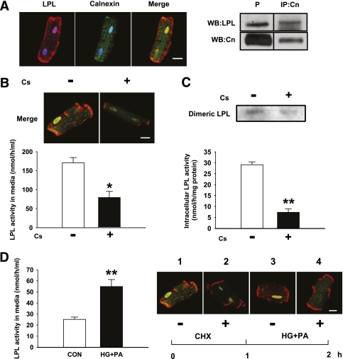
FIG. 3.
LPL maturation requires calnexin and is enhanced by HG and PA. Plated myocytes from control (CON) were immunostained with LPL (red) and calnexin (green). DAPI was used to stain the nuclei (blue), and colocalization of LPL and calnexin was visualized (Merge, yellow) (A). Cells were lysed with CHAPS lysis buffer (50 mmol/L HEPES buffer, pH 7.5, containing 1% CHAPS, 200 mmol/L NaCl, and protease inhibitor). Cell lysate of cardiomyocytes from control animals were immunoprecipitated using anti-calnexin (IP:Cn) antibody overnight at 4°C. The immunocomplex was captured by anti-rabbit Ig IP beads and immunoblotted for LPL. Direct Western blot (WB) of cell lysate was carried out as a positive control (P) (A, inset). Dividing lines on Western blot images describe where bands from the same blot have been juxtaposed. Isolated myocytes were incubated in the presence or absence of Cs for 2 h. At the end of treatment, colocalization of LPL and calnexin was visualized (B, inset). LPL activity released into the medium (B) and remaining in the myocytes (C) was measured. Treated cardiomyocytes were also loaded onto a heparin–sepharose column to determine the amount of dimeric LPL (C, inset). Results are the mean ± SE of three repeated experiments using different animals. *Significantly different from control, P < 0.05. **Significantly different from control, P < 0.01. Isolated myocytes were preincubated with 50 μmol/L CHX for 1 h. After sufficient washing to remove CHX, cells were treated with HG+PA or BSA only (CON) for another hour. LPL activity released into the media within this hour was measured (D). Results are the mean ± SE of three repeated experiments using different animals. **Significantly different from control, P < 0.05. Cells were also fixed with 4% paraformaldehyde before (D, inset 1) or after (inset 2) preincubation with CHX, and also after treatment with (inset 3) or without (inset 4) HG+PA. Colocalization of LPL and calnexin (yellow) in these conditions was visualized by immunofluorescence. Scale bar, 25 µm. (A high-quality digital representation of this figure is available in the online issue.)

