Abstract
The hamstring tendons are an increasingly popular graft choice for anterior cruciate ligament reconstruction due to preservation of quadriceps function and the absence of anterior knee pain post-operatively. Two commonly used hamstring grafts are a quadruple strand semitendinosus graft (4ST) and a double strand semitendinosus-double strand gracilis graft (2ST-2G). It has been suggested that concurrent harvest of the semitendinsous and gracilis tendons may result in sub-optimal hamstring strength recovery as the gracilis may play a role in reinforcing the semitendinosus particularly in deep knee flexion angles. The objective of this systematic review was to synthesize the findings of available literature and determine whether semitendinosus and gracilis harvest lead to post-operative hamstring strength deficits when compared to semitendinosus harvest alone. Seven studies were identified which compared hamstring strength outcomes between the common hamstring graft types. The methodological quality of each paper was assessed, and where possible effect sizes were calculated to allow comparison of results across studies. No differences were reported between the groups in isokinetic hamstring strength. Deficits in hamstring strength were reported in the 2ST-2G groups when compared to the 4ST groups in isometric strength testing at knee flexion angles ≥70°, and in the standing knee flexion angle. Preliminary evidence exists to support the hypothesis that harvesting the semitendinosus tendon alone is preferable to harvesting in combination with the gracilis tendon for minimizing post-operative hamstring strength deficits at knee flexion angles greater than 70°. However, due to the paucity of research comparing strength outcomes between the common hamstring graft types, further investigation is warranted to fully elucidate the implications for graft harvest.
Key words: anterior cruciate ligament, anterior cruciate ligament reconstruction, hamstring, strength testing, muscle strength
Introduction
Anterior cruciate ligament (ACL) reconstructive surgery is performed to correct instability and restore high level function following ACL rupture.1,2 Typically, autografts are harvested from the patella or hamstring tendons.3 Bone-patellar tendon-bone (BPTB) grafts provide strong graft fixation to the femur and tibia, and are widely accepted as the gold standard surgical option.4,5 However, the use of quadruple strand semitendinosus (4ST) or double strand semitendinosus-gracilis (2ST-2G) grafts in ACL reconstructive surgery is becoming increasingly common.6 This is primarily in an effort to combat post-operative anterior knee pain and persistent quadriceps weakness.7,8
Concerns regarding post-operative knee flexor weakness following hamstring ACL reconstruction have been raised, particularly when both the gracilis and semitendinosus tendons are harvested (2ST-2G).9 Although primarily considered a hip adductor, it has been hypothesized that the gracilis may act to reinforce the action of the hamstrings in deep knee flexion due to biomechanical alteration.10 This is thought to occur with a change in the line of pull of the muscle, as increasing knee flexion results in an alteration in the position of insertion of the gracilis with respect to the knee joint center.11,12 The gracilis may also undergo compensatory hypertrophy in response to semitendinosus graft harvest as well as facilitating more anatomic semitendinosus regeneration.11 These findings appear to suggest gracilis harvest may in fact be detrimental to post-surgical strength outcomes, and support the contention that semitendinosus harvest alone (4ST) is preferable.
While a large number of studies have compared the post-operative strength outcomes between patients receiving BPTB grafts with those receiving hamstring tendon grafts, few have compared outcomes between the 2ST-2G and 4ST groups. Some of these studies have suggested marked hamstring strength deficits exist at deep knee flexion angles (≥70° knee flexion) in the 2ST-2G group when compared to the 4ST group. However, much of the available literature is contradictory.9,11,13
Therefore, the aim of this review was to evaluate the literature comparing hamstring strength outcomes in patients following anterior cruciate ligament reconstruction to determine whether concurrent semitendinosus and gracilis (2ST-2G) harvest or semitendinosus (4ST) harvest alone produced optimal postoperative strength outcome.
Methods
Search strategy
Relevant articles were obtained through a search of the electronic databases CINAHL, Medline, Embase, The Cochrane Library, SPORTDiscus, PubMed, AMI, AusportMed, APAIS-Health and Meditext. Database entries were searched from the earliest reported date (January 1966 for Medline) to June 2008. Keywords used to produce the search strategy were entered in the database in three groups. Group 1: anterior cruciate ligament, anterior cruciate ligament autograft, semitendinosus graft, gracilis graft, autograft. Group 2: muscle strength, hamstring function, active knee flexion, knee flexor strength, hamstring torque, hamstring muscles. Group 3: isokinetic strength test, isometric strength test, dynamometry, muscle strength, biomechanics. The keywords were searched individually, with the results of each keyword search combined within each group. The results from each of the three groups were then combined between groups to produce the search strategy and final yield. To supplement the electronic database search the reference lists of relevant papers were also cross-checked. Citation tracking was also conducted via the electronic databases PubMed and Web of Science in order to identify any further relevant articles. Details from all studies identified in the literature search were exported to EndNote X to allow application of the selection criteria.
Inclusion criteria
The following criteria were applied to the final yield. All criteria must have been fulfilled to be included in the review:
English language report;
human subjects;
comparison of outcomes between groups having either a quadruple strand semitendinosus autograft (4ST) or a double strand semitendinosus-double strand gracilis autograft (2ST-2G) for their ACL reconstruction;
graft harvest from the ipsilateral limb to the ACL injury;
knee flexor strength of the harvest limb measured.
Exclusion criteria
review studies;
studies that did not report new data.
Studies were limited to English language reports due to translation costs. Human subjects were required as the review aimed to produce evidence to guide graft selection in humans undergoing ACL reconstruction.
Quality assessment
The included studies were assessed using Downs and Black's14 revised checklist for measuring study quality, which is appropriate for determining the quality of both randomized and non-randomized studies. The revised checklist for measuring study quality demonstrates high test-retest reliability (r = 0.88) and good inter-rater reliability (r = 0.75).14 The revised checklist for measuring study quality comprises 27 items all of which are applicable to comparative studies, and a maximum score of 27 is obtained if all criteria are met (see Appendix for item definitions).
Data extraction and synthesis
Data were obtained from each study using a data extraction form developed specifically for this review. The data extraction was conducted in order to assist collation of results across studies, allowing comparisons to be made between demographic data, methods and strength outcomes. A meta-analysis was not performed due to heterogeneity in the reporting of results and in study design and method. Effect sizes and 95% confidence intervals for the strength outcome measures (concentric knee flexion peak torque and standing knee flexion angle data) were determined where means and standard deviations were reported, using the G*Power web-based calculator.15 These results are presented in forest plots. The formula used to calculate effect size was:
d = M1 − M2/ σpooled where σpooled = √[( σ12 + σ22)/2] (M = mean and σ = standard deviation)
Results
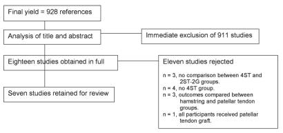
The literature search yielded 1,045 potential articles. Following deletion of duplicates the final yield was 928 articles. The title and abstract of each article were analyzed by the principal researcher (CLA) according to the inclusion and exclusion criteria, allowing immediate exclusion of 911 articles that did not fit the criteria. Full copies of articles were obtained where their appropriateness for inclusion was not able to be determined following review of the title and abstract. Eighteen articles were obtained in full for further evaluation by two reviewers (CLA and KEW). Following this evaluation eleven studies were excluded, leaving seven papers for full review. Figure 1 provides an outline of the process of study identification.
Figure 1.
Outline of the process of study identification.
Study quality
Overall the median quality assessment score was 21 out of a possible 27 (Table 1), with five out of the seven studies reviewed scoring 20 or above. No studies fulfilled the criteria assessing double blinding and reporting of statistical power. Allocation of participants to treatment groups may have been biased in five studies where allocation was conducted according to graft thickness or date of surgery.4,7,9,12,16 Description of the population from which their subjects were recruited was inadequate in all studies. Table 2 provides an overview of the allocation methods, length of follow-up and outcome measures employed by each study.
Table 1. Downs and Black's revised checklist for measuring study quality14 (scores by paper).
| Item | 1 | 2 | 3 | 4 | 5 | 6 | 7 | 8 | 9 | 10 | 11 | 12 | 13 | 14 | 15 | 16 | 17 | 18 | 19 | 20 | 21 | 22 | 23 | 24 | 25 | 26 | 27 | Total |
|---|---|---|---|---|---|---|---|---|---|---|---|---|---|---|---|---|---|---|---|---|---|---|---|---|---|---|---|---|
| Adachi et al. (2003)4 |
1 | 1 | 1 | 1 | 1 | 1 | 1 | 1 | 1 | 0 | 0 | 0 | 1 | 0 | 1 | 1 | 1 | 1 | 1 | 1 | 1 | 1 | 0 | 0 | 1 | 1 | 0 | 20 |
| Carter & Edinger (1999)16 |
1 | 1 | 1 | 1 | 1 | 1 | 1 | 1 | 1 | 0 | 1 | 0 | 1 | 0 | 0 | 1 | 1 | 1 | 1 | 1 | 1 | 0 | 0 | 0 | 1 | 1 | 0 | 19 |
| Gobbi et al. (2005)17 |
1 | 1 | 1 | 1 | 1 | 1 | 1 | 1 | 1 | 1 | 1 | 0 | 1 | 1 | 1 | 1 | 1 | 1 | 1 | 1 | 1 | 1 | 1 | 1 | 1 | 1 | 0 | 25 |
| Lipscomb et al. (1982)13 | 1 | 1 | 1 | 1 | 1 | 1 | 0 | 0 | 1 | 0 | 1 | 0 | 1 | 0 | 0 | 1 | 1 | 1 | 1 | 1 | 1 | 1 | 0 | 0 | 0 | 1 | 0 | 17 |
| Nakamura et al. (2002)9 |
1 | 1 | 1 | 1 | 1 | 1 | 1 | 1 | 1 | 1 | 1 | 0 | 1 | 0 | 0 | 1 | 1 | 1 | 1 | 1 | 1 | 1 | 0 | 0 | 1 | 1 | 0 | 21 |
| Segawa et al. (2002)7 |
1 | 1 | 1 | 1 | 1 | 1 | 1 | 1 | 1 | 1 | 1 | 0 | 1 | 0 | 0 | 1 | 1 | 1 | 1 | 1 | 1 | 1 | 0 | 0 | 1 | 1 | 0 | 21 |
| Tashiro et al. (2003)12 |
1 | 1 | 1 | 1 | 1 | 1 | 1 | 1 | 1 | 0 | 0 | 0 | 1 | 1 | 0 | 1 | 1 | 1 | 1 | 1 | 1 | 1 | 0 | 0 | 1 | 1 | 0 | 21 |
1 = yes; 0 = no, or not determinable from the information reported.
Table 2. Summary of research method by paper.
| Author | Allocation | Follow-up | Outcome measures |
|---|---|---|---|
| Adachi et al. (2003)4 | Non-random; if ST <7mm, G harvested to increase graft thickness | Mean: 35 months (24–58 months) | Knee laxity (KT-2000 knee arthrometer) Active and passive knee flexion angle Isokinetic strength testing (Cybex dynamometer) |
| Gobbi et al. (2005)17 | Random number generator | Examined by surgeon and physiotherapist at 3, 5 and 12 months post-operatively. Examined by independent, blinded examiner at mean 36 months post-operatively | Pre-operative questionnaire, IKDC form, Noyes, Tegner, Lysholm scores; satisfactionand return to sport with Single Assessment Numeric Evaluation (SANE) Clinical knee examination: crepitus, pain, sensation, ROM, kneeling Knee laxity (OSI CA4000) Isokinetic strength testing (Biodex dynamometer) Single leg hop, single leg vertical jump |
| Carter & Edinger (1999)16 | Every third patient consecutively had either PT, ST or ST and G harvest | 24 weeks (24–28 weeks) | Isokinetic strength testing (Cybex dynamometer) |
| Lipscomb et al. (1982)13 | Patients recalled at random from a sample of 482 who had an ACL reconstruction between 1975 and 1980 | 4ST = 34.6 months (18–52 months 2ST-2G = 17.4 months (12–26 months | Isokinetic strength testing (Cybex dynamometer) |
| Nakamura et al. (2002)9 |
Non-random, if ST <7mm, G harvested to increase graft thickness | 24 months | IKDC form Knee laxity (KT-1000 knee arthrometer) Isokinetic strength testing (Cybex dynamometer) Maximum standing knee flexion angle |
| Segawa et al. (2002)7 |
Non-random; if ST <7mm, G harvested to increase graft thickness | 24 months | IKDC form, Lysholm scores Knee laxity (KT-1000 knee arthrometer) Isokinetic strength testing (Cybex dynamometer) |
| Tashiro et al. (2003)12 |
Random; according to date of surgery | 0, 6, 12, 18 months | IKDC form Knee laxity (KT-1000 knee arthrometer) Isokinetic and isometric strength testing (Cybex dynamometer) |
ST = semitendinosus tendon; G = gracilis tendon; PT = patellar tendon.
Surgical procedure and post-operative rehabilitation
Six of the seven studies included for review described the use of a single incision arthroscopically guided procedure performed by the same surgeon for ACL reconstruction. The surgical procedure described by Lipscomb et al.13 was not arthroscopically assisted. This is likely due to the year the study was conducted (1982). The post-operative rehabilitation programs were comprehensively described in all studies. The post-operative rehabilitation protocol described by Lipscomb et al.13 is of note as it had a much longer period of knee joint immobilization (six weeks) when compared to the other studies in this review (usually one day). This difference also likely reflects the year the study was conducted.
Participants
In total, the seven studies reviewed evaluated 519 participants, 265 of whom had a 4ST graft, and 216 who had a 2ST-2G graft. Male participants numbered 246 over the six studies that reported the gender of their participants, while 167 female participants were evaluated. The age range of the participants in the six studies that reported this characteristic was 15–45 years, and the mean age of the participants was 24.8 years. Demographic data for all studies are presented in Table 3.
Table 3. Demographic data.
| Author | Participants | Gender (M:F) | Age (M ± SD) |
|---|---|---|---|
| Adachi et al. (2003)4 |
n=58 | ||
| 4ST=26 | 15:11 | 27.7±10.5yrs | |
| 2ST-2G=18 | 12:6 | 25.6±8.9yrs | |
| Carter & Edinger (1999)16 |
n=106 | Not reported | Not reported |
| 4ST=33 | |||
| 2ST-2G=35 | |||
| Gobbi et al. (2005)17 |
n=115 | 31:19 | Mean: 31.0yrs |
| 4ST=50 | 26:21 | Mean: 28.8yrs | |
| 2ST-2G=47 | |||
| Lipscomb et al. (1982)13 |
n= 51 | 45:6 | |
| 4ST=26 | Mean: 20.3yrs | ||
| 2ST-2G=25 | Mean: 19.5yrs(15–45yrs) | ||
| Nakamura et al. (2002)9 |
n= 74 | ||
| 4ST=49 | 28:21 | Mean: 24.3yrs | |
| 2ST-2G=25 | 6:19 | Mean: 25.7yrs | |
| Segawa et al.7 | n=62 | Mean: 20.8yrs(14–41yrs) | |
| 4ST=32 | 19:13 | ||
| 2ST-2G=30 | 15:15 | ||
| Tashiro et al. (2003)12 |
n=90 | 51:39 | |
| 4ST=49 | 30:19 | 24.5±7.7yrs | |
| 2ST-2G=36 | 19:17 | 24.8±6.4yrs |
Isokinetic strength testing
Significant variability existed in the strength testing protocols, particularly among the angular velocities and torque parameters used, and the ranges of motion through which strength was assessed. All studies considered isokinetic strength outcomes measured using a Cybex (Lumex, Ronkonkoma, New York) or Biodex (Shirley, New York) dynamometer.
Six studies included in this review measured concentric knee flexion peak torque at the 60° s−1 angular velocity4,7,9,12,13,17 and five studies recorded peak torque results at 180° s−1.4,9,12,16,17 Tashiro et al.12 reported a statistically significant reduction in peak torque at six months in the 2ST-2G group at the 180° s−1 angular velocity, compared to the 4ST group. However, this difference had resolved at the later 12 and 18 month reviews, and was not apparent at the 60° s−1 angular velocity. No further significant differences were observed in peak torque between the 4ST and 2ST-2G groups in any of the other six studies, at the 60° s−1 or 180° s−1 test conditions. The isokinetic peak torque results and effect sizes from final follow-up at all angular velocities are presented in Table 4.
Table 4. Isokinetic concentric knee flexor peak torque percentage strength deficit (involved/non-involved limb) and effect size for graft type comparison.
| Angular velocity | ||||||
|---|---|---|---|---|---|---|
| Author | 60° s−1 | 180° s−1 | 240° s−1 | 300° s−1 | ||
| Adachi et al. (2003)4 | 2ST-2G | 95.9% | 109.1% | |||
| 4ST | 98.3% | 101.9% | ||||
| Effect size (d) | 0.20 | 0.31 | ||||
| Carter & Edinger (1999)16 | 2ST-2G | 81.7% | 75.6% | |||
| 4ST | 80.6% | 79.1% | ||||
| Effect size (d) | 0.05 | 0.15 | ||||
| Lipscomb et al. (1982)13 | 2ST-2G | 97.5% | 101.3% | |||
| 4ST | 103.5% | 100.5% | ||||
| Nakamura et al. (2002)9 | 2ST-2G | 91.3% | 86.1% | |||
| 4ST | 93.7% | 89.4% | ||||
| Effect size (d) | 0.17 | 0.21 | ||||
| Segawa et al. (2002)7 | 2ST-2G | 93.6% | ||||
| 4ST | 93.9% | |||||
| Effect size (d) | 0.26 | |||||
| Tashiro et al. (2003)12 | 2ST-2G | 90% | 90% | |||
| 4ST | 93% | 95% | ||||
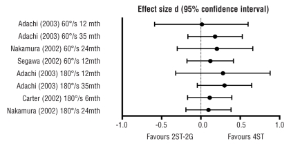
Effect sizes and 95% confidence intervals for the four studies that presented standard deviation data for peak torque,4,7,9,16 are presented in Figure 2. These results demonstrated that the differences between the groups were not statistically significant, although there was a trend in favor of the 4ST graft.
Figure 2.
Concentric knee flexion peak torque, comparison of 4ST and 2ST-2G groups (involved limb; side-to-side ratio).
Nakamura et al.9 also assessed the torque produced at 90° knee flexion at both angular velocities, and found no differences between groups. In contrast, Tashiro et al.12 obtained torques at 70°, 90° and 110° knee flexion from the torque curves recorded at 60° s−1, and found significant hamstring weakness (80% of contralateral in the 2ST-2G group vs. 90% in the 4ST group at 70°; 75% of contralateral in the 2ST-2G group vs. 85% in the 4ST group at 90°; 70% of contralateral in the 2ST-2G group vs. 82% in the 4ST group at 110°) in the 2ST-2G group at all angles at the 18-month review, compared to the 4ST group (p<0.05). The effect sizes and 95% confidence intervals were not able to be calculated as standard deviations for these data were not reported.
Isometric strength testing
Tashiro et al.12 were the only authors to consider isometric strength. They measured maximum isometric knee flexion torque at 70° and 90° in both seated and prone positions. Muscle strength was reported as a percentage of the contralateral limb for each group. The 2ST-2G group was significantly weaker than the 4ST group at 18 months, at angles of 70° (70% of contralateral in the 2ST-2G group vs. 80% in the 4ST group) and 90° (60% of contralateral in the 2ST-2G group vs. 75% in the 4ST group) in prone, and at 70° (80% of contralateral in the 2ST-2G group vs. 90% in the 4ST group) in sitting (p<0.05).
Maximum standing knee flexion angle
Adachi et al.4 and Nakamura et al.9 recorded post-operative active knee flexion range of motion (ROM). Measurements were taken in standing with the hip in neutral flexion/extension and the ankle in maximum plantarflexion. This testing position was chosen in an attempt to minimize the influence of the gastrocnemius and iliopsoas muscle on knee ROM. The authors hypothesized that this measure provided an indirect assessment of strength deficits in higher knee flexion angles.
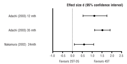
Adachi et al.4 found a significant difference in the loss of active knee flexion angle compared to the contralateral limb between the groups at 12 months (8.9° deficit in the 4ST group vs. 16.7° deficit in the 2ST-2G group; p<0.05), and at 35 months (7.7° deficit in the 4ST group vs. 17.1° deficit in the 2ST-2G group; p<0.05) in the 2ST-2G group when compared to the 4ST group. Nakamura et al.9 also reported a significant deficit in the loss of standing knee flexion angle compared to the contralateral in the 2ST-2G group (5.0° deficit in the 4ST group vs. 9.0° deficit in the 2ST-2G group; p=0.01) when compared to the 4ST group at 24 months. The effect sizes for the data presented in Figure 3 demonstrate the significant deficit in standing knee flexion angle with 2ST-2G harvest.
Figure 3.
Comparison of maximum standing knee flexion angle between graft types.
Clinical evaluation
Subjective outcome measures, post-operative knee laxity and the results of functional testing were reported in six of the included studies.4,7,9,12,13,16 The International Knee Documentation Committee subjective knee form (IKDC form) was the most commonly used post-operative subjective outcome measure. Gobbi et al.17 also reported Single Assessment Numeric Evaluation (SANE) method scores where participants were asked to evaluate their function and symptoms against their contralateral limb. No significant differences between groups were found in subjective outcome. Five studies measured post-operative knee laxity.4,7,9,12,17 No study reported significant differences in knee laxity between groups. Gobbi et al.17 measured each participant's functional ability with a single leg hop and a single limb vertical jump test. The authors found no significant differences between groups. Gobbi et al.17 and Segawa et al.7 also evaluated functional outcome using either the Noyes, Tegner or Lysholm scales. Again, no differences were found between the groups.
Discussion
Overall, the results from this review provide preliminary evidence that harvesting both the semitendinosus and gracilis tendons for ACL reconstruction may produce knee flexion strength deficits at deep knee flexion angles when compared to hamstring grafts where the semitendinosus tendon is harvested alone. Studies reporting no post-operative hamstring strength deficits were those that only examined concentric knee flexion peak torque. This variable has tended to be the most widely reported outcome measure used to evaluate post-operative hamstring strength in this literature. However, this approach may have produced a misleading means of strength evaluation as the knee flexion peak torque is typically generated at shallow knee flexion angles (around 15–30° into the flexion range), and is thought to be primarily a result of biceps femoris contraction.11 Strength testing protocols which assess this parameter in isolation are, therefore, unlikely to find differences in hamstring strength between groups as strength at deeper knee flexion angles, where there is thought to be a predominance of semitendinosus and gracilis activity, is not examined.
One study,12 reported significant isometric hamstring strength deficits in the 2ST-2G group in prone testing at 70° and 90° knee flexion. However, this study was the only study identified which evaluated post-operative isometric hamstring strength. Two studies4,9 have reported deficits in the 2ST-2G group when compared to the 4ST group in the standing knee flexion angle. Both the prone isometric strength and standing knee flexion angle testing protocols require the hip to be in the neutral position. The results of these studies appear to support the contention of Tashiro et al.12 that the semitendinosus and gracilis muscles may play a greater role in knee flexion while the hip is extended. Additionally, when the hip is in a relatively extended position, the biarticular hamstring muscles are approaching active insufficiency as they contract concentrically to simultaneously flex the knee. This may account for the significant differences reported between the groups in prone isometric strength and the standing knee flexion angle, despite no differences being observed in seated isokinetic strength where testing is performed with the hip in approximately 90° flexion. The position of the hip for isokinetic testing, therefore, may increase the mechanical advantage of the hamstrings as they are positioned in a range more conducive to maximal contraction. The gender implications of hamstring weakness in deep knee flexion angles, particularly when the hip is extended, are at present unclear. No study evaluated differences between genders in post-operative hamstring strength. As such, this is an issue requiring further investigation.
Carofino and Fulkerson11 suggest the standing knee flexion angle is sensitive to hamstring strength deficits post-ACL reconstruction. The standing knee flexion angle measure may allow an indirect measure of hamstring strength to be more readily obtained in the clinic without the need for time-consuming isokinetic testing. Furthermore, the results of the standing knee flexion angle may prove useful in guiding the prescription of appropriate post-operative hamstring rehabilitation programs. However, caution is required when interpreting the results of such a measure based on the evidence of two studies, in addition to the absence of evidence validating its sensitivity. It is possible the standing knee flexion angle may be measuring changes occurring due to alterations in the soft tissue architecture that result from tendon harvest and subsequent regeneration.8,18 Subsequently, this measure may not provide an accurate representation of post-operative hamstring strength recovery. The studies reporting significant differences between the groups appear to agree that the deficits observed in deeper angles of knee flexion may influence performance in sports where knee flexion strength is required at deep flexion angles, including gymnastics, judo and wrestling.4,9 Other authors18 have also raised concerns regarding the harvest of 2ST-2G grafts from athletes involved in these sports. Despite this, no study was identified that specifically examined the effects of hamstring tendon harvest in these sporting populations.
Considerable methodological variability exists within the reviewed literature regarding blinding, allocation of patients to intervention groups and inclusion criteria used. Adachi et al.4 and Gobbi et al.17 were the only authors to report assessment of participants by blinded assessors. Gobbi et al.17 and Lipscomb et al.13 also randomly allocated patients to groups; however, the remaining five studies allocated patients according to graft thickness4,7,9 or date of surgery.12,16 The non-random methods of group allocation introduce a selection bias which may restrict result comparability. Bias also arises with differences in the inclusion criteria for patients with concurrent knee pathology. Preferably, only patients with isolated unilateral ACL insufficiency would be included, to eliminate extraneous variables arising from differences in surgical and rehabilitation management. Five of the reviewed studies4,9,12,13,17 excluded patients with history of previous knee injury or knee surgery; however, five studies7,9,12,16,17 included those with concomitant meniscal injury, or failed to adequately report the inclusion criteria applied.
There are three main concerns with the current literature on hamstring tendon harvest for ACL reconstruction. The first is that only one study has reported a direct measure of a reduction in hamstring muscle strength at deep knee flexion angles in a 2ST-2G group when compared to a 4ST group.12 There is a need to replicate these results to further clarify the effects of hamstring graft harvest on post-operative hamstring strength. The second issue is the use of the standing knee flexion angle as a surrogate measure of hamstring strength. It is yet to be determined whether this measure can be used to represent the construct of strength. Thus there is the need for further investigation to explore the association between the standing knee flexion angle and direct measures of hamstring strength obtained from fixed dynamometry. Third, while concern has been raised regarding 2ST-2G graft harvest on the post-operative hamstring strength recovery of patients participating in sports such as gymnastics, judo and wresting, no study has specifically examined these populations. Therefore, a sport-specific analysis of hamstring strength recovery is required to guide graft selection in these patient groups.
Conclusion
This review highlights the limited evidence available to guide hamstring graft selection where preservation of hamstring strength is a priority. Preliminary evidence has been identified to support the harvest of a single semitendinosus tendon rather than in combination with the gracilis tendon where hamstring strength in deep knee flexion angles (≥70° knee flexion) is required. There are a limited number of studies comparing post-operative hamstring strength outcomes obtained between groups following hamstring tendon ACL reconstruction using either a 4ST graft or a 2ST-2G graft. Most of these studies have focused on measuring isokinetic knee flexion peak torque as the main outcome. However, it is likely that this method of evaluation is biased toward assessment of strength in the biceps femoris and semimembranosus muscles, not the semitendinosus and gracilis muscles. Thus the impact of hamstring tendon harvest for ACL reconstruction on post-operative recovery of hamstring muscle strength has not been resolved. In addition, two studies have attempted to use the standing knee flexion angle as a representation of post-operative hamstring strength deficit. While it is possible that this measure may in fact represent a surrogate measure of hamstring strength, its association with hamstring strength is still yet to be determined.
Appendix
Item description for Downs and Black's revised checklist for measuring study quality.14
Item Description
Aims clearly described
Outcomes clearly described
Subject characteristics clearly described
Interventions clearly described
Distribution of principal confounders clearly defined
Main findings clearly described
Estimates of variability provided for main outcomes
Important adverse effects reported
Characteristics of subjects lost to follow-up reported
Actual probabilities reported
Source population identified
Included subjects representative of source population
Intervention representative of that used in source population
Subjects blinded to intervention
Assessors blinded to groups
Use of ‘data dredging’ made clear if used
Same length of follow-up for all subjects
Appropriate statistical tests used
Reliable compliance with intervention
Main outcome measures reliable and valid
Subjects recruited from same population
Subjects recruited over same time
Subjects randomized to intervention groups
Randomization concealed until recruitment complete
Adequate adjustment for confounding
Losses of subjects to follow-up accounted for
Power analysis conducted
References
- 1.Cole BJ, Ernlund LS, Fu FH. Soft tissue problems of the knee. In: Baratz ME, Watson AD, Imbriglia JE, editors. Orthopaedic surgery: The essentials. New York: Thieme Medical Publishers Inc; 1999. pp. 551–60. [Google Scholar]
- 2.Starman JS, Ferretti M, Järvelä T, et al. Anatomy and biomechanics of the anterior cruciate ligament. In: Prodromos CC, editor. The anterior cruciate ligament: Reconstruction and basic science. Philadelphia: Saunders Elsevier; 2008. pp. 3–11. [Google Scholar]
- 3.Sherman OH, Banffy MB. Anterior cruciate ligament reconstruction: Which graft is best? Arthroscopy. J Arthroscopic Related Surg. 2004;20:974–80. doi: 10.1016/j.arthro.2004.08.001. [DOI] [PubMed] [Google Scholar]
- 4.Adachi N, Ochi M, Uchio Y, et al. Harvesting hamstring tendons for ACL reconstruction influences postoperative hamstring muscle performance. Arch Orthop Trauma Surg. 2003;123:460–5. doi: 10.1007/s00402-003-0572-2. [DOI] [PubMed] [Google Scholar]
- 5.Brown CH, Steiner ME, Carson EW. The use of hamstring tendons for anterior cruciate ligament reconstruction. Clinics in Sports Medicine. 1993;12:723–56. [PubMed] [Google Scholar]
- 6.Feller JA, Cooper R, Webster KE. Current Australian trends in rehabilitation following anterior cruciate ligament reconstruction. The Knee. 2002;9:121–6. doi: 10.1016/s0968-0160(02)00009-1. [DOI] [PubMed] [Google Scholar]
- 7.Segawa H, Omori G, Koga Y, et al. Rotational muscle strength of the limb after anterior cruciate ligament reconstruction using semitendinosus and gracilis tendon. Arthroscopy: J Arthroscopic Related Surg. 2002;18:177–82. doi: 10.1053/jars.2002.29894. [DOI] [PubMed] [Google Scholar]
- 8.Makihara Y, Nishino A, Fukubayashi T, Kanamori A. Decrease of knee flexion torque in patients with ACL reconstruction: combined analysis of the architecture and function of the knee flexor muscles. Knee Surgery Sports Traumatology Arthroscopy. 2006;14:310–7. doi: 10.1007/s00167-005-0701-2. [DOI] [PubMed] [Google Scholar]
- 9.Nakamura N, Horibe S, Sasaki S, et al. Evaluation of active knee flexion and hamstring strength after anterior cruciate ligament reconstruction using hamstring tendons. Arthroscopy: J Arthroscopic Related Surg. 2002;18:598–602. doi: 10.1053/jars.2002.32868. [DOI] [PubMed] [Google Scholar]
- 10.Kapandji IA. 5th ed. Edinburgh: Churchill Livingstone; 1987. The physiology of the joints. [Google Scholar]
- 11.Carofino B, Fulkerson J. Medial hamstring tendon regeneration following harvest for anterior cruciate ligament reconstruction: Fact, myth and clinical implication. Arthroscopy: J Arthroscopic Related Surg. 2005;21:1257–64. doi: 10.1016/j.arthro.2005.07.002. [DOI] [PubMed] [Google Scholar]
- 12.Tashiro T, Kurosawa H, Kawakami A, et al. Influence of medial hamstring tendon harvest on knee flexor strength after anterior cruciate ligament reconstruction. J Sports Med. 2003;31:522–9. doi: 10.1177/31.4.522. [DOI] [PubMed] [Google Scholar]
- 13.Lipscomb AB, Johnston RK, Snyder RB, et al. Evaluation of hamstring strength following use of semitendinosus and gracilis tendons to reconstruct the anterior cruciate ligament. J Sports Med. 1982;10:340–2. doi: 10.1177/036354658201000603. [DOI] [PubMed] [Google Scholar]
- 14.Downs SH, Black N. The feasibility of creating a checklist for the assessment of the methodological quality both of randomised and non-randomised studies of health care interventions. J Epidemiol Comm Health. 1998;52:377–84. doi: 10.1136/jech.52.6.377. [DOI] [PMC free article] [PubMed] [Google Scholar]
- 15.Erdfelder E, Faul F, Buchner A. GPOWER: A general power analysis program. Behav Res Methods, Instruments Computers. 1996;28:1–11. [Google Scholar]
- 16.Carter TR, Edinger S. Isokinetic evaluation of anterior cruciate ligament reconstruction: Hamstring versus patellar tendon. Arthroscopy: J Arthroscopic Related Surg. 1999;15:169–72. doi: 10.1053/ar.1999.v15.0150161. [DOI] [PubMed] [Google Scholar]
- 17.Gobbi A, Domzalski M, Pascual J, Zanazzo M. Hamstring anterior cruciate ligament reconstruction: is it necessary to sacrifice the gracilis? Arthroscopy: J Arthroscopic Related Surg. 2005;21:275–80. doi: 10.1016/j.arthro.2004.10.016. [DOI] [PubMed] [Google Scholar]
- 18.Irie K, Tomatsu T. Atrophy of semitendinosus and gracilis and flexor mechanism function after hamstring tendon harvest for anterior cruciate ligament reconstruction. Orthopedics. 2002;25:491–5. doi: 10.3928/0147-7447-20020501-15. [DOI] [PubMed] [Google Scholar]


