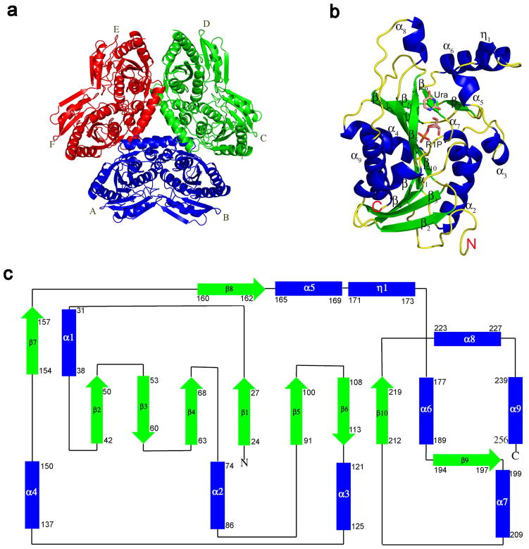
Figure 2.
Structures of SpUP. (a) Hexameric quaternary structure of SpUP color-coded to emphasize the 32 point symmetry or a trimer of dimers. Six subunits are labeled A to F. (b) SpUP protomer with the bound products, R1P and Ura, shown in the active site. The helices are colored blue, the strands green, and the loops yellow. The N-terminus and the C-terminus are also indicated with letters N and C. (c) Topology diagram of SpUP protomer. The colors of the helices and strands are the same as Figure 1b, except the loops are depicted as black lines. The first and last residues of each secondary structure are indicated by numbers.

