Abstract
Barbels are skin sensory appendages found in fishes, reptiles and amphibians. The zebrafish, Danio rerio, develops two pairs of barbels- a short nasal pair and a longer maxillary pair. Barbel tissue contains cells of ectodermal, mesodermal and neural crest origin, including skin cells, glands, taste buds, melanocytes, circulatory vessels and sensory nerves. Unlike most adult tissue, the maxillary barbel is optically clear, allowing us to visualize the development and maintenance of these tissue types throughout the life cycle.
This video shows early development of the maxillary barbel (beginning approximately one month post-fertilization) and demonstrates a surgical protocol to induce regeneration in the adult appendage (>3 months post-fertilization). Briefly, the left maxillary barbel of an anesthetized fish is elevated with sterile forceps just distal to the caudal edge of the maxilla. A fine, sterile spring scissors is positioned against the forceps to cut the barbel shaft at this level, establishing an anatomical landmark for the amputation plane. Regenerative growth can be measured with respect to this plane, and in comparison to the contralateral barbel. Barbel tissue regenerates rapidly, reaching maximal regrowth within 2 weeks of injury.
Techniques for analyzing the regenerated barbel include dissecting and embedding matched pairs of barbels (regenerate and control) in the wells of a standard DNA electrophoresis gel. Embedded specimens are conveniently photographed under a stereomicroscope for gross morphology and morphometry, and can be stored for weeks prior to downstream applications such as paraffin histology, cryosectioning, and/or whole mount immunohistochemistry. These methods establish the maxillary barbel as a novel in vivo tissue system for studying the regenerative capacity of multiple cell types within the genetic context of zebrafish.
Keywords: Developmental Biology, Issue 33, zebrafish, regeneration, barbel, surgery, vasculature, circulation, imaging, agar, embedding, microscopy
Protocol
Zebrafish husbandry
Zebrafish are housed and reared according to standard methods 1. Past the late larval period, the developmental stage of a zebrafish can be measured by its standard length (SL), or the straight line distance in millimeters from the anteriormost point of the upper lip to the base of the caudal fin (posteriormost hypural plate)2. At approximately one month post fertilization (10-12 mm SL), the nasal and maxillary barbels appear as transparent epithelial buds near the olfactory pits and on the posterior corners of the maxillae, respectively. As the zebrafish matures, the barbels extend into narrow, whisker-like appendages. Because the maxillary barbel is larger (2-3 mm) and easier to manipulate, our protocol applies only to this particular appendage; similar techniques, however, could be adapted to the smaller nasal barbel.
Barbel Clipping
Anaesthetize adult zebrafish at the desired stage (e.g., 1.5-2.5 cm SL, ~3-6 months post-fertilization) by immersion in 0.015% MS-222 (ethyl 3-aminobenzoate methanesulfonate) buffered to pH 7.0 in system water. Observe until swim movements stop and gill ventilation becomes slow and regular. Depending on fish size, complete anesthesia takes approximately 2-5 minutes.
Using an aquarium fishnet, transfer 1-3 fish at a time to a folded piece of wet paper towel in a Petri dish. Using a blunt spatula, orient the fish so they are parallel to each other and left lateral side up. Fold part of the wet paper towel over the caudal part of the fish, covering the entire body up to the operculum (gill covers) to keep the skin and gills moist during the procedure.
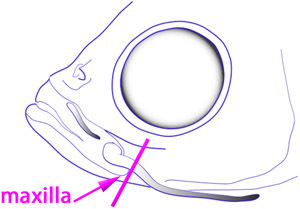
View the Petri plate under the stereomicroscope, using low-angle incident light to illuminate the head region. Focus on the base of the left maxillary barbel, which protrudes from the posterior ventral corner of the maxilla (Fig. 1).
 Figure 1.
Tightly grasp the shaft of the barbel slightly distal to the edge of the maxilla. Elevate the barbel shaft and insert the jaws of a fine-tipped spring scissors just proximal to the forceps. The scissors can be touched to the forceps for stability. Slide the jaws of the scissors along the shaft of the barbel until the cutting edge approaches the edge of the maxilla. Close the scissors to cut through the barbel shaft. The amputated barbel, still gripped in the forceps, can be removed for fixation or further observation.
Immediately transfer the fish to a small tank of clean system water (~500 mL) containing one drop of methylene blue to control superficial fungal infection. Allow the fish to recover overnight. The next morning, return the fish to the rearing system. Barbel regeneration is maximal after 2 weeks.
Agar Embedding of Matched Barbel Pairs
Collect fish by appropriate euthanasia technique and fix in a 50 mL tube of 4% paraformaldehyde-phosphate buffered saline (PBS) with agitation at 4°C overnight. The fixative volume should be at least ten times the volume of the tissue. After fixation, dispose of paraformaldehyde waste in an approved container and rinse the tissue thoroughly in several changes of PBS.
Prepare in a 250 mL glass flask 150 mL of 2% agarose (Tm ~37°C) in distilled water. Heat with a microwave or hot plate, agitating periodically until completely dissolved. Equilibrate the molten solution in a 50-60°C water bath.
Assemble a standard gel electrophoresis rig by placing a small gasketed gel mold (10 x 10 cm; ~100 mL gel volume) firmly against the sides of the buffer chamber. Add 2-4 small-toothed sample combs (4 x 1.5 mm).
On a level surface, pour the melted agarose into the mold to just cover the base of the combs, forming very shallow wells. Immediately put the leftover agarose back into the warm water bath; this will be used later to embed the tissue (Steps 9-12). Put a long glass Pasteur pipette into the flask to keep the pipette to the same temperature as the agarose.
Allow the gel to harden for 20-30 minutes. Remove the combs, then remove the gel mold containing the hardened gel from the buffer chamber. Firmly tape the open sides of the gel mold with laboratory tape so the gel does not slide out. This tape should extend several millimeters over the surface of the agarose so that additional agarose can be added later (Step 13).
Position the gel mold on the stage of a stereomicroscope. Magnify an empty well and bring the edges into focus.
Onto the surface of the agarose adjacent to the well, pipette a drop of water or buffer containing the fixed barbel specimens (e.g., regenerate and control). Using bent insect pins, drag the moist specimens into the well and push them to the bottom. Orient the barbel shafts parallel to each other and aligned against opposite long edges of the well. Surface tension will help hold the tissue in position.
When ready to embed, use a fine pipette tip or the corner of a laboratory tissue to remove excess fluid from the well. Work quickly so as not to let the tissue dry out.
Using the warmed glass Pasteur pipette, gently pipette fresh molten agarose in and around the specimens to seal the barbel tissue in place. Do not overfill. Before the agarose hardens, make last-minute adjustments with the insect pins.
Place a numbered paper specimen label next to the well and secure it with a small drop of agarose.
Repeat steps 6 through 10 for each well of specimens.
If desired for image calibration, insert a piece of paper with a printed micrometer scale (e.g., http://incompetech.com/graphpaper/multiwidth/) into the bottom of an empty well. Flatten the paper to the bottom of the well and cover it with warm agarose.
After all of the specimens are embedded, the surface of the gel will be irregular, with hardened drops of agarose. Place the gel on a flat surface and gently pour in additional melted agarose until the upper surface is uniform. Use the smallest amount of agarose necessary to obtain a smooth surface; too much agarose will obscure transmitted-light photography. Allow this top layer to harden.
The gel can now be photographed on the stage of a stereomicroscope to record gross morphology for each barbel pair. Image calibration is obtained by photographing the embedded micrometer scale.
The entire gel can be stored wrapped in wet paper towels and sealed in a plastic bag at 4° for several weeks.
For further analysis, agar blocks containing matched pair of barbels can be cut out of the gel with a scalpel or razor blade and processed for paraffin histology or cryosectioning by standard methods3,4. Alternatively, individual barbels can be dissected out of the agarose with fine needles, rinsed in water, and processed for other downstream applications (e.g., whole-mount immunohistochemistry or confocal microscopy).
Discussion
The zebrafish maxillary barbel is an underutilized tissue system for studying the growth, maintenance, and regeneration of several cell types in zebrafish. Although the barbel appendage has no human analog, the cell types it contains are highly conserved, making it possible to study skin, glands, melanocytes, circulatory vessels and nerves in an optically clear and anatomically simple cylindrical structure. Similar to the well-studied caudal fin, barbel tissue can be induced to regenerate by amputation. Using the border of the maxilla as an anatomical landmark, the amputation plane can be placed precisely, facilitating the measurement of barbel regrowth. For an experienced operator, each surgery takes only a few seconds. Recovery is rapid, and we have so far detected no short or long-term effects on fish behavior. Zebrafish with one maxillary barbel swim, eat and breed as effectively as non-surgical controls, and have comparable survival to the time of tissue collection, up to 6 months after surgery. The physiological impact of this surgery predicted to be minimal because 1) the extraoral taste buds carried on the barbel are also found on many other parts of the fish epithelium, including the lips, cheeks and head5, and 2) differentiating taste buds appear on the regenerating barbel within 72 hours (LeClair et al., unpublished data.) This makes the maxillary barbel a minimally invasive system for studying wound healing, revascularization, and reinnervation within the context of an adult vertebrate.
After surgical induction of regeneration, barbels can be collected at intervals for morphometric measurement and/or microscopic analysis of fixed tissue. Conveniently, the maxillary barbel is approximately the length (2-3 mm) and diameter (100-200 mm) of a zebrafish embryo, facilitating the application of many standard protocols, including paraffin histology, cryosectioning, whole-mount immunohistochemistry, and in situ hybridization. Taken together, these features make the maxillary barbel a highly feasible in vivo model for studying tissue repair and regeneration.
Acknowledgments
These methods were developed by E.E.L. during a research leave in the lab of J.T. Support was provided by the DePaul University Research Council and an NIH/NIDCR R01 grant to J.T. (DE016678). We gratefully acknowledge the efforts of Caroline Hunter, animal care technician in the CMRC zebrafish core facility and Paulina Pawluczuk, our undergraduate laboratory assistant.
References
- Westerfield M. The zebrafish book. A guide for the laboratory use of zebrafish (Danio rerio) 4th ed. Eugene: Univ. of Oregon Press; 2000. Chapter 1 General Methods for Zebrafish Care. [Google Scholar]
- Howe JC. Standard length: Not quite so standard. Fisheries Research (Amsterdam) 2002;56:1–7. [Google Scholar]
- Fischer AH, Jacobson KA, Rose J, Zeller R. Paraffin Embedding Tissue Samples for Sectioning. Cold Spring Harbor Protocols. 2008;6 doi: 10.1101/pdb.prot4989. [DOI] [PubMed] [Google Scholar]
- Westerfield M. The zebrafish book. A guide for the laboratory use of zebrafish (Danio rerio) 4th ed. Eugene: Univ. of Oregon Press; 2000. Ch 8.3 Agar Embedding for Cryostat Sectioning of Embryos or Larvae. [Google Scholar]
- Hansen A, Reutter K, Zeiske E. Taste bud development in the zebrafish, Danio rerio. Dev Dyn. 2002;223:483–496. doi: 10.1002/dvdy.10074. [DOI] [PubMed] [Google Scholar]


