

Abstract
Most of sequenced West Nile virus (WNV) genomes encode a single N-linked glycosylation site on their envelope (E) proteins. We previously found that WNV lacking the E protein glycan was severely inhibited in its ability to replicate and spread within two important mosquito vector species, Culex pipiens and Cx. tarsalis. However, recent work with a closely related species, Cx. pipiens pallens, found no association between E protein glycosylation and either replication or dissemination. To examine this finding further, we expanded upon our previous studies to include an additional Culex species, Cx. quinquefasciatus. The non-glycosylated WNV-N154I virus replicated less efficiently in mosquito tissues after intrathoracic inoculation, but there was little difference in replication efficiency in the midgut after peroral infection. Interestingly, although infectivity was inhibited when WNV lacked the E protein glycan, there was little difference in viral spread throughout the mosquito. These data indicate that E protein glycosylation affects WNV–vector interactions in a species-specific manner.
Introduction
West Nile virus (WNV) is a member of the family Flaviviridae and genus Flavivirus.1,2 This virus family includes several other medically important arboviruses, including the mosquito-borne viruses St. Louis encephalitis virus, Yellow fever virus, and Dengue virus (DENV), and the tick-borne viruses Tick-borne encephalitis virus and Powassan virus. West Nile virus is naturally maintained through an enzootic transmission cycle between Culex mosquito vectors and avian reservoir hosts. Although small mammals can become infected with WNV and outbreaks in humans and horses regularly occur, mammalian infection is not thought to substantially contribute to viral maintenance in nature.3–7
The envelope (E) protein of most flaviviruses contains an N-linked glycosylation site at amino acids 153/154; DENV contains a second site at E protein amino acid 67. Glycosylation of the E protein is not required for virion formation or infectivity because naturally non-glycosylated isolates of St. Louis encephalitis virus, yellow fever virus, and WNV have been identified.8–15 However, the presence or absence of a glycan on the E protein can affect the viral phenotype. E protein glycosylation increases in vitro infectivity of DENV and WNV, although particle release is significantly inhibited.16,17 In vivo studies have demonstrated that E protein glycosylation enhances WNV virulence in mice and chickens.18–20 Additionally, we have previously found that WNV lacking the E protein glycan replicated less efficiently in vivo and was almost completely defective in spread beyond the mosquito midgut in two important Culex vector species (Cx. pipiens and Cx. tarsalis).21 However, a recent study found no significant differences in WNV titer after intrathoracic inoculation and dissemination after peroral infection of another member of the Cx. pipiens complex, Cx. pipiens pallens.19
Our current study further addresses the possibility that E protein glycosylation affects WNV–vector interactions differently depending on the specific mosquito species, by examining the interactions of previously characterized recombinant viruses in which the E protein glycosylation site is either present or absent with Cx. quinquefasciatus, an important vector species common in the southern United States, South America, Africa, and Asia,22 and comparing these results with our previous results in Cx. pipiens and Cx. tarsalis.21 We first determined whether WNV replication in vivo was affected by the presence of the E protein glycan. We then examined the effects of E protein glycosylation on Cx. quinquefasciatus vector competence. The results of these studies will help clarify the effects of WNV glycosylation on virus-vector relationships and shed additional light on the potential for individual vector species to interact differently with variant WNV strains.
Materials and Methods
Viruses, cells, and mosquitoes.
Baby hamster kidney cells were used for viral growth. Viruses were titrated on African green monkey kidney (Vero) cells. Construction and recovery of the viruses used for these studies (WNV-WT and WNV-N154I) have been described.21,23 WNV-WT is a recombinant virus based on a NY99 genotype strain isolated in New York City.23 WNV-N154I contains an asparagine to isoleucine mutation at amino acid 154 in the E protein, but is otherwise identical to WNV-WT.21 All experiments involving infectious WNV were performed in the BioSafety Level 3 Laboratories at Wadsworth Center Arbovirus Laboratories.
Culex quinquefasciatus mosquitoes were derived from a laboratory colony (F30+) kindly provided by Dina Fonseca, who originally received egg rafts obtained in the southern United States from Benzon Research, Inc. (Carlisle, PA). Colonized mosquitoes were maintained on goose blood (for egg laying) and given 10% sucrose ad libitum. Larvae were reared and adults maintained under controlled conditions of temperature (27°C), humidity (70% relative humidity), and light (16:8 hour light:dark diurnal cycle) in 12² × 12² × 12² cages. All mosquito experiments involving infectious WNV were conducted in insectaries in the BioSafety Level Laboratories.
Viral replication in mosquito bodies after intrathoracic exposure.
WNV-WT and WNV-N154I were diluted in mosquito diluent (MD) (20% heat-inactivated fetal bovine serum in Dulbecco's phosphate-buffered saline plus 50 μg/mL of penicillin/streptomycin, 50 μg/mL of gentamicin, and 2.5 μg/mL of fungizone) to a titer of 105 plaque-forming units (PFU)/mL. Three-to-five day–old mosquitoes were inoculated intrathoracically with 10 PFU of either WNV-WT or WNV-N154I under CO2 anesthesia and held at 27°C for a 16:8 hour light:dark photoperiod for up to 10 days post-inoculation. At daily intervals from 0 to 10 days post-inoculation, 10 mosquitoes inoculated with each virus were removed to individual aliquots of 1 mL of MD. All samples were stored at –80°C. Mosquitoes were homogenized by using a mixer mill (Qiagen, Valencia, CA) and clarified by centrifugation. The viral loads of infected mosquitoes were determined by plaque titration on Vero cells and compared by using analysis of variance (ANOVA).
Viral replication in mosquito salivary glands after intrathoracic exposure.
Three-to-five day–old mosquitoes were intrathoracically inoculated with either WNV-WT or WNV-N154I, as described above, and held for up to 10 days post-inoculation. At 2, 4, 6, 8, and 10 days post-inoculation, salivary glands from 5–10 mosquitoes per virus were removed, washed in MD to remove hemolymph, and individually stored in 0.5 mL of MD at –80°C. Salivary glands were homogenized by using a mixer mill and clarified by centrifugation. Viral loads of infected salivary glands were determined by plaque titration on Vero cells and compared by using ANOVA. Salivary gland infection rates were compared by using Fisher's exact test.
Viral replication in mosquito midguts after peroral exposure.
Five-to-seven day–old adult female mosquitoes were deprived of sucrose for 48 hours before feeding. Either WNV-WT or WNV-N154I was added to 5 mL of defibrinated goose blood containing 2.5% sucrose to a final titer of 108 PFU/mL. Mosquitoes were fed for 1–2 hours by using a Hemotek membrane feeding apparatus (Discovery Workshops, Accrington, United Kingdom) as recommended by the manufacturer. After feeding, fully engorged mosquitoes were separated under CO2 anesthesia into 0.5-liter cartons supplied with 10% sucrose ad libitum, and held at 27°C for a 16:8 hour light-dark photoperiod for up to 14 days post-feeding. At 2, 4, 6, 8, and 10 days post-feeding, midguts from 10–20 mosquitoes per virus were removed, washed in MD to remove blood and/or hemolymph, and individually stored in 0.5 mL of MD at –80°C. Midguts were homogenized by using a mixer mill and clarified by centrifugation. The viral loads of infected midguts were determined by plaque titration on Vero cells and compared by using ANOVA.
Vector competence of mosquitoes.
Five-to-seven day–old adult female mosquitoes were fed blood meals containing either WNV-WT or WNV-N154I, as described above. After feeding, fully engorged mosquitoes were separated under CO2 anesthesia into 0.5-liter cartons supplied with 10% sucrose ad libitum, and held at 27°C for a 16:8 hour light:dark photoperiod for up to 20 days post-feeding. Transmission was evaluated at 7, 9, 14, and 20 days post-feeding in vitro, essentially as described.24 Briefly, 50–75 mosquitoes per virus were anesthetized with triethylamine (Sigma, St. Louis, MO), and their legs were removed and placed in 1 mL of MD. Mosquito mouthparts were placed into a capillary tube containing approximately 10 μL of a 1:1 mixture of fetal bovine serum and 50% sucrose for approximately 30 minutes, after which the contents of the capillary tube were expelled into 0.3 mL of MD. Mosquito bodies were placed in 1 mL of MD, and all samples were stored at –80°C. Mosquito bodies and legs were homogenized by using a mixer mill and clarified by centrifugation. The proportion of mosquitoes with infected bodies, legs, and salivary secretions was determined by plaque assay on Vero cells. Infection, dissemination, and transmission were defined as the proportion of mosquitoes with infected bodies, legs, and salivary secretions, respectively. Proportions were compared by using Fisher's exact test.
Sequencing of mosquito-derived viral genomes.
Viral genomic RNA was purified from infected bodies, legs, and salivary secretions of mosquitoes that fed on WNV-N154I using RNeasy spin columns (Qiagen) according to the manufacturer's instructions. A one-step reverse transcription–polymerase chain reaction (PCR) (Qiagen) was performed by using primers designed to amplify the 500-basepair region of the E gene surrounding the glycosylation site (nucleotides 1,208–1,700). PCR products were purified and visualized by electrophoresis on 1% agarose gels. Automated sequencing of purified PCR products was performed at the Wadsworth Center Applied Genomics Technology Core.
Results
Replicative ability of WNV-WT and WNV-N154I in vivo.
Adult female Cx. quinquefasciatus were intrathoracically inoculated with either WNV-WT or WNV-N154I to examine whether the glycosylation state of the E protein affected viral replication in this mosquito species. Titers in whole mosquitoes inoculated with WNV-N154I were significantly lower than those inoculated with WNV-WT beginning at 1 day post-inoculation and continuing throughout the experiment (P < 0.01; Figure 1A). The infection rate after inoculation was also significantly higher over the course of the experiment in mosquitoes inoculated with WNV-WT than in those inoculated with WNV-N154I (92% versus 66%; P < 0.0001).
Figure 1.
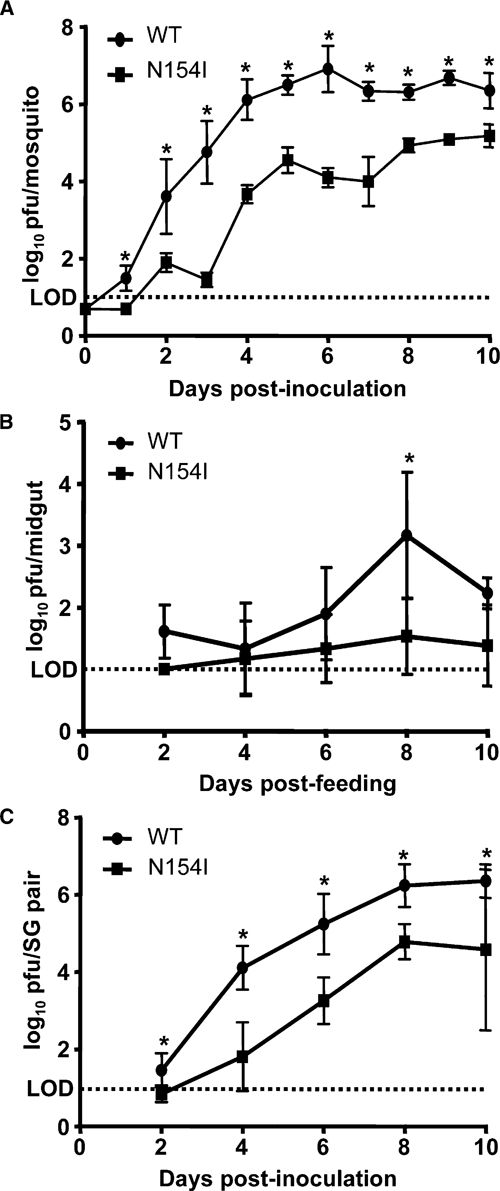
Viral replication of West Nile virus WNV-WT and WNV-N154I in Culex quinquefasciatus. A, Mosquitoes were intrathoracically inoculated with 10 plaque-forming units (PFU) of either WNV-WT or WNV-N154I and held for up to 10 days. At daily intervals, 10 mosquitoes per virus were removed and the viral titers present in their bodies were determined by plaque titration on Vero cells. Mean viral titers from infected mosquitoes at each time point are shown as log10 PFU/mosquito. B, Mosquitoes were fed on defibrinated goose blood containing 108 PFU/mL of either WNV-WT or WNV-N154I and held for up to 10 days post-feeding. At the indicated time points, midguts were removed from 20 mosquitoes per virus and their viral titers were determined by plaque titration on Vero cells. Mean viral titers from infected midguts at each time point are shown as log10 PFU/midgut. C, Mosquitoes were intrathoracically inoculated with 10 PFU of either WNV-WT or WNV-N154I and held for up to 10 days. At the indicated time points, salivary glands were removed from 10 mosquitoes per virus and their viral titers were determined by plaque assay on Vero cells. Mean viral titers from infected salivary gland pairs at each time point are shown as log10 PFU/salicary gland pair. LOD = limit of detection. *P ≤ 0.05.
We also examined the effect of E protein glycosylation on WNV replication in Cx. quinquefasciatus midguts after peroral exposure and salivary glands after inoculation. Mosquitoes exposed perorally to WNV-WT had slightly higher viral loads in their midguts than those exposed to WNV-N154I. However, the differences were only significant at 8 days post-feeding (P = 0.02; Figure 1B). WNV-WT replicated to significantly higher titers in mosquito salivary glands at days 4, 6, and 8 post-intrathoracic inoculation (P < 0.01; Figure 1C).
Vector competence of Cx. quinquefasciatus.
To assess the importance of E protein glycosylation on the ability of WNV to infect and spread within Cx. quinquefasciatus mosquitoes after peroral exposure, we determined their vector competence for WNV-WT and WNV-N154I. Infection rates of WNV-WT were significantly higher than those of WNV-N154I at all days post-feeding (Table 1). However, viral loads in the bodies of mosquitoes infected with WNV-WT were similar to those observed in those of WNV-N154I-infected mosquitoes at 7, 14, and 20 days post-feeding (Table 1).
Table 1.
Vector competence of Culex quinquefasciatus after peroral infection with West Nile virus*
| Virus | Days post-feeding | |||||||||||||||
|---|---|---|---|---|---|---|---|---|---|---|---|---|---|---|---|---|
| 7 | 9 | 14 | 20 | |||||||||||||
| I | D | T | Titer (95% CI) | I | D | T | Titer (95% CI) | I | D | T | Titer (95%CI) | I | D | T | Titer (95% CI) | |
| WNV-WT | 39 (n = 123) | 35 (n = 48) | 21 | 3.2 (2.7–3.8) | 37 (n = 100) | 38 (n = 37) | 22 | 3.5 (3.0 –4.0) | 43 (n = 100) | 30 (n = 43) | 19 | 3.5 (3.0–4.1) | 51 (n = 96) | 43 (n = 49) | 22 | 3.1 (2.7–3.6) |
| WNV-N154I | 16 (n = 125) | 75 (n = 20) | 30 | 3.9 (3.1–4.7) | 15 (n = 125) | 32 (n = 19) | 21 | 2.4 (1.7–3.1) | 13 (n = 125) | 53 (n = 16) | 35 | 3.4 (2.7–4.1) | 16 (n = 117) | 42 (n = 19) | 21 | 3.1 (2.4–3.7) |
| P† | < 0.0001 | 0.0037 | 0.532 | 0.160 | 0.0002 | 0.772 | 1.00 | 0.008 | < 0.0001 | 0.139 | 0.19 | 0.789 | < 0.0001 | 1.00 | 1.00 | 0.857 |
I = % infected; D = % disseminated (of infected); T = % transmitting (of infected); Titer = mean log10 plaque-forming units/mosquito body; CI = confidence interval.
By Fisher's exact test. Statistically significant values are shown in bold.
Viral dissemination in WNV-WT–infected mosquitoes was significantly lower than in mosquitoes infected with WNV-N154I at 7 days post-feeding, but no significant differences in dissemination were observed at 9, 14, or 20 days post-feeding (Table 1). No significant differences were found in transmission of WNV-WT or WNV-N154I at any individual day post-feeding (Table 1) or in overall transmission over the course of the experiment. We sequenced a 500-basepair region of the E gene from viruses present in the salivary secretions of Cx. quinquefasciatus that fed on WNV-N154I (n = 20); in all cases, the virus in the salivary secretions was the virus on which the mosquito had fed.
Discussion
Although glycosylation of flavivirus premembrane proteins appears to be absolutely required for efficient virion assembly and maturation,16,25–28 WNV strains lacking a glycosylation site in the E protein have been isolated from naturally infected vertebrates and mosquitoes.8–13 Previously, we have shown that WNV lacking the E protein glycan is attenuated in the natural vector species Cx. pipiens and Cx. tarsalis.21 In the current study, we expanded upon our previous work to examine the effect of E protein glycosylation on WNV interactions with a third important vector species in the United States (Cx. quinquefasciatus) because vector competence differences have been noted among members of the Cx. pipiens species complex.
We first examined whether E protein glycosylation affected WNV replication in Cx. quinquefasciatus mosquitoes. WNV-WT replicated to significantly higher titers than WNV-N154I in the bodies and salivary glands of intrathoracically inoculated mosquitoes (Figure 1A and C), similar to previous results for Cx. pipiens and Cx. tarsalis.21 However, results in perorally infected mosquitoes differed strikingly from our previous data. Although WNV-WT titers were slightly higher than those of WNV-N154I in Cx. quinquefasciatus midguts, the differences were only significant at 8 days post-feeding (Figure 1B), whereas in Cx. pipiens and Cx. tarsalis WNV-WT titers were significantly higher at all days post-feeding.21 Overall viral loads in mosquitoes infected with WNV-WT and WNV-N154I through feeding were also similar in Cx. quinquefasciatus (Table 1), and significant differences were observed in Cx. pipiens and Cx. tarsalis.21
We then examined the effect of E protein glycosylation on Cx. quinquefasciatus vector competence after peroral infection. Although the infection rate of WNV-N154I was significantly lower than that of WNV-WT at all days post-feeding, dissemination and transmission of both viruses were similar (Table 1). These results also differ strikingly from our previous data, in which dissemination and transmission in Cx. pipiens and Cx. tarsalis were significantly inhibited by the lack of an E protein glycan.21
Because of the contrast between these results, we sequenced the E genes of viruses present in the salivary secretions of Cx. quinquefasciatus that had fed on WNV-N154I to determine if they had reverted to a wild-type glycosylation phenotype. In our previous study, WNV presence in the salivary secretions of Cx. pipiens and Cx. tarsalis that had fed on WNV-N154I was strongly associated with reversion to a wild-type sequence at the E protein glycosylation site.21 However, in the current study, all of the viral E genes sequenced contained the N154I mutation that had been engineered into the virus on which the mosquitoes had fed. Our results in this study indicate that although E protein glycosylation has significant effects on WNV replication in and infectivity for Cx. quinquefasciatus mosquitoes, it has little to no effect on viral spread within this vector species.
Our results appear to contradict recent work by Murata and others that found no detectable difference between in vivo replication or disseminated infection of glycosylated and non-glycosylated WNV.19 Several differences between our studies and the study of Murata and others could contribute to the apparent discrepancy between our results. The viruses used in the study of Murata and others contained genome changes in addition to those that affect E protein glycosylation, whereas our viruses differed by a single amino acid. The mutation resulting in the loss of the E protein glycosylation site differed between our studies and the study of Murata and others, which could have affected the results. However, other groups have used several different amino acid mutations to remove the E protein glycosylation site and have obtained similar results in vitro and in mice.16,20,29 Therefore, would not expect that our results were caused solely by the presence of an isoleucine at anino acid position 154, rather than the lack of the E protein glycan. Additionally, if an isoleucine at amino acid position 154 was universally detrimental to the virus, we would expect to see reversion to the wild-type asparagine in Cx. quinquefasciatus, as we had previously seen in other Culex species. However, no reversion was detected in any Cx. quinquefasciatus that fed on WNV-N154I.
Perhaps most importantly, the study of Murata and others used Cx. pipiens pallens mosquitoes, another member of the Cx. pipiens species complex. As we describe in this report, WNV–vector interactions may differ significantly depending on the vector species, even among members of the Cx. pipiens species complex, such as Cx. pipiens and Cx. quinquefasciatus. Therefore the inconsistency between these results is not entirely surprising. It would be interesting to determine the genetic basis for the differences in WNV–vector interactions between closely related mosquito species. The differences in infectivity and spread in individual Culex species also suggest that WNV midgut infection and escape barriers may differ in a species-specific manner. Future work examining the impact of viral sequence changes and vector species on these barriers should yield additional information regarding how WNV interacts with its various vector mosquito species.
Although WNV strains lacking a glycosylation site on their E proteins have been isolated in nature, their frequency of isolation is fairly low.8–13 Multiple studies have found that WNV strains lacking the E protein glycosylation site are less infectious for mammalian and avian hosts than glycosylated strains.13,18–20 Additionally, our data indicate that the lack of E protein glycosylation decreases WNV infectivity and/or spread within several species of vector mosquitoes, both of which have the overall effect of decreasing the likelihood that a mosquito will transmit a non-glycosylated strain (Table 1).21 It is interesting to note that there is significant overlap between the worldwide Cx. quinquefasciatus distribution and the regions from which non-glycosylated WNV strains have been isolated.8–13 Because transmission of WNV by some vector species, including Cx. quinquefasciatus, does not appear to be affected by E protein glycosylation, this finding could indicate that low level transmission of non-glycosylated strains by permissive mosquito species enables this subset of viruses to persist in nature. However, even in regions where Cx. quinquefasciatus is the predominant WNV vector, such as in the southern United States, non-glycosylated strains are only isolated intermittently, which is likely caused by the infectivity advantage of glycosylated strains for avian hosts.19 The combination of decreased transmission of non-glycosylated strains by mosquito vectors and low infectivity of these strains for vertebrate hosts could explain the apparent dearth of naturally non-glycosylated WNV isolates.
ACKNOWLEDGMENTS
We thank Pamela Chin for excellent technical assistance, Dina Fonseca for providing mosquitoes to establish the Cx. quinquefasciatus colony, the Wadsworth Center Tissue Culture Core facility for providing Vero cells, and the Wadsworth Center Applied Genomics Technologies Core facility for providing DNA sequencing services.
Footnotes
Financial support: This study was supported by National Institute of Allergy and Infectious Disease contract NO1-AI25490. Robin M. Moudy was supported by a Ruth L. Kirschstein National Research Service Award from the National Institute of Allergy and Infectious Disease, National Institutes of Health (1F32-AI074238).
Authors' addresses: Robin M. Moudy, Anne F. Payne, Brittany L. Dodson, and Laura D. Kramer, Arbovirus Laboratories, Wadsworth Center, New York State Department of Health, Slingerlands, NY, E-mails: rmoudy@wadsworth.org, apayne@wadsworth.org, bdodson@wadsworth.org, and kramer@wadsworth.org.
References
- 1.Gubler D, Kuno G, Markoff L. In: Fields Virology. Fifth edition. Knipe DM, Howley PM, editors. Philadelphia, PA: Lippincott William and Wilkins; 2007. pp. 1153–1252. (Flaviviruses). [Google Scholar]
- 2.Lindenbach BD, Thiel HJ, Rice CM. Fields Virology. Fifth edition. Lippincott William and Wilkins; 2007. pp. 1101–1152. (Flaviviridae: The Virus and Their Replication). [Google Scholar]
- 3.Gomez A, Kramer LD, Dupuis AP, Kilpatrick AM, Davis LJ, Jones MJ, Daszak P, Aguirre AA. Experimental infection of eastern gray squirrels (Sciurus carolinensis) with West Nile virus. Am J Trop Med Hyg. 2008;79:447–451. [PMC free article] [PubMed] [Google Scholar]
- 4.Platt KB, Tucker BJ, Halbur PG, Tiawsirisup S, Blitvich BJ, Fabiosa FG, Bartholomay LC, Rowley WA. West Nile virus viremia in eastern chipmunks (Tamias striatus) sufficient for infectiong different mosquitoes. Emerg Infect Dis. 2007;13:831–837. doi: 10.3201/eid1306.061008. [DOI] [PMC free article] [PubMed] [Google Scholar]
- 5.Root JJ, Oesterle PT, Nemeth NM, Klenk K, Gould DH, McLean RG, Clark L, Hall JS. Experimental infection of fox squirrels (Sciurus niger) with West Nile virus. Am J Trop Med Hyg. 2006;75:697–701. [PubMed] [Google Scholar]
- 6.Weaver SC, Barrett ADT. Transmission cycles, host range, evolution and emergence of arboviral disease. Nat Rev Microbiol. 2004;2:789–801. doi: 10.1038/nrmicro1006. [DOI] [PMC free article] [PubMed] [Google Scholar]
- 7.Root JJ, Bentler KT, Nemeth NM, Gidlewski T, Spraker TR, Franklin AB. Experimental infection of raccoons (Procyon lotor) with West Nile virus. Am J Trop Med Hyg. 2010;83:803–807. doi: 10.4269/ajtmh.2010.10-0173. [DOI] [PMC free article] [PubMed] [Google Scholar]
- 8.Adams SC, Broom AK, Sammels LM, Hartnett AC, Howard MJ, Coelen RJ, MacKenzie JS, Hall RA. Glycosylation and antigenic variation among Kunjin virus isolates. Virology. 1995;206:49–56. doi: 10.1016/s0042-6822(95)80018-2. [DOI] [PubMed] [Google Scholar]
- 9.Berthet FX, Zeller HG, Drouet MT, Rauzier J, Digoutte JP, Deubel V. Extensive nucleotide changes and deletions within the envelope glycoprotein gene of Euro-African West Nile viruses. J Gen Virol. 1997;78:2293–2297. doi: 10.1099/0022-1317-78-9-2293. [DOI] [PubMed] [Google Scholar]
- 10.Davis CT, Ebel GD, Lanciotti RS, Brault AC, Guzman H, Siirin M, Lambert A, Parsons RE, Beasley DW, Novak RJ, Elizondo-Quiroga D, Green EN, Young DS, Stark LM, Drebot MA, Artsob H, Tesh RB, Kramer LD, Barrett AD. Phylogenetic analysis of North American West Nile virus isolates, 2001–2004: evidence for the emergence of a dominant genotype. Virology. 2005;342:252–265. doi: 10.1016/j.virol.2005.07.022. [DOI] [PubMed] [Google Scholar]
- 11.Ebel GD, Carricaburu J, Young D, Bernard KA, Kramer LD. Genetic and phenotypic variation of West Nile virus in New York, 2000–2003. Am J Trop Med Hyg. 2004;71:493–500. [PubMed] [Google Scholar]
- 12.Lanciotti RS, Ebel GD, Deubel V, Kerst AJ, Murri S, Meyer R, Bowen M, McKinney N, Morrill WE, Crabtree MB, Kramer LD, Roehrig JT. Complete genome sequences and phylogenetic analysis of West Nile virus strains isolated from the United States, Europe, and the Middle East. Virology. 2002;298:96–105. doi: 10.1006/viro.2002.1449. [DOI] [PubMed] [Google Scholar]
- 13.Beasley DW, Davis CT, Estrada-Franco J, Navarro-Lopez R, Campomanes-Cortes A, Tesh RB, Weaver SC, Barrett AD. Genome sequence and attenuating mutations in West Nile virus isolate from Mexico. Emerg Infect Dis. 2004;10:2221–2224. doi: 10.3201/eid1012.040647. [DOI] [PMC free article] [PubMed] [Google Scholar]
- 14.Vorndam V, Mathews JH, Barrett ADT, Roehrig JT, Trent DW. Molecular and biological characterization of a non-glycosylated isolate of St. Louis encephalitis virus. J Gen Virol. 1993;74:2653–2660. doi: 10.1099/0022-1317-74-12-2653. [DOI] [PubMed] [Google Scholar]
- 15.Post PR, Santos CN, Carvalho R, Cruz AC, Rice CM, Galler R. Heterogeneity in envelope protein sequence and N-linked glycosylation among yellow fever virus vaccine strains. Virology. 1992;188:160–167. doi: 10.1016/0042-6822(92)90745-b. [DOI] [PubMed] [Google Scholar]
- 16.Hanna SL, Pierson TC, Sanchez MD, Ahmed AA, Murtadha MM, Doms RW. N-linked glycosylation of west nile virus envelope proteins influences particle assembly and infectivity. J Virol. 2005;79:13262–13274. doi: 10.1128/JVI.79.21.13262-13274.2005. [DOI] [PMC free article] [PubMed] [Google Scholar]
- 17.Lee E, Leang SK, Davidson A, Lobigs M. Both E protein glycans adversely affect dengue virus infectivity but are beneficial for virion release. J Virol. 2010;84:5171–5180. doi: 10.1128/JVI.01900-09. [DOI] [PMC free article] [PubMed] [Google Scholar]
- 18.Beasley DW, Whiteman MC, Zhang S, Huang CY, Schneider BS, Smith DR, Gromowski GD, Higgs S, Kinney RM, Barrett AD. Envelope protein glycosylation status influences mouse neuroinvasion phenotype of genetic lineage 1 West Nile virus strains. J Virol. 2005;79:8339–8347. doi: 10.1128/JVI.79.13.8339-8347.2005. [DOI] [PMC free article] [PubMed] [Google Scholar]
- 19.Murata R, Eshita Y, Maeda A, Maeda J, Akita S, Tanaka T, Yoshii K, Kariwa H, Umemura T, Takashima I. Glycosylation of the West Nile virus envelope protein increases in vivo and in vitro viral multiplication in birds. Am J Trop Med Hyg. 2010;82:696–704. doi: 10.4269/ajtmh.2010.09-0262. [DOI] [PMC free article] [PubMed] [Google Scholar]
- 20.Shirato K, Miyoshi H, Goto A, Ako Y, Ueki T, Kariwa H, Takashima I. Viral envelope protein glycosylation is a molecular determinant of the neuroinvasiveness of the New York strain of West Nile virus. J Gen Virol. 2004;85:3637–3645. doi: 10.1099/vir.0.80247-0. [DOI] [PubMed] [Google Scholar]
- 21.Moudy RM, Zhang B, Shi PY, Kramer LD. West Nile virus envelope protein glycosylation is required for efficient viral transmission by Culex vectors. Virology. 2009;387:222–228. doi: 10.1016/j.virol.2009.01.038. [DOI] [PMC free article] [PubMed] [Google Scholar]
- 22.Peng Z, Li H, Simons FE. Immunoblot analysis of salivary allergens in 10 mosquito species with worldwide distribution and the human IgE responses to these allergens. J Allergy Clin Immunol. 1998;101:498–505. doi: 10.1016/S0091-6749(98)70357-4. [DOI] [PubMed] [Google Scholar]
- 23.Shi PY, Tilgner M, Lo MK, Kent KA, Bernard KA. Infectious cDNA clone of the epidemic West Nile virus from New York City. J Virol. 2002;76:5847–5856. doi: 10.1128/JVI.76.12.5847-5856.2002. [DOI] [PMC free article] [PubMed] [Google Scholar]
- 24.Aitken TH. An in vitro feeding technique for artificially demonstrating virus transmission by mosquitoes. Mosq News. 1977;37:130–133. [Google Scholar]
- 25.Goto A, Yoshii K, Obara M, Ueki T, Mizutani T, Kariwa H, Takashima I. Role of the N-linked glycans of the prM and E envelope proteins in tick-borne encephalitis virus particle secretion. Vaccine. 2005;23:3043–3052. doi: 10.1016/j.vaccine.2004.11.068. [DOI] [PubMed] [Google Scholar]
- 26.Konishi E, Mason PW. Proper maturation of the Japanese encephalitis virus envelope glycoprotein requires cosynthesis with the premembrane protein. J Virol. 1993;67:1672–1675. doi: 10.1128/jvi.67.3.1672-1675.1993. [DOI] [PMC free article] [PubMed] [Google Scholar]
- 27.Lorenz IC, Allison SL, Heinz FX, Helenius A. Folding and dimerization of tick-borne encephalitis virus envelope proteins prM and E in the endoplasmic reticulum. J Virol. 2002;76:5480–5491. doi: 10.1128/JVI.76.11.5480-5491.2002. [DOI] [PMC free article] [PubMed] [Google Scholar]
- 28.Ocazionez Jimenez R, Lopes da Fonseca BA. Recombinant plasmid expressing a truncated dengue-2 virus E protein without co-expression of prM protein induces partial protection in mice. Vaccine. 2000;19:648–654. doi: 10.1016/s0264-410x(00)00247-4. [DOI] [PubMed] [Google Scholar]
- 29.Beasley DW, Whiteman MC, Zhang S, Huang CY, Schneider BS, Smith DR, Gromowski GD, Higgs S, Kinney RM, Barrett AD. Envelope protein glycosylation status influences mouse neuroinvasion phenotype of genetic lineage 1 West Nile virus strains. J Virol. 2005;79:8339–8347. doi: 10.1128/JVI.79.13.8339-8347.2005. [DOI] [PMC free article] [PubMed] [Google Scholar]