Abstract
Herpesvirus of turkey (HVT) is being widely used as a vector for development of recombinant vaccines and US2 and US10 genes are often chosen as insertion sites for targeted gene expression. However, the different effects of the two genes for generation of recombinant HVT vaccines were unknown. In order to compare the effects of inserted genes in the two sites on the efficacy of the recombinant vaccines, host-protective haemagglutinin (HA) gene of the highly pathogenic avian influenza virus (HPAIV) H5N1 was inserted into either US2 or US10 gene locus of the HVT. The resulting US2 (rHVT-US2-HA) or US10 (rHVT-US10-HA) recombinant HVT viruses were used to infect chicken embryo fibroblasts. Plaques and the growth kinetics of rHVT-US2-HA-infected chicken embryo fibroblasts were similar to those of parental HVT whereas rHVT-US10-HA infected chicken embryo fibroblasts had different growth kinetics and plaque formation. The viremia levels in rHVT-US10-HA virus-infected chickens were significantly lower than those of rHVT-US2-HA group on 28 days post infection. The vaccine efficacy of the two recombinant viruses against H5N1 HPAIV and virulent Marek's disease virus was also evaluated in 1-day-old vaccinated chickens. rHVT-US2-HA-vaccinated chickens were better protected with reduced mortality than rHVT-US10-HA-vaccinated animals following HPAIV challenge. Furthermore, the overall hemaglutination inhibition antibody titers of rHVT-US2-HA-vaccinated chickens were higher than those of rHVT-US10-HA-vaccinated chickens. Protection levels against Marek's disease virus challenge following vaccination with either rHVT-US2-HA or rHVT-US10-HA, however, were similar to those of the parental HVT virus. These results, for the first time, indicate that US2 gene provides a favorable foreign gene insertion site for generation of recombinant HVT vaccines.
Introduction
Herpesvirus of turkey (HVT) is a naturally occurring, non-pathogenic alphaherpesvirus originally isolated from domestic turkeys in the late of 1960s [1]. HVT is a member of the genus Mardivirus and is antigenically and genetically related to Marek's disease (MD) virus (MDV), the etiologic agent of the globally and economically significant Marek's disease in chickens [2], [3]. MDV is a chicken pathogen that results in the development of T-cell lymphomas and mononuclear infiltration of peripheral nerves in a matter of weeks following infection [2]. Since antigenic similarities between MDV and HVT have been documented, these similarities have been exploited in the context of vaccination strategies, that is, HVT vaccination of chickens has resulted in long-lasting, protective immunity against MD [4], [5]. Since the early 1970s chicken vaccinations with HVT have dramatically reduced MD-related losses [6].
HVT not only serves as a viable vaccine option for prevention of MD but can also be used as a vector for development of recombinant vaccines. Specifically, HVT provides an efficient delivery system for immunogenic genes that can facilitate the control of multiple poultry-related diseases. HVT possesses some ideal characteristics: (1) HVT is a herpesvirus that infects chickens persistently, resulting in continuous immune system stimulation that helps maintain protective antibody levels elevated, (2) HVT vaccine is also available in a cell-free ‘dry’ (lyophilized) form that is convenient for long-term storage and transport [7], [8] and (3) MDV genome is large enough to accommodate multiple foreign genes. Recombinant HVT (rHVT) vaccine has been proven to be one of useful viral vectors of targeted gene expression and developed for the prevention of diseases caused by infections with various fowl disease-associated viruses [7], [9]–[13].
Some genes in some alphaherpesviruses have been reported as ‘nonessential’ for viral growth in cell culture, but ‘nonessential’ genes can be used in the context of specific in vitro systems and do not necessarily suggest that a respective gene product is nonessential in all in vitro or in vivo models. Nevertheless, nonessential genes are usually the targets of foreign gene insertions for design of alphaherpesvirus vectors [1], [10]. In the context of the herpesvirus genome, the unique short (US) 1, US2, US10 and thymidine kinase genes have been defined as ‘nonessential’ for growth in cell cultures [11], [14], [15] and the US2 and US10 genes have been used as insertion sites for foreign genes in development of recombinant HVT or MDV. For example, when a recombinant CVI-988 (rCVI-988) expressing infectious bursal disease virus (IBDV) VP2 at the US2 site was engineered, vaccination with this recombinant vaccine conferred partial protection against virulent IBDV (>55%) and full protection against vvMDV challenge [16]. Baigent et al. constructed a full-length infectious bacterial artificial chromosome (BAC) clone consisting of HVT (HVT-BAC) following insertion into the US2 locus and these HVT-BAC clones conferred 100% protection against vMDV challenge [1]. In addition, rHVT expressing Newcastle disease virus (NDV) fusion protein (F) at the US10 site conferred 90% protection against velogenic NDV and effective protection against vvMDV in various studies [10], [17]. In these studies US2 and US10 gene loci were often chosen as insertion sites and the results demonstrated that insertion of foreign genes into the US2 and US10 gene loci did not impair recombinant virus replication rates in vivo [10], [18], [19]. Although US2 and US10 genes have been used in turn as insertion sites for generating vaccines against the same or various diseases, the different effects of the two genes for the generation of rHVT vaccines were unknown. Therefore, our goal was to find which gene, US2 or US10, would be a more suitable insertion site for foreign genes in the development of recombinant alphaherpesvirus vaccines.
Avian influenza (AI) is a highly contagious, re-emerging infectious disease affecting poultry worldwide. Highly pathogenic avian influenza (HPAI) viruses (HPAIV) are comprised of a particular avian influenza virus (AIV) subtype H5 and H7 by the World Organization for Animal Health (OIE) [20]. Conventional inactivated vaccines have been considered to be effective in the control and prevention of avian influenza outbreaks but the difficulty in differentiating infected birds from vaccinated ones limits their use [21]. The basis of protective humoral responses is contingent on the development of neutralizing antibodies against the haemagglutinin protein and a variety of vaccines under development derived from the AIV HA gene product, including recombinant virus vaccines and DNA vaccines, have shown effective protection against challenge with homologous strains [22]–[25].
In this study, we describe construction of two rHVT viruses expressing the AIV H5 HA gene at the US2 and US10 sites, respectively. The abilities of the rHVTs to replicate in vitro and vivo and elicit protective immunity in chickens following challenge with either AIV or MDV were assessed. We demonstrated that the HVT-based recombinant vaccine expressing the AIV HA gene at the US2 site conferred more effective protection against challenge with AIV when compared to protection conferred by rHVT expressing the AIV HA gene at the US10 site.
Results
Construction and purification of recombinant viruses
rHVT viruses expressing the AIV H5 gene at either US2 or US10 site were generated by constructing two recombinant plasmids following a series of functional fragment insertions containing the left-and right-side homologies of US2 or US10 gene in addition to both the Eco-gpt cassette (for selection purposes) and the HA cassette. Transfection of chicken embryo fibroblasts (CEFs) with wild-type HVT (wtHVT) DNA together with pGAB-gpt-HA or pUAB-gpt-HA, respectively, resulted in viral replication demonstrating the viability of the respective recombinant viruses. After eight rounds of selection process in selection medium, purified rHVT-US2-HA and rHVT-US10-HA recombinants were obtained, respectively.
Southern blotting hybridization
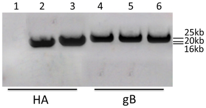
A major 16 kb fragment from the BamHI-digested rHVT-US2-HA DNA and another major 20 kb fragment from the BamHI-digested rHVT-US10-HA DNA were detected with HA probe by Southern blotting hybridization, respectively, indicating that the transfer vectors were correctly inserted in the US2 region or the US10 region. We also carried out Southern hybridization to confirm the existence of gB from the wtHVT and recombinant viruses. As expected, a single band of the predicted size of about 25 kb was detected in DNA extracted from rHVT-US2-HA or rHVT-US10-HA-infected cells. A similar-size band was also detected in the DNA samples extracted from wtHVT-infected cells (Figure 1).
Figure 1. Southern blotting hybridization of BamHI-digested DNA extracted from cells infected with rHVT-US2-HA, rHVT-US10-HA or wtHVT virus.
HA specific band was detected with HA probe in the DNA from rHVT-US2-HA (Lane 2) or rHVT-US10-HA (Lane 3). In contrast, no band was dectected in the DNA from wtHVT (Lane 1). With gB probe, DNA of cells infected with rHVT-US2-HA, rHVT-US10-HA or wtHVT virus all contained gB specific band (Lane 4–6).
Immunofluorescence staining and Western blot analysis
CEFs were infected with either rHVT-US2-HA, rHVT-US10-HA or wtHVT virus. HA expression was detected by immunofluorescence staining using chicken anti-H5 serum and fluorescein isothiocyanate (FITC)-labeled rabbit anti-chicken IgG. Fluorescence was detected in recombinant virus-infected cells following microscopic analysis in contrast to cells infected with wtHVT (Figure 2). These results indicated that rHVT-US2-HA and rHVT-US10-HA were successfully expressed in CEFs.
Figure 2. Immunofluorescence staining of cells expressing HA antigen of the highly pathogenic avian influenza virus H5N1 in turkey herpesvirus.
Confluent CEFs were infected with either (A) rHVT-US2-HA, (B) rHVT-US10-HA or (C) wtHVT. Cells were incubated with polyclonal chicken antiserum against HA antigen H5 of avian influenza virus, stained with rabbit anti-chicken IgG-FITC conjugate and then examined under fluorescence microscopy 48 hours after infection. Magnification 20×.
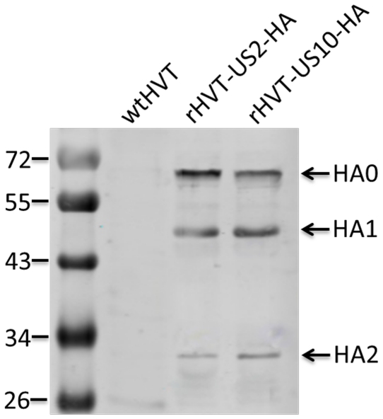
Three bands were detected by Western blot analysis using chicken anti-H5 AIV HA serum and IR dye 800-labeled rabbit anti-chicken IgY in lysates of cells infected with rHVT-US2-HA or rHVT-US10-HA after incomplete treatment with trypsin, which represented the intact HA precursor HA0, the cleaved products HA1 and HA2. As expected, HA specific band was not detected in CEFs infected with wtHVT (Figure 3).
Figure 3. Western blot analysis.
CEFs inoculated with recombinant HVT vaccines were subjected to SDS-PAGE followed by transfer onto a nitrocellulose membrane. Blots were incubated with chicken anti-HA antiserum and rabbit anti-chicken IgG conjugate. HA-specific bands corresponding to the cleaved HA1 and HA2 were detected in preparations of rHVT-US2-HA or rHVT-US10-HA-infected cells, but not wtHVT. No staining was observed after incubating blots with conjugate only (data not shown).
Haemagglutination assay
Haemagglutination assays using 0.5% chicken red blood cells demonstrated that the HA antigen produced on each of the two recombinant viruses agglutinated chicken red blood cells in contrast to wtHVT. The haemagglutination titers of rHVT-US2-HA, rHVT-US10-HA and wtHVT were 3log2, 2log2 and 0, respectively.
Biological characterization of the recombinant viruses
After recombinant viruses were confirmed to express HA, we determined whether the growth curves of the US2 and US10 gene-deleted recombinant viruses were comparable to that of wtHVT. This was carried out three times by infecting CEFs with 100 plaque forming units (p.f.u.) of either wtHVT or one of the two recombinant viruses and assessing plaque formation at 0, 24, 36, 48, 54, 72, 96 and 120 h post infection.
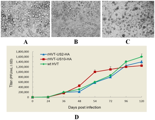
Plaque morphology and sizes of wtHVT and rHVT-US2-HA virus infected CEFs were indistinguishable from each other at 96 h post infection, and rHVT-US2-HA viruses had very similar in vitro replication kinetics to wtHVT. However, extensive syncytia were formed in HVT-US10-HA infected cells and it replicated at a higher rate than wtHVT between 48–54 h post infection and grew slowly 54 h post infection (Figure 4).
Figure 4. Plaque morphology and growth rates.
CEFs were incubated with either (A) wtHVT, (B) rHVT-US2-HA or (C) rHVT-US10-HA for 96 h and their respective morphology was assessed. The growth rates of the recombinant and wild-type viral isolates were compared over time (D).
Viremia levels of chickens infected with HVT-US2-HA or HVT-US10-HA
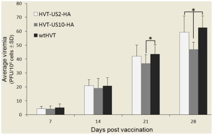
The viremia levels in five birds from each group were determined on 7, 14, 21, 28 days post infection. As indicated in Figure 5, the viremia levels in rHVT-US10-HA virus-infected chickens were slightly lower than those of rHVT-US2-HA group on 14 and 21 days post infection, but were significantly lower than those of rHVT-US2-HA group on 28 days post infection (P<0.05). The viremia levels were indistinguishable between wtHVT virus infected chickens and rHVT-US2-HA virus infected chickens during the whole experimental period.
Figure 5. Comparison of viremia levels between rHVT-US2-HA, rHVT-US10-HA or wtHVT.
Day old chicks were vaccinated with either wtHVT, rHVT-US2-HA or rHVT-US10-HA and bled on 7, 14, 21, 28 days post infection for determination of viremia. Stars indicate that the differences were significant between groups (P<0.05).
Evaluation of protection against AIV challenge
To determine the protective efficacy of the recombinant viruses against challenge with HPAIV, chickens were infected with 105 EID50 of HPAIV H5N1 A/Goose/HLJ/QFY/2003 4 weeks post vaccination. Only 4/15 (26.7%) chickens vaccinated with rHVT-US10-HA were protected whereas 9/15 (60%) of the chickens vaccinated with rHVT-US2-HA were protected (Table 1). In contrast to the vaccine groups, control chickens injected with wtHVT died within two days post challenge. Furthermore, we were able to isolate viruses from chickens vaccinated with rHVT-US2-HA or rHVT-US10-HA on 3 and 5 days post challenge but virus shedding was undetectable at 1 week post challenge.
Table 1. Protective efficacy of the recombinant vaccines against HPAIV H5 challenge in chickens.
| Vaccine formulation tested | Virus isolated from collected swabs (shedding/total [log10 EID50])a | ||||
| Day 3 p.c. | Day 5 p.c. | Survival/total | |||
| Oropharyngeal | Cloacal | Oropharyngeal | Cloacal | ||
| rHVT-US2-HA | 4/13(2.4±0.7) | 1/13(2.8±0.4) | 1/10(2.1±0.2) | 1/10(1.8±0.3) | 9/15A |
| rHVT-US10-HA | 4/9(2.1±0.3) | 5/9(1.5±0.5) | 3/7(2.8±0.3) | 2/7(2.4±0.6) | 4/15B |
| HVT Fc 126 | —b | —b | —b | —b | 0/15 |
Oropharyngeal and cloacal swabs were collected on days 3, 5, and 7 post challenge and titrated in SPF eggs. All control group chickens died before day 7. No virus was detected in the vaccinated chickens. For this reason day 7 data are excluded.
All chickens in this group died before day 3.
Different uppercase superscript letters indicate a significant difference (P<0.05) between groups on respective rows.
HA antibodies constitute one of the major defenses against viral infections, therefore, we examined the relative capacities of the rHVT-HA vaccines to elicit protective humoral immune responses (Table 2). Although chickens vaccinated with either rHVT-US2-HA or rHVT-US10-HA elicited HA antibody responses 2 weeks post vaccination, the antibody levels detected were low. However, haemagglutination inhibition (HI) antibody titers increased 3–4 weeks post vaccination and the increase of mean HI antibody titers in chickens vaccinated with rHVT-US2-HA was statistically significant (P<0.05) when compared to chickens vaccinated with rHVT-US10-HA on 21 and 28 days post challenge. The control group inoculated with wtHVT showed no evidence of HI antibody responses.
Table 2. Results of haemagglutination inhibition test of chickens vaccinated with recombinant vaccines.
| Vaccine formulation tested | Log2 HI titer at different days post-vaccination(mean±SD) | ||||
| 7 | 14 | 21 | 28 | 35 | |
| rHVT-US2-HA | 0 | 1.6A±0.5 | 3.05A±0.61 | 4.05A±0.69 | 14.3±1.11 |
| rHVT-US10-HA | 0.3±0 | 0.9B±0.3 | 2.2B±0.35 | 3.15B±0.70 | 13.5±1.04 |
| HVT Fc 126 | ND | ND | ND | ND | —a |
ND = not determined.
Negative control; all died.
Different uppercase superscript letters indicate a significant difference (P<0.05) between groups on respective rows.
Evaluation of protection against MDV challenge
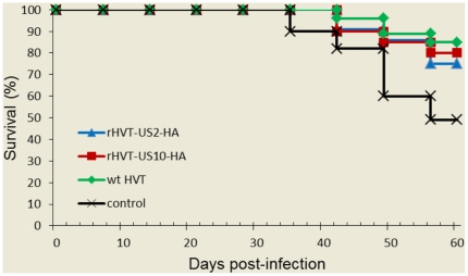
The protective efficacy against Marek's disease of the rHVT vaccines was determined by assessing cumulative survival rates and gross/histological lesions in vaccinated animals post challenge with MDV. Evidence of MD was observed in control animals 4 weeks post challenge with vMDV strain J-1and 95% of the chickens in this group developed MD during the 60-day experimental period (Table 3). Lymphoid tumors were observed in several visceral organs of the dead chickens, particularly in hearts and kidneys. In contrast, the vaccine groups had survival rates above 70% (Figure 6); 20% of wtHVT and rHVT-US10-HA vaccinated animals presented with MD (PI value of 78.9) and rHVT-US2-HA vaccinated animals had an MD incidence rate of 33% (PI value of 65.3). The PI values of the three vaccine groups were not statistically different (P>0.05).
Table 3. Protective efficacy of recombinant HVTs and wtHVT against MDV challenge.
| Vaccine | Number of chickens/group | MD mortality | MD% | Protection Index (PI) % |
| rHVT-US2-HA | 15 | 4 | 33 | 65.3 |
| rHVT-US10-HA | 15 | 3 | 20 | 78.9 |
| HVT Fc126 | 15 | 3 | 20 | 78.9 |
| Control | 20 | 11 | 95 | 0 |
MD (%) indicates the percentage of MDV-infected chickens that died after challenge with MDV strain vJ-1 or developed gross tumors prior to experimental termination.
Figure 6. Characterization of vaccine efficacy.
Day old chickens were vaccinated with either wtHVT, rHVT-US2-HA or rHVT-US10-HA and challenged 14 days later with MDV J-1. Data are expressed as percentage survival of birds from unvaccinated or vaccinated groups.
Discussion
In this study, two recombinant HVTs (rHVT-US2-HA and rHVT-US10-HA) were generated, and preliminary experiments indicated that the respective insertions did not affect the rHVT growth in CEFs as previously described [10], [19]. Results from in vitro viral growth curve analysis demonstrated that replication rates and plaque sizes of rHVT-US2-HA infected CEFs were similar to those of parental HVT. In contrast, plaque morphology and replication rates of rHVT-US10-HA infected CEFs were different to those of wtHVT [10]. Our in vivo experiments have shown the different growth rates of the recombinant HVT viruses and wtHVT in chickens by viremia analysis. The viremia levels in rHVT-US10-HA virus-infected chickens were significantly lower than those of rHVT-US2-HA group on 28 days post infection and there was no obvious difference on growth rate between rHVT-US2-HA and wtHVT group, revealing a similar in vitro growth rate. However, rHVT-US10-HA replicated at a higher rate than wtHVT between 48–54 h post vaccination, indicating a different in vivo growth rate. The viremia levels in rHVT-US10-HA group were lower than those in rHVT-US2-HA group or wtHVT group during the whole experimental period.
The virus challenge experiments showed that rHVT-US2-HA and rHVT-US10-HA vaccinated chickens were 60 and 26.7% protected against HPAIV challenge and virus shedding data were consistent with mortality, respectively. Serological data suggested that rHVT-vaccinated chickens persistently stimulated their host-immune systems. Chickens vaccinated with rHVT-US2-HA had significantly higher HI antibody titer levels compared to rHVT-US10-HA vaccinated chickens on 21 and 28 days post infection. These results indicated that HVT expressing the HA gene at the US2 position was significantly more effective in conferring protective immunity than the virus expressing HA at the US10 site.
Both rHVTs conferred effective protection against vMDV J-1 challenge. rHVT-US10-HA and parental HVT conferred the same level of protection against MDV challenge (PI value 78.9) and there were no significant differences between the two groups whereas protection conferred by rHVT-US2-HA was slightly less effective (PI value 65.3). These results demonstrated that HA insertion into either the US2 or US10 gene positions did not significantly affect the rHVT inducted protection against MDV, confirming the previous findings [1], [10].
When HVT infects CEFs, the cytopathic effect (CPE) was commonly observed first as round cells and then their fusion (syncytia) that formed cell foci. The degenerated cells eventually detached from the dish, producing plaques on the monolayer cell sheet. Previous studies showed that particular deletion mutations could alter viral plaque morphology. Mutation in gB gene of HSV-1 caused extensive syncytia formation in infected cells, rather than rarely caused cell-cell fusion as previously reported [26]. Obvious and extensive syncytia were formed after CEFs were infected with rHVT-US10-HA. It is assumed that US10 gene was responsible for plaque morphology of HVT. In previous MDV/HVT US10 gene relevant studies, the corruption of this site did not affect plaque morphology, but all these studies adopted insertion method for interruption and part of US10 gene could still be expressed [10], [27]. We replaced HVT US10 gene with targeted gene in our study, which perhaps can explain why we obtained different results. Nonetheless, we cannot rule out the possibility that HA protein expression has impact on viral plaque morphology. Additional studies are currently underway to define factors involved in mediating these effects.
Previously, recombinant fowlpox virus (rFPV) or Newcastle disease virus (rNDV) expressing the AIV HA gene were generated and vaccination data using these recombinant viruses demonstrated that the rFPV and rNDV conferred significant levels of protection against AIV challenge, indicating that recombinant viral vector expressing AIV gene was a good candidate for control of avian influenza [21], [25], [28]–[31]. In this study, the two rHVTs conferred only partial protection against AIV challenge. However, beyond our expectation, almost 90% of chickens in control group died within 24 h post challenge and the rest of them died two days post challenge. In previous studies, the cases like this high mortality of chickens in control group were rare. Therefore, the reason for the slightly lower protection levels conferred by the rHVT vaccines was likely due to either low immunization dose or high dose of challenge with HPAIV, overwhelming protective immune responses. Most of HVT-vector based antigen delivery systems have been developed for making recombinant viral vaccines. Studies on recombinant HVT vaccines have been attempted to develop as bivalent vaccines against NDV, IBDV or AIV, besides protection from MDV [7], [9], [10], [16]. Remarkably, the resulting vaccine VaxxitekR HVT+IBD was licensed and commercialized as an animal herpesvirus vector based product. Therefore, HVT-vector is a good potential candidate for vaccine development. However, current MDV vaccines including HVT cannot stop viral replication and shedding in chicken although they can protect against tumour formation and hence mortality [1]. The continued evolution of field viruses towards pathotypes of greater virulence is attributed to the selection pressures imposed on these virulent viruses in vaccinated chicken. Therefore, HVT vaccine may not effectively protect against MDV in the future.
Our study, for the first time, describes different effects of US2 and US10 insertion sites in the development of recombinant HVTs and the US2 gene locus as an insertion site for the expression of vaccine targets is more effective than US10 site in construction of an rHVT vaccine for use in chickens.
Materials and Methods
Ethics Statement
Animal experiments were approved by Harbin Veterinary Research Institute of the Chinese Academy of Agricultural Sciences and performed in accordance with animal ethics guidelines and approved protocols. The Animal Ethics Committee approval number was Heilongjiang-SYXK 2006-032.
Viruses and cells
The wtHVT FC126 strain (twelfth duck embryo fibroblast passage stock) was used for construction of the recombinant viruses. MDV strain J-1 is a reference virulent MDV strain isolated from Beijing district in China [32] and is a standard virulent reference challenge strain in MDV research (tenth duck embryo fibroblast passage stock) [33], [34]. Both viral strains were obtained from the Avian Infectious Diseases Laboratory of the Harbin Veterinary Research Institute of the Chinese Academy of Agricultural Sciences and were propagated in primary CEFs prepared from 10-day-old, specific-pathogen-free embryos.
HPAIV H5N1 virus A/goose/Guangdong/3/96 [35] and A/Goose/HLJ/QFY/2003 [36] shares approximately 97% identity in HA gene. They were propagated in the allantoic cavities of 10-day-old SPF chicken embryonated eggs and then kept at −70°C before RNA extraction or use in challenge studies.
Cloning of the AIV HA gene and HA cassette
cDNA corresponding to the A/goose/Guangdong/3/96 HA open reading frame (1,716 nucleotides) was PCR-amplified using specific primers (Table 4) that included Kozak consensus sequences [37]. The PCR products of cDNA HA were digested with HindIII and SalI and cloned into the pN1-EGFP-derived pN1 vector (deficient in EGFP) (Clontech, Tokyo, Japan). The HA cassette containing the HCMV immediate-early promoter and the SV40 poly-adenylation signal were PCR-amplified using primers containing a PacI restriction site (Table 4).
Table 4. PCR primer sequences used in amplification experiments.
| Primer | Sequence | Target |
| HA-upper | AGGAAGCTTTACCATGGAGAGAATAGTGCTTC | HA open reading frame |
| HA-lower | CGGGTCGACTTAAATGCAAATTCTGCATTGTA | |
| HA cassette-upper | CGGCGGTTAATTAACGCCATGCATTAGTTATT | HA cassette |
| HA cassette-lower | CGG CGG TTAATTAACGCTTACAATTTACGCCT | |
| gpt cassette-upper1 | ATTAAGCTTCGCCATGCATTAGTTATTAATAGT | gpt cassette with HindIII |
| gpt cassette-lower1 | ATCAAGCTTCGCTTACAATTTACGCCTTAAGAT | |
| US10 left-upper | GAGCTCGGGTCCGGGAGGAAGTGA | 2.6 kb left region |
| US10 left-lower | GCGGCCGCTAATCAACATATATTGTAT | |
| US10 right-upper | TAAGCGGCCGCTTAATTAACATAGGCACGCTCTGATGT | 2.6 kb right region |
| US10 right-lower | CGGAAGCTTAGATTAGCAGATTTTCTGG | |
| gpt cassette-upper2 | ATTGCGGCCGCCGCCATGCATTAGTTATTAATAGT | gpt cassette with NotI |
| gpt cassette-lower2 | ATCGCGGCCGCCGCTTACAATTTACGCCTTAAGAT | |
| HA test-upper | ATGGAGAGAATAGTGCTTCTCC | HA probe |
| HA test-lower | CAAATTCTGCATTGTAACGAT | |
| gB-upper | AGGGAAAGTAGTAGTCGGGGCTGCAGGG | gB probe |
| gB-lower | TTCATCATCCGTCTCAGAATCCGTGTCG |
Construction of the US2 (pGAB-gpt-HA) deletion plasmid
For construction of the US2 gene transfer vector, guanine phosphoribosyl transferase gene (Eco-gpt) was PCR-amplified from the pEco-gpt plasmid using primers containing HindIII restriction enzyme sites (Table 4) and then cloned into pGAB that included 2.0 and 2.7 kilo base pair (kb) fragments flanking the HVT US2 gene to obtain the pGAB-gpt plasmid [38]. The pEco-gpt plasmid was constructed in our laboratory, which contained the E. coli selective gpt marker under the control of the HCMV immediate-early promoter [36]. The HA cassette was cut using PacI and inserted into pGAB-gpt to obtain the transfer plasmid pGAB-gpt-HA.
Construction of the US10 deletion plasmid (pUAB-gpt-HA)
The US10 gene transfer vector was constructed by PCR-amplification of two fragments (2.6 kb and 2.6 kb) mapping to each side of the HVT US10 open reading frame using primers containing appropriate restriction enzyme sites (Table 4). Both fragments were subsequently cloned into the pUC119 vector (TaKaRa, Dalian, China) to generate pUAB. The Eco-gpt gene was then PCR amplified from the pEco-gpt plasmid using primers containing NotI restriction site (Table 4) and ligated with pUAB to obtain the plasmid pUAB-gpt. The HA cassette was then cut by PacI and inserted into the pUAB-gpt to obtain the recombination plasmid pUAB-gpt-HA.
Transfection and isolation of HVT recombinants
Recombinant HVT viruses were generated as described previously [1], [38]. Briefly, primary CEFs were co-transfected with 1 µg of each of pGAB-gpt-HA and 5 µg HVT DNA using liposome [39]. The transfected cells were maintained in selective medium containing mycophenolic acid (350 µg/ml), xanthine (70 µg/ml) and hypoxanthine (100 µg/ml), and monitored daily for CPE. To purify rHVT-US2-HA, virus-containing cells were passaged in selection medium for eight rounds until no cells with CPE were observed in selection medium suspensions. The rHVT-US10-HA virus was obtained by the same method.
Southern blot analysis
Total viral (rHVT-US2-HA, rHVT-US10-HA or wtHVT) DNA was extracted using the sodium dodecyl sulfate (SDS)-protinase K-phenol protocol and analysed by restriction digestion with BamHI. For Southern hybridization, DNA was separated by 1.0% agarose gel electrophoresis, transferred to membranes and probed with DIG-labeled probes (Roche Molecular Biochemicals, Mannheim, Germany) specific for glycoprotein B (gB) and HA according to the manufacturer's protocols. The sequences of the primers used to synthesize the DIG-labeled probes are shown in Table 4.
Indirect immunofluorescence assay (IFA) and Western blot analysis
Chicken embryo fibroblast monolayers (80–90% confluent) were infected with rHVT-US2-HA, rHVT-US10-HA or wtHVT respectively, and then washed 3 times with phosphate buffered saline (PBS) and fixed with ice-cold ethanol for 15 min following the appearance of CPE. The wells were overlaid with polyclonal chicken antibodies produced by vaccination with the H5 AIV HA gene DNA vaccine (1∶100), and incubated at 37°C for 1 h. The wells were washed 3 times with PBS and incubated with anti-chicken IgY (IgG) (whole molecule)-FITC antibody produced in rabbit (1∶300) (Sigma, Shanghai, China) at 37°C for 1 h. Wells were then washed as above, dried and analyzed using an inverted phase contrast microscope with fluorescence light and a 20×ELWD objective (Nikon, Tokyo, Japan).
Western blot analysis was carried out as described previously [40]. Briefly, primary CEFs were infected by PCR-positive recombinant viruses, respectively, and cells collected when CPE was detected. Total cell lysates were prepared following incubation with lysis buffer (10 mM Tris-Cl, pH 7.4, 1 mM MgCl2, 0.5% NP40, 20 µg/ml DNase I), followed by 0.1% trypsin for 30 min, subjected to 10% SDS-PAGE and then transferred to nitrocellulose. For HA protein detection, membranes were incubated with chicken H5-AIV HA specific antiserum (1∶100) followed by detection with IR dye 800-labeled polyclonal rabbit anti-chicken IgG (Li-Cor, Lincoln, NE, USA) (1∶2000) and analyzed using an Odyssey infrared imager (Li-Cor). Cells infected with wtHVT were used as negtive control.
Haemagglutination assay
CEFs were infected with rHVT-US2-HA and rHVT-US10-HA, respectively, and the rHVT expressing AIV H5 HA in the media concentrated following precipitation with 10% PEG-8000. The precipitant was resuspended in PBS for the HA assay. Chicken red blood cells were washed three times with PBS and resuspended (5%, v/v) in PBS for use in the HA assay. Haemagglutination tests were performed in 96-well round-bottom microtiter plates. To determine whether recombinant viruses possessed chicken red blood cell haemagglutination properties, 50 µl of concentrated cell supernatants and an equal volume of 0.5% chicken red blood cells were added to each well and the plates incubated for 30 min at 25°C. Chicken red blood cell suspensions were mixed and allowed to settle for 30 to 45 min. The HA titers were described as the reciprocal of the highest virus dilution with 100% HA.
Comparison of in vitro growth rates between wtHVT and recombinant viruses
The rates of in vitro growth of the viruses on CEFs were studied by counting the p.f.u. at various time points. For each virus 100 p.f.u. was inoculated onto tissue-culture dishes with 2×10⁶ CEFs. At various hours post inoculation, virus-infected CEFs were harvested and serial 10-fold dilutions were added in triplicate onto the 48-well plates of CEFs. After three days, the titers of the virus at each time point were calculated from the number of p.f.u. from each of the dilutions and the growth curves of rHVT-US2-HA, rHVT-US10-HA and wtHVT determined.
Determination of viremia
One-day-old SPF chicks were assigned to three groups of 20 chickens each and vaccinated intramuscularly with a total of 3000 p.f.u. of either rHVT-US2-HA, rHVT-US10-HA or wtHVT. After vaccination, five chickens were removed weekly from each of these groups, and the levels of HVT viremia were determined for individual chickens as reported previously [10]. Briefly, blood samples in anticoagulants were collected from five birds of each group and 5 ml blood from each bird was mixed with 5 ml RPMI 1640 medium and 3 ml Histopaque 1077 (Sigma, Shanghai, China). Samples were centrifuged at 1000× g for 30 min. The leukocytes were recovered and counted. Co-cultivations were done in duplicate by seeding 2×10⁶ leukocytes onto 60-mm plates with CEF monolayers. After 5 days, dishes were stained with crystal violet and plaques were counted.
AIV challenge experiment
One-day-old chicks were randomly divided into groups of 15 and vaccinated intramuscularly with a total of 3000 p.f.u. of either rHVT-US2-HA or rHVT-US10-HA. Another group was inoculated with wtHVT as a negative control. Day-old chicks were vaccinated only once and then bled via wing veins weekly for five times. Chick sera were tested for antibody-mediated haemagglutination inhibition using the OIE standard method. Four-weeks post vaccination chicks in each group were challenged intranasally with 0.1 ml of 106 ELD50 of highly pathogenic H5N1 A/Goose/HLJ/QFY/2003 virus. Oropharyngeal and cloacal swabs from respective chickens were collected on days 3, 5, and 7 post challenge for virus titration and chickens were observed for disease presentation and mortality.
MDV challenge experiment
A total of 65 1-day-old SPF chicks were vaccinated intramuscularly with 3000 p.f.u. of either rHVT-US2-HA, rHVT-US10-HA or wtHVT. Twenty negative control chicks were inoculated with non-infected CEFs. On 14 days of age birds were challenged intra-abdominally with 1000 p.f.u. virulent MDV J-1 virus. Mortality during the course of the experiment was recorded and chickens were examined for gross MD lesions. On 60 days post challenge, all surviving birds were euthanized and examined for gross and histopathological lesions. The percentage of gross MD was calculated for each test group as the number of chickens with gross MD lesions divided by number at risk (survivors plus MD deaths)×100 [41]. Vaccinal immunity to MD was expressed as a protective index (PI) calculated as the percentage of gross MD in non-vaccinated challenged control chickens minus the percentage of gross MD in vaccinated, challenged chickens divided by the percentage of gross MD in non-vaccinated challenged control chickens and multiplied by 100.
Statistical analyses
Comparisons of single treatment among vaccinated groups were performed using a nonparametric one-way ANOVA followed by LSD's multiple comparison post test (for Table 2). Comparison of PI and protective rate was performed using χ2 analysis. The analyses were done by using SPSS version 17.0 for Windows (SPSS, Chicago, IL, USA). P values less than 0.05 were considered statistically significant in all cases.
Acknowledgments
We are grateful to Hualan Chen, Ming Liu and Guobin Tian for providing AIV strains and Changjun Liu for MDV strains.
Footnotes
Competing Interests: The authors have declared that no competing interests exist.
Funding: The study was supported by a grant from the National Key Laboratory of Veterinary Biotechnology as Basic Research Programs of China (NKLVBP200803). The funders had no role in study design, data collection and analysis, decision to publish, or preparation of the manuscript.
References
- 1.Baigent SJ, Petherbridge LJ, Smith LP, Zhao Y, Chesters PM, et al. Herpesvirus of turkey reconstituted from bacterial artificial chromosome clones induces protection against Marek's disease. J Gen Virol. 2006;87:769–776. doi: 10.1099/vir.0.81498-0. [DOI] [PubMed] [Google Scholar]
- 2.Calnek BW, Witter RL, editors. Marek's disease, 9th ed. Iowa: Iowa State University Press; 1991. [Google Scholar]
- 3.Fauqet CM, Mayo MA, Maniloff J, Desselberger U, Ball LA, editors. Virus Taxonomy, VIIIth Report of the ICTV. London: Elsevier; 2005. [Google Scholar]
- 4.Okazaki W, Purchase HG, Burmester BR. Protection against Marek's disease by vaccination with a herpesvirus of turkeys. Avian Dis. 1970;14:413–429. [PubMed] [Google Scholar]
- 5.Purchase HG, Okazaki W, Burmester BR. Long-term field trials with the herpesvirus of turkeys vaccine against Marek's disease. Avian Dis. 1972;16:57–71. [PubMed] [Google Scholar]
- 6.Witter RL. Control strategies for Marek's disease: a perspective for the future. Poult Sci. 1998;77:1197–1203. doi: 10.1093/ps/77.8.1197. [DOI] [PubMed] [Google Scholar]
- 7.Reddy SK, Sharma JM, Ahmad J, Reddy DN, McMillen JK, et al. Protective efficacy of a recombinant herpesvirus of turkeys as an in ovo vaccine against Newcastle and Marek's diseases in specific-pathogen-free chickens. Vaccine. 1996;14:469–477. doi: 10.1016/0264-410x(95)00242-s. [DOI] [PubMed] [Google Scholar]
- 8.Witter RL, Nazerian K, Purchase HG, Burgoyne GH. Isolation from turkeys of a cell-associated herpesvirus antigenically related to Marek's disease virus. Am J Vet Res. 1970;31:525–538. [PubMed] [Google Scholar]
- 9.Darteil R, Bublot M, Laplace E, Bouquet JF, Audonnet JC, et al. Herpesvirus of turkey recombinant viruses expressing infectious bursal disease virus (IBDV) VP2 immunogen induce protection against an IBDV virulent challenge in chickens. Virology. 1995;211:481–490. doi: 10.1006/viro.1995.1430. [DOI] [PubMed] [Google Scholar]
- 10.Morgan RW, Gelb J, Jr, Schreurs CS, Lutticken D, Rosenberger JK, et al. Protection of chickens from Newcastle and Marek's diseases with a recombinant herpesvirus of turkeys vaccine expressing the Newcastle disease virus fusion protein. Avian Dis. 1992;36:858–870. [PubMed] [Google Scholar]
- 11.Morgan RW, Gelb J, Jr, Pope CR, Sondermeijer PJ. Efficacy in chickens of a herpesvirus of turkeys recombinant vaccine containing the fusion gene of Newcastle disease virus: onset of protection and effect of maternal antibodies. Avian Dis. 1993;37:1032–1040. [PubMed] [Google Scholar]
- 12.Bacon LD, Witter RL. Influence of turkey herpesvirus vaccination on the B-haplotype effect on Marek's disease resistance in 15.B-congenic chickens. Avian Dis. 1992;36:378–385. [PubMed] [Google Scholar]
- 13.Rauw F, Gardin Y, Palya V, Anbari S, Lemaire S, et al. Improved vaccination against Newcastle disease by an in ovo recombinant HVT-ND combined with an adjuvanted live vaccine at day-old. Vaccine. 2010;28:823–833. doi: 10.1016/j.vaccine.2009.10.049. [DOI] [PubMed] [Google Scholar]
- 14.Ross LJ, Binns MM, Tyers P, Pastorek J, Zelnik V, et al. Construction and properties of a turkey herpesvirus recombinant expressing the Marek's disease virus homologue of glycoprotein B of herpes simplex virus. J Gen Virol. 1993;74(Pt 3):371–377. doi: 10.1099/0022-1317-74-3-371. [DOI] [PubMed] [Google Scholar]
- 15.Zelnik V, Tyers P, Smith GD, Liang C, Ross NL. Structure and properties of a herpesvirus of turkeys recombinant in which US1, US10 and SORF3 genes have been replaced by a lacZ expression cassette. J Gen Virol. 1995;76(Pt 11):2903–2907. doi: 10.1099/0022-1317-76-11-2903. [DOI] [PubMed] [Google Scholar]
- 16.Tsukamoto K, Kojima C, Komori Y, Tanimura N, Mase M, et al. Protection of chickens against very virulent infectious bursal disease virus (IBDV) and Marek's disease virus (MDV) with a recombinant MDV expressing IBDV VP2. Virology. 1999;257:352–362. doi: 10.1006/viro.1999.9641. [DOI] [PubMed] [Google Scholar]
- 17.Sakaguchi M, Nakamura H, Sonoda K, Okamura H, Yokogawa K, et al. Protection of chickens with or without maternal antibodies against both Marek's and Newcastle diseases by one-time vaccination with recombinant vaccine of Marek's disease virus type 1. Vaccine. 1998;16:472–479. doi: 10.1016/s0264-410x(97)80001-1. [DOI] [PubMed] [Google Scholar]
- 18.Cantello JL, Anderson AS, Francesconi A, Morgan RW. Isolation of a Marek's disease virus (MDV) recombinant containing the lacZ gene of Escherichia coli stably inserted within the MDV US2 gene. J Virol. 1991;65:1584–1588. doi: 10.1128/jvi.65.3.1584-1588.1991. [DOI] [PMC free article] [PubMed] [Google Scholar]
- 19.Parcells MS, Anderson AS, Cantello JL, Morgan RW. Characterization of Marek's disease virus insertion and deletion mutants that lack US1 (ICP22 homolog), US10, and/or US2 and neighboring short-component open reading frames. J Virol. 1994;68:8239–8253. doi: 10.1128/jvi.68.12.8239-8253.1994. [DOI] [PMC free article] [PubMed] [Google Scholar]
- 20.Alexander DJ. A review of avian influenza in different bird species. Vet Microbiol. 2000;74:3–13. doi: 10.1016/s0378-1135(00)00160-7. [DOI] [PubMed] [Google Scholar]
- 21.Nayak B, Rout SN, Kumar S, Khalil MS, Fouda MM, et al. Immunization of chickens with Newcastle disease virus expressing H5 hemagglutinin protects against highly pathogenic H5N1 avian influenza viruses. PLoS One. 2009;4:e6509. doi: 10.1371/journal.pone.0006509. [DOI] [PMC free article] [PubMed] [Google Scholar]
- 22.Veits J, Wiesner D, Fuchs W, Hoffmann B, Granzow H, et al. Newcastle disease virus expressing H5 hemagglutinin gene protects chickens against Newcastle disease and avian influenza. Proc Natl Acad Sci U S A. 2006;103:8197–8202. doi: 10.1073/pnas.0602461103. [DOI] [PMC free article] [PubMed] [Google Scholar]
- 23.Veits J, Romer-Oberdorfer A, Helferich D, Durban M, Suezer Y, et al. Protective efficacy of several vaccines against highly pathogenic H5N1 avian influenza virus under experimental conditions. Vaccine. 2008;26:1688–96. doi: 10.1016/j.vaccine.2008.01.016. [DOI] [PubMed] [Google Scholar]
- 24.Schroer D, Veits J, Grund C, Dauber M, Keil G, et al. Vaccination with Newcastle disease virus vectored vaccine protects chickens against highly pathogenic H7 avian influenza virus. Avian Dis. 2009;53:190–197. doi: 10.1637/8416-072308-Reg.1. [DOI] [PubMed] [Google Scholar]
- 25.Webster RG, Kawaoka Y, Taylor J, Weinberg R, Paoletti E. Efficacy of nucleoprotein and haemagglutinin antigens expressed in fowlpox virus as vaccine for influenza in chickens. Vaccine. 1991;9:303–308. doi: 10.1016/0264-410x(91)90055-b. [DOI] [PubMed] [Google Scholar]
- 26.Gage PJ, Levine M, Glorioso JC. Syncytium-inducing mutations localize to two discrete regions within the cytoplasmic domain of herpes simplex virus type 1 glycoprotein B. J Virol. 1993;67:2191–2201. doi: 10.1128/jvi.67.4.2191-2201.1993. [DOI] [PMC free article] [PubMed] [Google Scholar]
- 27.Purchase HG, Burmester BR, Cunningham CH. Pathogenicity and Antigenicity of Clones from Strains of Marek's Disease Virus and the Herpesvirus of Turkeys. Infect Immun. 1971;3:295–303. doi: 10.1128/iai.3.2.295-303.1971. [DOI] [PMC free article] [PubMed] [Google Scholar]
- 28.Chen SJ, Sun L, Liu WJ, Sun XH, Liu XF. [Recombinant fowlpox virus coexpressing HA and NA gene from subtype H5N1 of avian influenza virus and its protective efficacy]. Wei Sheng Wu Xue Bao. 2006;46:111–114. [PubMed] [Google Scholar]
- 29.Cheng J, Liu X, Pen D, Liu H. [Recombinant fowlpox virus expressing HA from subtype H9N2 of avian influenza virus and its protective immunity against homologous challenge in chickens]. Wei Sheng Wu Xue Bao. 2002;42:442–447. [PubMed] [Google Scholar]
- 30.Ge J, Deng G, Wen Z, Tian G, Wang Y, et al. Newcastle disease virus-based live attenuated vaccine completely protects chickens and mice from lethal challenge of homologous and heterologous H5N1 avian influenza viruses. J Virol. 2007;81:150–158. doi: 10.1128/JVI.01514-06. [DOI] [PMC free article] [PubMed] [Google Scholar]
- 31.Mingxiao M, Ningyi J, Zhenguo W, Ruilin W, Dongliang F, et al. Construction and immunogenicity of recombinant fowlpox vaccines coexpressing HA of AIV H5N1 and chicken IL18. Vaccine. 2006;24:4304–4311. doi: 10.1016/j.vaccine.2006.03.006. [DOI] [PubMed] [Google Scholar]
- 32.Xia CY, Guan YT, Nan X, Yu WT, Chen HY. Test of virulent Marek's disease virus Jing-1 strain infection in BWEL-SPF chicks. Chinese J Vet Sci Tech. 2000;30:24–25. [Google Scholar]
- 33.Liu AL, Liu CJ, Zhang YP, Li JM, Shi WS, et al. Mutational analysis of Meq, RLORF4, RLORF12 and 132bpr genes of epidemic Marek's disease virus strains highly passaged on chicken embryo fibroblast. Bing Du Xue Bao. 2009;25:368–375. [PubMed] [Google Scholar]
- 34.Shi WS, Liu CJ, Zhang YP, Qin YA, Zhang XW, et al. Cloning and sequence analysis of the Meq gene of 4 Marek's disease virus isolates from China. Bing Du Xue Bao. 2008;24:117–125. [PubMed] [Google Scholar]
- 35.Cui S, Tong G. A chromatographic strip test for rapid detection of one lineage of the H5 subtype of highly pathogenic avian influenza. J Vet Diagn Invest. 2008;20:567–571. doi: 10.1177/104063870802000505. [DOI] [PubMed] [Google Scholar]
- 36.Liu CN, Liu M, Zhang Y, Liu CG, Gao XL. Pathogenicity of H5N1 Influenza Virus for Ducks. Scientia Agricultura Sinica. 2006;39:412–417. [Google Scholar]
- 37.Kozak M. At least six nucleotides preceding the AUG initiator codon enhance translation in mammalian cells. J Mol Biol. 1987;196:947–950. doi: 10.1016/0022-2836(87)90418-9. [DOI] [PubMed] [Google Scholar]
- 38.Lan D, Shi X, Wang Y, Cui H, Liu C, et al. [Reconstituting turkey herpesvirus with bacterial artificial chromosome clones]. Wei Sheng Wu Xue Bao. 2008;48:811–817. [PubMed] [Google Scholar]
- 39.Cui HY, Wang YF, Shi XM, An TQ, Tong GZ, et al. Construction of an infectious Marek's disease virus bacterial artificial chromosome and characterization of protection induced in chickens. J Virol Methods. 2009;156:66–72. doi: 10.1016/j.jviromet.2008.10.021. [DOI] [PubMed] [Google Scholar]
- 40.Sambrook J, Fritsch EF, Maniatis T, editors. Molecular cloning: a laboratory manual. 2nd ed. Cold Spring Harbor, NY: Cold Spring Harbor Laboratory Press; 1989. [Google Scholar]
- 41.Lee LF, Lupiani B, Silva RF, Kung HJ, Reddy SM. Recombinant Marek's disease virus (MDV) lacking the Meq oncogene confers protection against challenge with a very virulent plus strain of MDV. Vaccine. 2008;26:1887–1892. doi: 10.1016/j.vaccine.2008.01.046. [DOI] [PubMed] [Google Scholar]


