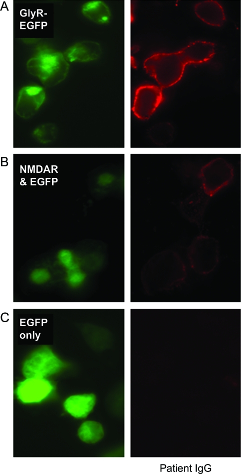
Figure 2. Glycine receptor (GlyR) and NMDA receptor (NMDAR) antibodies.
HEK cells transfected with EGFP-tagged GlyR cDNA (GlyR-EGFP; A) or cotransfected with NMDARs (NR1 and NR2B subunits) plus EGFP (NMDAR and EGFP; B). EGFP fluorescence shown in green. Patient immunoglobulin G (IgG) binding shown in red: score of 3 for GlyR antibody and 2 for NMDAR antibody (scoring system as in Irani et al.1). Patient IgG did not bind to HEK cells transfected with EGFP only (C).

