Abstract
The mutational status of the immunoglobulin heavy-chain variable region (IGHV) genes utilized by chronic lymphocytic leukemia (CLL) clones defines two disease subgroups. Patients with unmutated IGHV have a more aggressive disease and a worse outcome than patients with cells having somatic IGHV gene mutations. Moreover, up to 30% of the unmutated CLL clones exhibit very similar or identical B cell receptors (BcR), often encoded by the same IG genes. These “stereotyped” BcRs have been classified into defined subsets. The presence of an IGHV gene somatic mutation and the utilization of a skewed gene repertoire compared with normal B cells together with the expression of stereotyped receptors by unmutated CLL clones may indicate stimulation/selection by antigenic epitopes. This antigenic stimulation may occur prior to or during neoplastic transformation, but it is unknown whether this stimulation/selection continues after leukemogenesis has ceased. In this study, we focused on seven CLL cases with stereotyped BcR Subset #8 found among a cohort of 700 patients; in six, the cells expressed IgG and utilized IGHV4-39 and IGKV1-39/IGKV1D-39 genes, as reported for Subset #8 BcR. One case exhibited special features, including expression of IgM or IgG by different subclones consequent to an isotype switch, allelic inclusion at the IGH locus in the IgM-expressing cells and a particular pattern of cytogenetic lesions. Collectively, the data indicate a process of antigenic stimulation/selection of the fully transformed CLL cells leading to the expansion of the Subset #8 IgG-bearing subclone.
INTRODUCTION
CLL is the most common leukemia in the Western world and is characterized by a monoclonal accumulation in the blood and in peripheral lymphoid organs of B lymphocytes with a characteristic surface phenotype, that is, CD5+, CD23+, CD22− and low levels of surface Ig (1). In the past, it was generally accepted that this CLL cell accumulation could be attributed to defective in vivo apoptosis; however, more recent evidence indicates that leukemic cells are capable of active proliferation in vivo, which largely compensates for the cell loss occurring by apoptosis (2). Such evidence is provided by in vivo labeling studies with deuterated cells and also is corroborated by observations on the CLL cell apoptotic capacities and on telomere length and telomerase activity (3,4). Proliferation of the CLL clones in vivo is likely sustained by the intrinsic cytogenetic alterations of the cells and also promoted through stimulation by certain antigens with the help of accessory cells and/or cytokines, although the relative contribution of the two phenomena and their timing remains to be ascertained (5–7).
Different studies indicate that antigenic stimulation plays a role in promoting the onset of CLL cells (8,9). A substantial proportion of CLL clones utilize somatically mutated IGHV and IGKV/ IGLV genes. Since somatic mutations occur during antigenic stimulation, these leukemic cells are clearly antigen- experienced (10–13). Furthermore, CLL clones utilizing unmutated IGHV and IGKV/IGLV genes exhibit a skewed BcR repertoire compared with normal, virgin B cells, a finding which implies antigenic stimulation/selection (11,14–17). Finally, up to 30% of CLL clones utilize “stereotyped BcR” (11,14–17), defined as BcR expressed by different CLL clones sharing the same IGHV and IGKV/IGLV genes and very similar or identical CDR3s. Again, this would indicate a strong selective pressure imposed by a seemingly restricted set of antigens or antigenic determinants (18–20).
The above evidence indicates that antigens may play a fundamental role in expanding B cells prior to transformation and in sustaining survival/expansion of the cells in the early steps of leukemogenesis, when they are not capable of independent growth (21,22), but does not tell whether antigenic stimulation/ selection contributes to the expansion of fully transformed CLL clones. However, the observation that CLL patients, whose leukemic clones express self-reactive BcR, have a more downhill clinical course, constitutes circumstantial evidence in favor of the latter hypothesis (23).
In this study, we provide evidence indicating that stimulation/selection occurs on fully blown leukemic cells and contributes to the shaping of the CLL clone. Our observations were made on a CLL case expressing a stereotyped BcR of the Subset #8, characterized by the utilization of genes IGHV4-39 and IGKV1-39/IGKV1D-39 (16,24). This CLL case was found with six other similar cases during the screening process of 700 CLL patients recruited mainly through an observational study organized by Gruppo Italiano Studio Linfomi (GISL). Unlike the others, this particular CLL case had special features supporting the notion that antigenic stimulation continues after the process of leukemogenesis is completed and leads to the selective expansion of particular subclones.
MATERIALS AND METHODS
B-CLL Samples
A cohort of 700 CLL patients enrolled in an observational study organized by GISL or seen at our clinics was screened for IgV gene sequences. Inclusion criteria consisted of a diagnosis of typical CLL based on the National Cancer Institute (NCI) Working Group criteria and confirmed by flow-cytometry analysis of neoplastic cells (25). The study was approved by the Institutional Review Board and informed consent was obtained consistently. Most patients were at Binet Stage A and were untreated.
Flow-cytometry studies and IGHV-IGHD-IGHJ and IGKV-IGKJ and IGLV-IGLJ gene sequence determinations were carried out in a single laboratory in Genoa (11). Sequence data were analyzed using the IMGT/V-Quest tools (26) and submitted to EMBL (Accession numbers FR820867 → FR820882). Fragments length IGHV-IGHD-IGHJ analyses were obtained by PCR amplification with primers specific for FR1 IGHV2 gene subgroup and FR1 IGHV4 gene subgroup (11) in conjunction with 6FAM-5′-CTG A(AG)G AGA C(AG)G TGA CC IGHJ gene-specific primers. Subsequently, products were run on 3130XL Genetic analyzer (Applied Biosystems, Monza, Italy). FISH analyses for detection of trisomy 12, del(17p13.1), del(11q22.3) and del(13q14) were performed as described (27).
Phenotype Analysis and Cell Fractionation Procedures
The following antibodies were used: FITC-conjugated anti-IgD (DAKO, Glostrup, Denmark), PE-Cy5 conjugated anti-IgM (BD, Franklin Lakes, New Jersey, USA), APC-conjugated anti-IgG (BD), PE- conjugated anti-CD3 (BD), APC-H7 anti-CD19 (BD), PE-conjugated anti-CD19 and FITC-conjugated anti-CD5 (BD). FITC Anti-κ-PE anti-λ and RPE-Cy5 anti-CD19 triple color combination (DAKO) was used for the identification of Ig Light chain. FACS sorting experiments were performed with a precalculated six-color automatic compensation by FacsDiva (BD) software. To isolate IgD-positive and IgG-positive CLL cells, leukemic B cells were identified with anti-CD19-PE-Cy7 (BD) and anti-CD5-PE (BD). CD19-CD5 double positive cells were gated and sorted based on the expression of IgD or IgG molecules.
PROTEIN TYROSINE PHOSPHORYLATION
Protein tyrosine phosphorylation was measured as described (28). Briefly, about 4 × 10⁶ B cells were exposed to aμ-Ab, aδ-Ab, aγ-Ab, or aδ-Ab + aμ-Ab for 1, 5 or 10 min at 37°C and lysed as described (28). Nuclei-free cell extracts from 106 cells were fractionated by sodium dodecylsulfate electrophoresis on 10% polyacrylamide gels under reducing conditions, and then transferred electrophoretically onto nitrocellulose membranes (Hybond C Extra; Amersham, UK). Tyrosine phosphorylated proteins were detected by incubating the membranes with 0.2 μg/mL biotin-conjugated anti-phosphotyrosine antibody (clone PY99, Santa Cruz Biotechnology Inc, Santa Cruz, CA, USA) in 1% milk-TBST, followed by 0.3 μg/mL peroxidase-labeled streptavidin (Dako-Cytomation, Milan, Italy) in 1% milk-TBST. SYK and LYN proteins were identified using mouse anti-SYK (Upstate-Millipore, Temecla, CA, USA) and rabbit anti-LYN (Upstate) specific antibodies. Data were then expressed as a ratio between a region of interest (ROI) and its reference value. In detail, the ROI of each band observed following aμ-Ab, aδ-Ab, aγ-Ab, or aδ-Ab + aμ-Ab stimulation at 1 min time point was compared with the corresponding ROI of the unstimulated sample (negative). The total amount of phosphorylation was calculated by summing the phosphorylation of the single bands corresponding to 120 kDa, 105 kDa, 75 kDa and 55 kDa (1D Image Analysis Software version 3.5, Kodak). The fold increase was considered respective to the sum of the bands of the un-stimulated sample.
RESULTS AND DISCUSSION
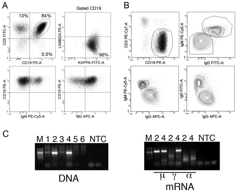
Seven cases expressing Subset #8 BcRs were identified. They utilized the IGHV4-39, IGHD6-13 and IGHJ5 genes, and shared very similar or identical VH CDR3 and a high degree of homology with those reported in the literature (15,24; Table 1). IGHV-IGHD-IGHJ rearrangements did not have somatic mutations with the exception of CA101 that exhibited a single nucleotide difference from germ line (Table 1). The cells from all seven CLL cases expressed surface κ chains as assessed by flow-cytometry (not shown) and utilized the IGKV1-39/ IGK1D-39 gene in the six cases studied by sequence analysis (CA101 could not be investigated). Again VK CDR3 were very similar (Table 1) and there was the presence of arginine (R) at the IGKV-IGKJ junction (position L96) in 3 of 6 of the patients. A frequent presence of R in this position is a characteristic feature of Subset #8 CLL (15,24,29). Case GE146 exhibited a second productive IGKV-IGKJ gene rearrangement which utilized an un-mutated IGKV4-1 gene. Since this rearrangement was detected both in the DNA and cDNA, these findings are suggestive of lack of allelic exclusion at the IGK locus (Table 1). Six out of seven of our cases expressed IgG only as assessed by both flow-cytometry for surface Ig isotypes and cDNA studies, which is in-line with the findings in all Subset #8 cases. CLL BR001 represented a remarkable exception, since IgM- and IgD-expressing cells were detected in addition to IgG-bearing cells (Figure 1A). IgM and IgD were coexpressed on the same cells as assessed by multicolor flow cytometry (Figure 1B). The presence of the same L chain type and of the same IGKV-IGKJ gene rearrangement clearly indicated that both IgM-IgD- and IgG-bearing cells belonged to the same neoplastic BR001 clone.
Table 1.
IGHV-IGHD-IGHJ and IGKV-IGKJ genes used by B-CLL cases of the Subset #8 group.
| Sample | IGHV | IGHD | IGHJ | % Homology | VH CDR3 amino acids |
|---|---|---|---|---|---|
| MS0115 | IGHV4-39 | IGHD6-13 | IGHJ5 | 100.00 | ARRSGYSSSWYDGVNWFDP |
| CA101 | IGHV4-39 | IGHD6-13 | IGHJ5 | 99.66 | ARQLGYSSSWYRNNWFDP |
| GE401 | IGHV4-39 | IGHD6-13 | IGHJ5 | 100.00 | ARRMGYSSNWYVGVNWFDP |
| NI99 | IGHV4-39 | IGHD6-13 | IGHJ5 | 100.00 | ARSSGYSSSWYSQYNWFDP |
| RC25 | IGHV4-39 | IGHD6-13 | IGHJ5 | 100.00 | ASLIGYSSSWYGGYNWFDP |
| GE146 | IGHV4-39 | IGHD6-13 | IGHJ5 | 100.00 | ARRLGYSSSWYGTYNWFDP |
| BR001 | IGHV4-39 | IGHD6-13 | IGHJ5 | 100.00 | ARRHGYSSSWYGVDWFDP |
| BR001 | IGHV2-5 | IGHD3-3 | IGHJ6 | 100.00 | AHSDTYYDFWSGYYSRTVGMDV |
| Sample | IGKV | IGKJ | % Homology | VK CDR3 amino acids |
|---|---|---|---|---|
| MS0115 | IGKV1-39/IGKV1D-39 | IGKJ1 | 100.00 | QQSYSTPQT |
| CA101 | Not Assigned | |||
| GE401 | IGKV1-39/IGKV1D-39 | IGKJ2 | 99.64 | QQSYSTPPYT |
| NI99 | IGKV1-39/IGKV1D-39 | IGKJ1 | 100.00 | QQSYSTPRT |
| RC25 | IGKV1-39/IGKV1D-39 | IGKJ1 | 100.00 | QQSYSTPRT |
| GE146 | IGKV1-39/IGKV1D-39 | IGKJ4 | 100.00 | QQSYSTPLT |
| GE146 | IGKV4-1 | IGKJ4 | 100.00 | QQYYSTPLT |
| BR001 | IGKV1-39/IGKV1D-39 | IGKJ1 | 99.64 | QQSYSTPRT |
Figure 1.
Main cellular and molecular features of the BR001 patient from Subset #8. (A) Staining of BR001 cells with anti-isotype reagents shows monoclonal cells expressing IgM and IgG. (B) IgM-expressing cells coexpress IgD, while IgG is found on a different cell sub-population. (C) Agarose gel resolution of IGHV-IGHD-IGHJ rearrangements expressed by BR001 is shown. M indicates the DNA ladder marker; numbers 1–6 indicate the six IGHV gene subgroups investigated; NTC indicates no template control. Two monoclonal IGH VDJ rearrangements in the IGHV2 and IGHV4 gene subgroup respectively, were amplified from DNA. Both the rearrangements were present in μcDNA, but only one rearrangement (IGHV4) was found in the γcDNA.
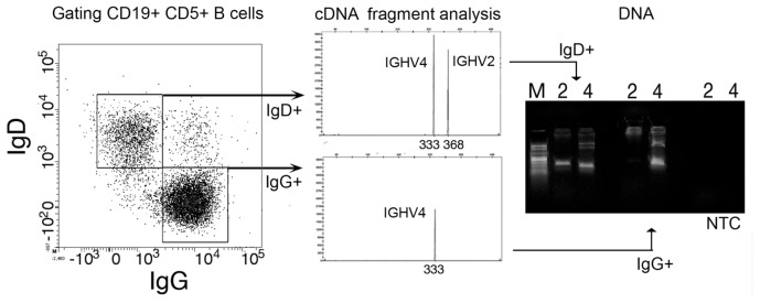
Another productive IGHV-IGHD-IGHJ rearrangement carrying the IGHV2-5, IGHD3-3 and the IGHJ6 genes was detected in BR001 cells (Table 1). This rearrangement was not somatically mutated and was present in the cell DNA and in μcDNA, but not in the γ and αcDNA (Figure 1C) suggesting that the IgM-IgD-bearing cells had two potentially productive allelic rearrangements, whereas only one rearrangement remained in IgG-bearing cells. BR001 cells were stained for CD19, CD5 and surface IgG and IgD and sorted (Figure 2). Both the DNA and the μcDNA from IgM-IgD-bearing cells had the two IGHV-IGHD-IGHJ rearrangements observed in unfractionated cells, whereas IgG-bearing cells had only the rearrangement carrying the IGHV4-39 gene in both γcDNA and DNA (see Figure 2). All cell fractions exhibited the same productive rearrangement carrying the IGKV1-39/IGKV1D-39 gene detected in the unfractionated cells (data not shown).
Figure 2.
IG gene expression by BR001 CLL subclones. CLL cells were stained for CD19, CD5 and surface IgD and IgG. CD19-CD5 positive cells were gated and IgD-positive and IgG-positive cells were sorted (left). cDNA fragment analysis (middle) shows the presence of two IGHV-IGHD-IGHJ rearrangements in IgD-expressing cells and of a single IGHV-IGHD-IGHJ rearrangement in IgG-positive cells. Gel agarose analysis of PCR-amplified DNA demonstrates a single rearrangement of IGHV4 gene subgroup in IgG-expressing cells (right).
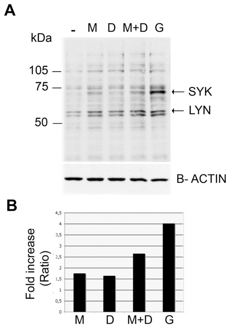
Collectively, the data indicate the following scenario: BR001 clone was comprised initially of IgM-IgD-bearing cells which were allelically included; because of an isotype switch, an IgG-bearing sub-clone subsequently was selected and expanded. This subclone utilized solely the rearranged IGHV4-39 gene, whereas the rearranged sequence containing the IGHV2-5 gene was deleted. The IgG- expressing subclone became predominant, since the ratio of IgG- over IgM- expressing cells ranged consistently around 4:1 in the second year elapsed between diagnosis, at Binet Stage A, and treatment, at Binet Stage C. The reasons for this selective expansion of the IgG-bearing subclone may be manifold and generally imply the expression of the products of both rearranged alleles at the IGH locus by the IgM-IgD-bearing cells, a fact that cannot be tested experimentally at present. For example, the concomitant expression of two H chains with different antibody-combining sites, and the consequent BcR heterogeneity of IgM-IgD-expressing cells, could have prevented the optimal binding by the stimulating antigen(s). This hypothesis is sustained by recent findings showing the presence of B cells capable of producing two functional immunoglobulin molecules (30,31). Indeed, the binding affinity for the self antigens used in this mouse model was reduced strongly in cells expressing heterogeneous BcRs at the surface (30). In contrast, the homogeneity of BcR on IgG-bearing cells and perhaps the combination of an IGHV4-39 gene with a γ chain gene could have contributed to the expression of a BcR endowed with a higher binding affinity for the stimulating antigen(s). Indeed, Subset #8 CLL cells utilize a BcR of the IgG isotype which has the ability of recognizing non-muscle myosin heavy chain IIA. This antigen molecule is expressed by normal and neoplastic cells following apoptosis (20). A prerequisite for the hypothesis that BcR stimulation plays a role in shaping subclone composition is that the neoplastic cell BcR is capable of delivering activating signals. This is particularly true in the case of CLL cells, which appear anergic to BcR stimulation in many patients (28,32). This issue was explored by exposing BR001 cells to specific aμ or aγ mAbs in vitro for 1, 5 or 10 min and measuring protein tyrosine phosphorylation. A substantial protein tyrosine phosphorylation in general, and of SYK in particular, was consistently observed after 1 min exposure to aμ or aγ mAbs (Figure 3) and this phosphorylation remained high at later times (not shown). The more efficient phosphorylation observed following exposure of the unfractionated BR001 cells to aγ Ab compared with that obtained with aμ Ab was most likely related to the proportion of IgG-versus IgM-IgD-bearing cells. Cell separation according to isotype expression was not feasible since exposure to mAb would have resulted in cell preactivation, creating difficulties for the interpretation of the results.
Figure 3.

Protein tyrosine phosphorylation following surface Ig cross-linking. (A) Tyrosine phosphorylation at 1 min following the indicated stimuli (M,D and G indicate IgM, IgD and IgG antibodies respectively) was analyzed by Western blotting. Arrows indicate the bands corresponding to LYN and SYK proteins. (B) The pixel intensity of the pooled bands at 120, 105, 75 and 55 kDa was measured and plotted as the fold increase observed following aμ-Ab (M), aδ-Ab (D), aμ-Ab + aδ-Ab (M + D) or aγ-Ab (G) stimulation compared with that of the nonstimulated cells.
FISH analyses of BR001 cells at different intervals consistently demonstrated Del 17p13.1 and Del 13q14 in virtually 100% of the IgG-expressing cells and in 60% to 70% of the IgM-IgD-bearing cells. In the latter cells, those having one deletion also exhibited the other (not shown). No trisomy 12 or Del 11q22 were detected. These findings again make BR001 different from the other cases utilizing Subset #8 BcR characterized by trisomy 12 without additional chromosomal abnormalities (29). Trisomy 12 was observed in all three patients with Subset #8 BcR that we could investigate by FISH in our cohort.
BR001 CLL provides evidence for intra clonal selection occurring following transformation which is likely to depend at least in part on the cell BcR specificity as discussed above. The presence of shared chromosomal abnormalities in the different subclones indicates that these were not a major cause for subclonal selection. However, it cannot be ruled out that different cytogenetic abnormalities, not detected by conventional FISH methodologies, and distinguishing IgM-IgD-expressing cells from those expressing IgG could have contributed to subclonal selection. It is of interest that the sub-clone expressing IgM-IgD remained of the same size throughout the course of the disease, suggesting a selective pressure to maintain the IgM-IgD-expressing subclone, the nature of which is however unknown.
In conclusion, the studies on IGHV and IGKV gene expression of BR001 cells provides evidence for continuing stimulation/selection of fully transformed CLL cells. This is in agreement with other observations on CLL clones characterized by an intraclonal diversification of IGHV genes and accumulation of somatic mutations (33,34). Although the nature of the stimulating antigen(s) was unknown, the analysis of such mutations and of their location on the gene segments was clearly favoring the hypothesis of a continuous selection by a stimulating antigenic determinant probably aimed at preserving the antigenic combining site (35–37).
ACKNOWLEDGMENTS
The authors would like to thank Massimo Federico, Fortunato Morabito and all the staff of GISL for the help in organizing the GISL observational study. The authors would like to thank Fondazione Internazionale in Medicina Sperimentale (FIRMS) for providing financial and administrative assistance and Laura Veroni for excellent administrative help.
This work was supported in part by Associazione Italiana Ricerca sul Cancro (to M Ferrarini) and in part by Compagnia di San Paolo 4824 SD/CV, 2007.2880 (to G Cutrona, F Fais). M Colombo was supported by a fellowship of Compagnia di San Paolo.
Footnotes
Online address: http://www.molmed.org
DISCLOSURE
The authors declare that they have no competing interests as defined by Molecular Medicine, or other interests that might be perceived to influence the results and discussion reported in this paper.
REFERENCES
- 1.Swerdlow SH, et al., editors. WHO Classification of Tumours of Haemapoietic and Lymphoid Tissues. 4th edition. Lyon (France): International Agency for Cancer Research; 2008. pp. 180–2. [Google Scholar]
- 2.Chiorazzi N. Cell proliferation and death: forgotten features of chronic lymphocytic leukemia B cells. Best Pract Res Clin Haematol. 2007;20:399–413. doi: 10.1016/j.beha.2007.03.007. [DOI] [PubMed] [Google Scholar]
- 3.Messmer BT, et al. In vivo measurements document the dynamic cellular kinetics of chronic lymphocytic leukemia B cells. J Clin Invest. 2005;115:755–64. doi: 10.1172/JCI23409. [DOI] [PMC free article] [PubMed] [Google Scholar]
- 4.Damle RN, et al. Telomere length and telomerase activity delineate distinctive replicative features of the B-CLL subgroups defined by immunoglobulin V gene mutations. Blood. 2004;103:375–82. doi: 10.1182/blood-2003-04-1345. [DOI] [PubMed] [Google Scholar]
- 5.Caligaris-Cappio F, Ghia P. Novel insights in chronic lymphocytic leukemia: are we getting closer to understanding the pathogenesis of the disease. J Clin Oncol. 2008;26:4497–503. doi: 10.1200/JCO.2007.15.4393. [DOI] [PubMed] [Google Scholar]
- 6.Chiorazzi N, Ferrarini M. Cellular origin(s) of chronic lymphocytic leukemia: cautionary notes and additional considerations and possibilities. Blood. 2010;117:1781–91. doi: 10.1182/blood-2010-07-155663. [DOI] [PMC free article] [PubMed] [Google Scholar]
- 7.Herishanu Y, et al. The lymph node microenvironment promotes B-cell receptor signaling, NF-{kappa}B activation, and tumor proliferation in chronic lymphocytic leukemia. Blood. 2011;117:563–74. doi: 10.1182/blood-2010-05-284984. [DOI] [PMC free article] [PubMed] [Google Scholar]
- 8.Chiorazzi N, Ferrarini M. B cell chronic lymphocytic leukemia: lessons learned from studies of the B cell antigen receptor. Annu Rev Immunol. 2003;21:841–94. doi: 10.1146/annurev.immunol.21.120601.141018. [DOI] [PubMed] [Google Scholar]
- 9.Stevenson FK, Caligaris-Cappio F. Chronic lymphocytic leukemia: revelations from the B-cell receptor. Blood. 2004;103:4389–95. doi: 10.1182/blood-2003-12-4312. [DOI] [PubMed] [Google Scholar]
- 10.Damle RN, et al. B-cell chronic lymphocytic leukemia cells express a surface membrane phenotype of activated, antigen-experienced B lymphocytes. Blood. 2002;99:4087–93. doi: 10.1182/blood.v99.11.4087. [DOI] [PubMed] [Google Scholar]
- 11.Fais F, et al. Chronic lymphocytic leukemia B cells express restricted sets of mutated and un-mutated antigen receptors. J Clin Invest. 1998;102:1515–25. doi: 10.1172/JCI3009. [DOI] [PMC free article] [PubMed] [Google Scholar]
- 12.Hamblin TJ, Davis Z, Gardiner A, Oscier DG, Stevenson FK. Unmutated Ig V(H) genes are associated with a more aggressive form of chronic lymphocytic leukemia. Blood. 1999;94:1848–54. [PubMed] [Google Scholar]
- 13.Schroeder HW, Jr, Dighiero G. The pathogenesis of chronic lymphocytic leukemia: analysis of the antibody repertoire. Immunol Today. 1994;15:288–94. doi: 10.1016/0167-5699(94)90009-4. [DOI] [PubMed] [Google Scholar]
- 14.Messmer BT, et al. Multiple distinct sets of stereotyped antigen receptors indicate a role for antigen in promoting chronic lymphocytic leukemia. J Exp Med. 2004;200:519–25. doi: 10.1084/jem.20040544. [DOI] [PMC free article] [PubMed] [Google Scholar]
- 15.Murray F, et al. Stereotyped patterns of somatic hypermutation in subsets of patients with chronic lymphocytic leukemia: implications for the role of antigen selection in leukemogenesis. Blood. 2008;111:1524–33. doi: 10.1182/blood-2007-07-099564. [DOI] [PubMed] [Google Scholar]
- 16.Stamatopoulos K, et al. Over 20% of patients with chronic lymphocytic leukemia carry stereotyped receptors: pathogenetic implications and clinical correlations. Blood. 2007;109:259–70. doi: 10.1182/blood-2006-03-012948. [DOI] [PubMed] [Google Scholar]
- 17.Widhopf GF, 2nd, et al. Chronic lymphocytic leukemia B cells of more than 1% of patients express virtually identical immunoglobulins. Blood. 2004;104:2499–504. doi: 10.1182/blood-2004-03-0818. [DOI] [PubMed] [Google Scholar]
- 18.Lanemo Myhrinder A, et al. A new perspective: molecular motifs on oxidized LDL, apoptotic cells, and bacteria are targets for chronic lymphocytic leukemia antibodies. Blood. 2008;111:3838–48. doi: 10.1182/blood-2007-11-125450. [DOI] [PubMed] [Google Scholar]
- 19.Catera R, et al. Chronic lymphocytic leukemia cells recognize conserved epitopes associated with apoptosis and oxidation. Mol Med. 2008;14:665–74. doi: 10.2119/2008-00102.Catera. [DOI] [PMC free article] [PubMed] [Google Scholar]
- 20.Chu CC, et al. Many chronic lymphocytic leukemia antibodies recognize apoptotic cells with exposed nonmuscle myosin heavy chain IIA: implications for patient outcome and cell of origin. Blood. 2010;115:3907–15. doi: 10.1182/blood-2009-09-244251. [DOI] [PMC free article] [PubMed] [Google Scholar]
- 21.Chiorazzi N, Rai KR, Ferrarini M. Chronic lymphocytic leukemia. N Engl J Med. 2005;352:804–15. doi: 10.1056/NEJMra041720. [DOI] [PubMed] [Google Scholar]
- 22.Ghia P, Chiorazzi N, Stamatopoulos K. Microenvironmental influences in chronic lymphocytic leukaemia: the role of antigen stimulation. J Intern Med. 2008;264:549–62. doi: 10.1111/j.1365-2796.2008.02030.x. [DOI] [PubMed] [Google Scholar]
- 23.Herve M, et al. Unmutated and mutated chronic lymphocytic leukemias derive from self-reactive B cell precursors despite expressing different antibody reactivity. J Clin Invest. 2005;115:1636–43. doi: 10.1172/JCI24387. [DOI] [PMC free article] [PubMed] [Google Scholar]
- 24.Ghiotto F, et al. Remarkably similar antigen receptors among a subset of patients with chronic lymphocytic leukemia. J Clin Invest. 2004;113:1008–16. doi: 10.1172/JCI19399. [DOI] [PMC free article] [PubMed] [Google Scholar]
- 25.Hallek M, et al. Guidelines for the diagnosis and treatment of chronic lymphocytic leukemia: a report from the International Workshop on Chronic Lymphocytic Leukemia updating the National Cancer Institute-Working Group 1996 guidelines. Blood. 2008;111:5446–56. doi: 10.1182/blood-2007-06-093906. [DOI] [PMC free article] [PubMed] [Google Scholar]
- 26.Brochet X, Lefranc MP, Giudicelli V. IMGT/V-QUEST the highly customized and integrated system for IG and TR standardized V-J and V-D-J sequence analysis. Nucleic Acids Res. 2008;36:W503–8. doi: 10.1093/nar/gkn316. [DOI] [PMC free article] [PubMed] [Google Scholar]
- 27.Fabris S, et al. Molecular and transcriptional characterization of 17p loss in B-cell chronic lymphocytic leukemia. Genes Chromosomes Cancer. 2008;47:781–93. doi: 10.1002/gcc.20579. [DOI] [PubMed] [Google Scholar]
- 28.Cutrona G, et al. Clonal heterogeneity in chronic lymphocytic leukemia cells: superior response to surface IgM cross-linking in CD38, ZAP-70-positive cells. Haematologica. 2008;93:413–22. doi: 10.3324/haematol.11646. [DOI] [PubMed] [Google Scholar]
- 29.Athanasiadou A, et al. Recurrent cytogenetic findings in subsets of patients with chronic lymphocytic leukemia expressing IgG-switched stereotyped immunoglobulins. Haematologica. 2008;93:473–4. doi: 10.3324/haematol.11872. [DOI] [PubMed] [Google Scholar]
- 30.Gaudin E, et al. Positive selection of B cells expressing low densities of self-reactive BCRs. J Exp Med. 2004;199:843–53. doi: 10.1084/jem.20030955. [DOI] [PMC free article] [PubMed] [Google Scholar]
- 31.Chu YP, Spatz L, Diamond B. A second heavy chain permits survival of high affinity autoreactive B cells. Autoimmunity. 2004;37:27–32. doi: 10.1080/08916930310001619481. [DOI] [PubMed] [Google Scholar]
- 32.Muzio M, et al. Constitutive activation of distinct BCR-signaling pathways in a subset of CLL patients: a molecular signature of anergy. Blood. 2008;112:188–95. doi: 10.1182/blood-2007-09-111344. [DOI] [PubMed] [Google Scholar]
- 33.Bagnara D, et al. IgV gene intraclonal diversification and clonal evolution in B-cell chronic lymphocytic leukaemia. Br J Haematol. 2006;133:50–8. doi: 10.1111/j.1365-2141.2005.05974.x. [DOI] [PubMed] [Google Scholar]
- 34.Kostareli E, et al. Intraclonal diversification of immunoglobulin light chains in a subset of chronic lymphocytic leukemia alludes to antigen-driven clonal evolution. Leukemia. 2010;24:1317–24. doi: 10.1038/leu.2010.90. [DOI] [PubMed] [Google Scholar]
- 35.Campbell PJ, et al. Subclonal phylogenetic structures in cancer revealed by ultra-deep sequencing. Proc Natl Acad Sci U S A. 2008;105:13081–6. doi: 10.1073/pnas.0801523105. [DOI] [PMC free article] [PubMed] [Google Scholar]
- 36.Gurrieri C, et al. Chronic lymphocytic leukemia B cells can undergo somatic hypermutation and intraclonal immunoglobulin V(H)DJ(H) gene diversification. J Exp Med. 2002;196:629–39. doi: 10.1084/jem.20011693. [DOI] [PMC free article] [PubMed] [Google Scholar]
- 37.Volkheimer AD, et al. Progressive immunoglobulin gene mutations in chronic lymphocytic leukemia: evidence for antigen-driven intraclonal diversification. Blood. 2007;109:1559–67. doi: 10.1182/blood-2006-05-020644. [DOI] [PMC free article] [PubMed] [Google Scholar]


