Abstract
The emissary veins are residual connections between intra-cerebral veins and their extra-cranial drainage. Mastoid emissary vein is a rare but definite entity which if not diagnosed preoperatively could be a cause of severe hemorrhage at the time of surgery which may prove to be life threatening. These veins may vary in size from that of a mere thread to that of a wax match or 1/8th–3/8th of an inch. We report one such case of a giant mastoid emissary vein which was opened while operating on mastoid and caused profuse bleeding which could only be controlled by surgical pack.
Keywords: Emissary vein, Mastoid, High resolution CT, Haemorrhage
Introduction
Emissary veins are residual connection between intra-cerebral veins and their extra-cranial drainage. Emissary vein originates from the sigmoid sinus and communicate with extra cranial veins. Emissary veins are usually <1 mm in diameter but can be as large as 4 mm [1]. The vein may be opened at its exit in making incision or stripping up the periosteum in various operative procedures or in its course in removing bone for extensive cellular infection. As a rule bleeding is very trivial and easily controlled by gel foam or bone wax but in large ones it may be troublesome. It can also cause difficulty in proper eradication of the disease especially in hands of beginners and inexperienced surgeons and can also be the source of infection or thrombosis. Preoperative imaging (HRCT) with or without contrast and magnetic resonance angiography can help to show the site and course of the vein.
Case Report
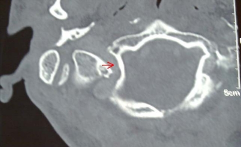
A 23 year male presented with complains of persistent ear discharge, decreased hearing, otalgia on the right side. Examination revealed muco-purulent discharge, perforated tympanic membrane and cholesteatoma. Pure tone audiometery showed conductive hearing loss. High resolution CT scan showed soft tissue mass filling Mastoid and middle ear cavity. Patient was taken for modified radical mastoidectomy. While elevating the periosteum from the mastoid, there was profuse bleeding near the mastoid tip anteriorly at the tympano-mastoid suture from a vessel which was coming out through the mastoid cortex. The bleeding could not be controlled by routine methods like, local pressure, gel foam pack and electric cauterization. Finally a surgical pack was kept in the lumen of the vessel which controlled the bleeding. The mastoidectomy was completed. The postoperative period was uneventfull. Postoperatively a review of the HRCT temporal bone was sought from the radiologist and it was reported as soft tissue mass in the middle ear with erosion of malleus, incus and stapes superstructure. The soft tissue in mastoid antrum also showed a giant vein postero-medial to the mastoid tip communicating with sub cranial portion of internal jugular vein and the adjacent sigmoid sinus (Fig. 1).This finding was missed preoperatively.
Fig. 1.
Showing vessel traversing through the mastoid cortex to the exterior communicating with the sigmoid sinus
Discussion
The cerebral venous drainage pathway is mainly formed by the internal jugular vein. During the development of embryo primary capillary plexus develops in three layers, superficial layer drains into the external jugular vein middle and deep layers drains into internal jugular vein [2]. Emissary veins consists of connections between the superficial and middle layers. They originate from the sigmoid sinus and communicate with extra cranial veins [2]. It may originate on the outer edge of the lateral sinus groove just below the bend and has a short course in the substance of the bone in an upward and backward direction. It is open on the surface just behind the upper posterior edge of the mastoid process at the level of the bony auditory meatus to enter the occipital or posterior auricular vein. These form a part of external jugular vein [3]. Emissary veins are usually <1 mm in diameter but can be as large as 4 mm [1].The sigmoid sinus may be smaller in presence of large mastoid emissary vein. Three emissary venous pathways have been described in humans. The lower or posterior condylar vein (it exits the sigmoid sinus above the jugular bulb and is usually of moderate size). The middle or mastoid emissary vein, this is the most constant of all the three veins (it crosses the mastoid foramen and unites the sigmoid sinus with the posterior auricular or occipital vein) and the upper petrosquamosal emissary vein, originating at the junction of the transverse and sigmoid sinus (it is usually very small) [2].
The vein may be opened at the exit in making incision or when striping up the periosteum in various operative procedures or in its course in removing bone for extensive cellular infections or for exploration and treatment of lateral sinus thrombosis in operation of decompression of facial nerve and in fracture of skull. As a rule bleeding is trivial and easily controlled by plugging with gauze or bone wax but in case of large ones it may be troublesome. The vein may also be infected from lateral sinus and would appear to be edematous swelling or may form abscess at the exit [4]. We experienced one such case of large mastoid emissary vein which was opened up while making the incision and caused severe hemorrhage which could only be controlled by surgical pack kept in the lumen of the vessel. Mastoid emissary vein is a rare but definite entity which if not diagnosed preoperatively could be a cause of severe hemorrhage at time of surgery which may prove to be life threatening. It can cause difficulty in proper eradication of the disease especially in hands of beginners and inexperienced surgeons. It can also be the source of infection or thrombosis. Preoperative imaging (HRCT) with or without contrast and magnetic resonance angiography can help to show the site and course of the vein.
References
- 1.Boyd GI. The emissary foramina of the cranium in man and the arthropods. J Anat. 1930;65:108–121. [PMC free article] [PubMed] [Google Scholar]
- 2.Marsot-Dupuch K, Elmaleh-Berges M, Bonneville F, Lasjaunias P. The petrosquamosal sinus: CT and MR findings of a rare emissary vein. Am J Neuroradiol. 2001;22:1186–1193. [PMC free article] [PubMed] [Google Scholar]
- 3.Cheatle AH (1992) Mastoid emissary vein and its surgical importance. Anatomy [PMC free article] [PubMed]
- 4.Smith JA, Danner CJ. Complication of chronic otitis media and cholesteatoma. Otolaryngol Clin N Am. 2006;39:1237–1255. doi: 10.1016/j.otc.2006.09.001. [DOI] [PubMed] [Google Scholar]


