Abstract
Esthesioneuroblastoma (ENB) also known as olfactory neuroblastoma is an uncommon malignant neoplasm arising in the roof of nasal cavity. It is now understood to originate from the olfactory epithelium. Case reports published worldwide have been very few. Common presenting symptoms of Esthesioneuroblastoma include nasal obstruction, epistaxis, facial pain, diplopia, proptosis, and anosmia. Apart from being locally aggressive, it metastasizes widely by both hematogenous and lymphatic routes.
Keywords: Esthesioneuroblastoma, Olfactory neuroblastoma, Malignant tumour of nasal cavity
Introduction
The incidence of Esthesioneuroblastoma is relatively low worldwide. A 1997 literature search identified 1,457 cases in the published literature [1]. The tumour was first described by Berger and Luc [2]. This tumor constitutes 3% of all intranasal neoplasms and can be seen in all ages, with a peak in the second and sixth decades of life and with equal distribution between the sexes [1, 3]. The tumour aggressively spreads submucosally to involve the orbits, anterior cranial fossa, and brain. The most common site of metastasis is the cervical lymph nodes (10–33% of patients), while sites of distant metastasis are the lungs, brain, and bone, but are less common [1, 3].
Case Report
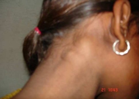
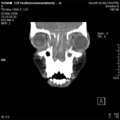
A female patient, Sonam age 12 years belonging to a poor migrant labour family from Uttar Pradesh presented to the ENT Department, Govt. Medical College, Amritsar as a patient of epistaxis in the emergency. The patient was giving a history of nasal obstruction along with nasal discharge from the left side for last 4 months. There was history of earlier episodes of self containing epistaxis, off and on for last 4 months. On examination of the patient, a thick blood stained foul smelling discharge was seen coming from the left nasal cavity. On cleaning the discharge, a pinkish-grey mass was seen filling the left nasal cavity. Consistency of the mass was firm and did not look like a polyp. The overlying surface of the mass was irregular. The mass was bleeding on manipulation which was done to clean and examine the lesion. The epistaxis with which the patient presented, was managed with packing over the lesion. There was proptosis and lateral displacement of left eyeball (Fig. 1). Cervical lymphadenopathy was seen; with bilateral multiple nodes, firm in consistency, each approximately 2–4 cm in size, non-tender but partially mobile (Figs. 2, 3). All required hematological and biochemical investigations were done. These were not indicative of any specific pathology. CT scan showed that there was a mass involving left nasal cavity, left maxillary sinus, and both side ethmoid sinuses. There was intraorbital extension of the mass with erosion of lamina papyracea and medial part of floor of orbit on the left side (Fig. 4). There was destruction of the uncinate process and medial wall of maxillary sinus on the left side. There was erosion of cribriform plate with intracranial extension in left frontal region (Fig. 5). There were lymph nodes in supra and infrahyoid regions with central hypodensity. The CT impression was suggestive of a Esthesioneuroblastoma but to be differentiated from a Lymphoma. Under general anesthesia, biopsy was taken from the nasal mass. The mass bled on biopsy but was controlled with packing. Simultaneously lymph node biopsy was taken from the neck node on the left side. The nasal mass on biopsy showed round cells, with minimal cytoplasm, round and hyperchromatic nuclei. The cells were arranged around blood vessels. There was necrosis and infiltration along with prominent peritheliomatous arrangement. The histopathology report was suggestive of a neuroblastoma. The histopathology was confirmed from two different centres and the report was similar from both. The Lymph node biopsy showed groups of cells with hyperchromatic nuclei and indistinct cytoplasm, and keeping in view the histopathology of biopsy from nasal mass; the lymph node biopsy report was suggestive of metastatic deposits of Neuroblastoma.
Fig. 1.
Proptosis and lateral displacement of left eyeball
Fig. 2.
Multiple neck nodes—Right
Fig. 3.
Multiple neck nodes—Left
Fig. 4.
Axial section of CT scan showing intraorbital extension
Fig. 5.
Coronal section of CT scan showing intracranial extension
The patient presented to us with extensive primary lesion and metastatic neck nodes. A combined craniofacial and neurosurgical intervention along with a neck dissection was advised to the patient to be followed by post operative radiotherapy. Unfortunately the patient was one of her many siblings and the family was not willing for an extensive treatment regime, hence she was given treatment in the form of external radiotherapy combined with chemotherapy. The patient died after a 2-year follow up.
Discussion
ENB is an uncommon tumour and there is no specific age, sex, or racial predilection. There are no known etiological factors. The tumour poses diagnostic difficulties for both the clinician as well as the pathologist. Well-differentiated ENBs exhibit homogenous small cells with uniform round-to-oval nuclei with rosette or pseudorosette formation and eosinophilic fibrillary intercellular background material, which appears clear on traditional pathological sections. In undifferentiated ENB characterized by anaplastic hyperchromatic small cells with numerous mitoses and scant cytoplasm, differentiation from other small-cell nasal neoplasms via light microscopy becomes difficult. Immunohistochemical staining and electron microscopy are essential for establishing the pathological diagnosis of sinonasal small cell neoplasms, which include malignant melanoma, embryonal rhabdomyosarcoma, malignant lymphoma, extramedullary plasmocytoma, sinonasal undifferentiated carcinoma and sinonasal neuroendocrine carcinoma. No specific immunocytologic stain identifies ENB [4].
Kadish et al. [5] were the first to propose a staging classification for ENB. Staging was given as per Group A: limited to tumors of the nasal fossa; Group B: extension to the paranasal sinuses; and Group C: extension beyond the paranasal sinuses. Biller et al. [6] gave their staging as: T1 indicates tumor involving the nasal cavity and paranasal sinuses, excluding the sphenoid, with or without erosion of the bone of the anterior cranial fossa; T2: tumor extending into the orbit or protruding into the anterior cranial fossa; T3: tumor involving the brain that is resectable with margins; and T4: unresectable tumor. Dulguerov and Calcaterra [7] further modified the grading to: T1 indicates tumor involving the nasal cavity and/or paranasal sinuses, excluding the sphenoid, sparing most superior ethmoidal cells; T2: tumor involving the nasal cavity and/or paranasal sinuses, including the sphenoid, with extension to or erosion of the cribriform plate; T3: tumor extending into the orbit or protruding into the anterior cranial fossa; and T4: tumor involving the brain.
Treatment modalities have included extracranial surgical excision, craniofacial surgical resection, radiotherapy and chemotherapy used alone or in combination. Loubutin et al. [8] observed that leptomeningeal infiltration by ENB carried a poor prognosis. Homzie and Elkon [9] included the presence of metastases and local extension of the tumor (e.g., ethmoidal, nasopharynx, orbital) as negative prognostic factors in their series. Resto et al. [10] found that the only predictor of survival was nodal metastases. Broich et al. [1] reviewed the literature and found a 72.5% 5-year survival rate in patients treated with combined therapies compared with 62.5% with surgery alone and 53.8% with Radiotherapy alone. Dulguerov and Calcaterra [7] observed in their study that 92% of patients treated with complete gross resection of the tumor plus Radiotherapy remained recurrence free, as opposed to 40% with Radiotherapy alone and 14% with surgery alone.
References
- 1.Broich G, Pagliari A, Ottaviani F. Esthesioneuroblastoma: a general review of the cases published since the discovery of the tumour in 1924. Anticancer Res. 1997;17(4A):2683–2706. [PubMed] [Google Scholar]
- 2.Berger L, Luc R. Esthésioneuroépithéliome olfactif. Bull Assoc Fr Etud Cancer. 1924;3:410–421. [Google Scholar]
- 3.Elkon D, Hightower SI, Lim ML, et al. Esthesioneuroblastoma. Cancer. 1979;44:1087–1094. doi: 10.1002/1097-0142(197909)44:3<1087::AID-CNCR2820440343>3.0.CO;2-A. [DOI] [PubMed] [Google Scholar]
- 4.Pavel D, Abdelkarim SA (2008) Esthesioneuroblastoma. http://www.emedicine.com/med/topic748.htm. Retrieved 19 July 2008
- 5.Kadish S, Goodman M, Wang CC. Olfactory neuroblastoma: a clinical analysis of 17 cases. Cancer. 1976;37:1571–1576. doi: 10.1002/1097-0142(197603)37:3<1571::AID-CNCR2820370347>3.0.CO;2-L. [DOI] [PubMed] [Google Scholar]
- 6.Biller HF, Lawson W, Sachdev VP, Som P. Esthesioneuroblastoma: surgical treatment without radiation. Laryngoscope. 1990;100:1199–1201. doi: 10.1288/00005537-199011000-00013. [DOI] [PubMed] [Google Scholar]
- 7.Dulguerov P, Calcaterra T. Esthesioneuroblastoma: the UCLA experience 1970–1990. Laryngoscope. 1992;102:843–849. doi: 10.1288/00005537-199208000-00001. [DOI] [PubMed] [Google Scholar]
- 8.Loubutin JP, Maugard-Loubutin C, Fumoleau P. Leptomeningeal infiltration in esthesioneuroblastoma: report of two cases with poor prognosis. Eur Neurol. 1994;34:236–238. doi: 10.1159/000117046. [DOI] [PubMed] [Google Scholar]
- 9.Homzie MJ, Elkon D. Olfactory esthesioneuroblastoma: variables predictive of tumor control and recurrence. Cancer. 1980;46:2509–2513. doi: 10.1002/1097-0142(19801201)46:11<2509::AID-CNCR2820461132>3.0.CO;2-Z. [DOI] [PubMed] [Google Scholar]
- 10.Resto VA, Eisele DW, Forastiere A, et al. Esthesioneuroblastoma: the Johns Hopkins experience. Head Neck. 2000;22:550–558. doi: 10.1002/1097-0347(200009)22:6<550::AID-HED2>3.0.CO;2-0. [DOI] [PubMed] [Google Scholar]


