Abstract
A case of epiglottic abscess in an otherwise healthy adult male is reported. Patient was initially managed as acute epiglottis with parenteral antibiotics, humidified air, and steroids close ICU monitoring. Failure of recovery and re-evaluation revealed an abscess of epiglottis. Management with intubation and incision and drainage of abscess lead to very rapid recovery and subsequent discharge of patient.
Keywords: Adult Epiglottic Abscess, Epiglottitis
Introduction
In epiglottis, or more correctly supraglottitis, the cellulites involves multiple areas of supraglottis. It is typically a disease of children between the ages of 2 and 6, although any age, including adults can be affected. Haemophillus influenzae type B (HIB) is the responsible pathogen in most cases. The introduction of HIB vaccine in most areas world over has reduced the incidence of supraglottitis by 90%. Epiglottic abscess is an uncommon sequelae of acute epiglottitis, occurring in fewer than 4% of cases and very few cases of the same have reported in literature. However, an increased incidence of epiglottic abscess and adult epiglottitis have been reported lately. This probably indicates increased resistance to conventional antibiotics used for epiglottitis or a changing microbiological profile.
Case Report
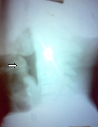
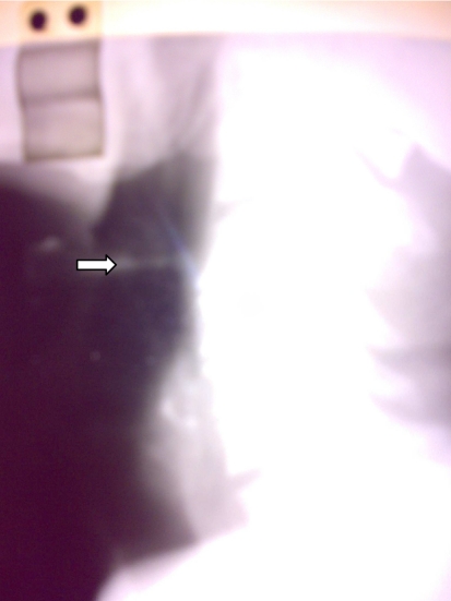
A 38 year old male presented with hoarseness of voice and breathing difficulty of two day duration. Patient was febrile and had anxious, worried look. Radiography in the form of soft tissue, lateral view of neck revealed the typical thumb sign of epiglottis (Fig. 1). Patient was admitted in ICU under close monitoring for spO2, aggravated breathing difficulty and provision for humidified air and parenteral antibiotics. Blood counts revealed a WBC count of 7,700 with 65% neutrophils. Ampicillin in dose of 1 gm every 6 h was started and constant vigil of the general condition kept. After a further of two day monitoring re-evaluation was deemed necessary. Indirect laryngoscopy revealed a congested swollen epiglottis covered with thick whitish slough on left side, with normal arytenoids mobility. Fiber-optic laryngoscopy revealed reduction in glottic chink by almost 50%. Patient was shifted to theater and incision with drainage of pus was done under general anaesthesia with uneventful extubation after 12 h of ICU monitoring. Radiograph neck post-op on second day showed only minimal oedema (Fig. 2). Microbiology revealed mixed flora with predominant gram positive cocci. Patient was discharged two days after surgery with almost normal voice on oral augmenting for 5 days.
Fig. 1.
Lateral view of neck revealed the typical thumb sign of epiglottis
Fig. 2.
Radiograph neck post-op on second day showed only minimal oedema
Discussion
Supraglottitis is a rare infection, but the awareness of the disease is important in view of high mortality and changing trends of the disease in the HIB vaccination era (e.g, an increase in the presenting age).
Symptoms of supraglottitis progress rapidly over a matter of hours. The onset in adults, however, is not that rapid. Because of the swollen airway in supraglottic region patients usually get a muffled voice. However, as the airway in adults is of larger diameter rapidly progressing breathing difficulty is not a usual feature. The main symptoms found are dysphagia/odynophagia (85%), followed by fever (55%) and pharyngocervical pain. Tenderness in the anterior neck in the region of hyoid serves as a quick aid in the diagnosis of the condition.
Besides the commoner Haemophilus influnzae B, many other pathogens exist which can cause epiglottitis and include various genera of streptococci, including Streptococcus pneumoniae, Staphyloccus aureus, Meningococci, Haemophilus parainfluenza, Kleibsella pneumoniae, viruses, and Candida albicans also.
Though the main complication of epiglottitis is respiratory obstruction, a few rare complications include epiglottic abscess and descending necrotizing mediastinitis can also occur in a case of epiglottitis that may warrant surgical intervention. This patient developed epiglottic abcess that was not responding to medical management and had to be drained surgically. The response after surgical drainage was dramatic with resultant resolution of symptoms [1–5].
References
- 1.Saeed I, Malik SM, Ahmad S. Acute epiglottic abscess in an adult. JCPSP. 2007;17(7):427–428. [PubMed] [Google Scholar]
- 2.Gilbert A, Timothy J. Epiglottic abscess. Ear Nose Throat J. 2004;83:154–155. [PubMed] [Google Scholar]
- 3.Frantz TD, Rasgon BM, Quesenberry CP., Jr Acute epiglottitis in adults. Analysis of 129 cases. JAMA. 1994;272:1358–1360. doi: 10.1001/jama.272.17.1358. [DOI] [PubMed] [Google Scholar]
- 4.Heeneman H, Ward KM. Epiglottic abscess: Its occurrence and management. J Otolaryngol. 1977;6:31–36. [PubMed] [Google Scholar]
- 5.Stack BC, Jr, Ridley MB. Epiglottic abscess. Head Neck. 1995;17:263–265. doi: 10.1002/hed.2880170316. [DOI] [PubMed] [Google Scholar]


