Abstract
Epidermoid cyst is usually due to infection of pilosebaceous gland or due to traumatic migration of epidermis to the deeper structure of skin. They may present in any place of body which is lined by squamous epithelium. They are rarely present in head neck and in oral cavity. We are presenting rare cases of epidermoid cyst presenting in post aural region and floor of mouth.
Keywords: Dermoid cyst, Epidermoid cyst, Post aural region, Floor of mouth, USG
Introduction
Epidermoid and dermoid cysts can be present any where in the body lined by squamous epithelium. About 7% occur in head and neck region, and they represent about 0.01% of all oral cavity cysts [1]. The cysts can be defined as epidermoid when the lining presents only epithelium, dermoid cysts when skin adnexa are found, and teratoid cysts when other tissue such as muscle, cartilage, and bone are present [2]. Because of the similar epithelium lining, all three of these cysts may have cheesy keratinaceous material within the lumen [3]. An epidermoid cyst can be differentiated with other sublingual cyst by histopathological examination only.
Case Report
Case 1
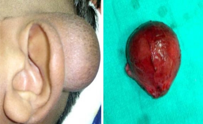
A 25 years male presented with a swelling on the left post aural region since 1 year. There was no history of ear complaint, any trauma or surgery and discharge from the swelling. On examination, about 3 × 3 cm swelling present in post aural region from the medial surface of conchal cartilage to the upper part of mastoid process. Swelling was soft, non tender, and freely mobile with defined margins. There was no sinus nor does it show any signs of inflammation. Swelling was non translucent and without any signs of intracranial extension. Extended post aural incision was given and mass was excised completely. It was lying above perichondrium without involving it and posteriorly above the temporalis muscle. Post operative histopathology report suggestive of epidermoid cyst. Patient is doing well without recurrence in follow-up (Fig. 1).
Fig. 1.
Pre-operative photograph showing post aural cyst and postoperative specimen
Case 2
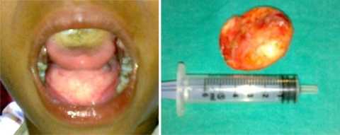
A 12 years male presented with swelling on the floor of mouth since 2 years, difficulty in swallowing since 6 months, unclear voice and occasional breathing difficulty. There was no history of trauma. On examination a large firm cystic lesion present on floor of mouth which displaced the tongue superiorly and posteriorly (Fig. 2). Bimanually it was not palpable. Clinically, it lies above the level of geniohyoid and myelohyoid muscle. USG showed a well defined mass on the floor of mouth above the previously mentioned muscles. Cyst was removed en mass intraorally. Histopathology report showed acidophilic stratum corneum and basophilic dot like staining of stratum granulosum. These findings were suggestive of epidermoid cyst.
Fig. 2.
Pre-operative photograph showing sublingual cyst and postoperative specimen
Discussion
Epidermoid cysts are lined by stratified squamous epithelium and contain keratin debris. Men are mostly affected and such cysts generally appear after puberty. It is common in young and old, but is rare in childhood [4]. The common sites are the face, neck, shoulders, chest areas, and oral cavity. They may be congenital or acquired type. Congenital type is due to entrapment of ectodermal substance between the midline fusion of first and second branchial arches during third and fourth intrauterine life [5]. Acquired type cysts usually occur due to infection around pilosebaceous follicle and sometime deep implantation of epidermis as a result of penetrating or blunt injury [6]. It is slow growing and non tender mass. When present in dermis, it raises epidermis to produce a firm elastic dome-shaped protuberance which is mobile over the deeper structures. They grow slowly and may become inflamed and firm time to time. Suppuration may occur. In the oral cavity, they displace tongue superiorly and present with dysphagia, dyspnoea, and dysphonia. The content of a cyst sometimes escape slowly from the duct orifices and dry in successive layers on the skin, forming a sebaceous horn. The differential diagnosis of sublingual lesions includes: infectious process, ranula, lymphatic malformation, dermoid cyst, epidermoid cyst, heterotopic gastrointestinal cyst, and duplication foregut cyst. Ultrasonography is the best investigation for these types of cyst. It is economical, reliable and without radiation exposure [7]. Epidermoid cysts have fluid attenuation on CT scans and are hypointense on T1-weighted images and hyperintense on T2-weighted images, following the signal intensity of fluid. Computed tomography and magnetic resonance imaging allow more precise localization of the lesion in relationship to geniohyoid and mylohyoid muscles, and they also enable the surgeon to choose the most appropriate surgical approach, especially for very large lesions [5]. Surgical excision of the cyst is often required and the entire cyst wall is removed to prevent recurrence [8]. Incomplete removal is common if attempted in the presence of recent infection.
References
- 1.Turetschek K, Hospodka H, Steiner E. Case report: epidermoid cyst of the floor of the mouth: diagnostic imaging by sonography, computed tomography and magnetic resonance imaging. Br J Radiol. 1995;68:205–207. doi: 10.1259/0007-1285-68-806-205. [DOI] [PubMed] [Google Scholar]
- 2.Calderon S, Kaplan I. Concomitant sublingual and submental epidermoid cysts: a case report. J Oral Maxillofac Surg. 1993;51:790–792. doi: 10.1016/S0278-2391(10)80425-2. [DOI] [PubMed] [Google Scholar]
- 3.Hunter T, Paplanus S, Chernin M, Coulthard S. Dermoid cyst of the floor of the mouth: CT appearance. Am J Roentgenol. 1983;141:1239–1240. doi: 10.2214/ajr.141.6.1239. [DOI] [PubMed] [Google Scholar]
- 4.Weiss RA. Orbital disease. In: McCord CD, Tanenbacum M, Nunery WR, editors. Oculoplastic surgery, 3rd edn. New York: Raven Press; 1995. pp. 417–476. [Google Scholar]
- 5.Longo F, Maremonti P, Mangone GM, Maria G, Califano L. Midline (dermoid) cysts of the floor of the mouth: report of 16 cases and review of surgical techniques. Plast Reconstr Surg. 2003;112:1560–1565. doi: 10.1097/01.PRS.0000086735.56187.22. [DOI] [PubMed] [Google Scholar]
- 6.Legent F, Beauvillain C, Gaillard F, Leroy G. Peri-auricular epidermal cysts secondary to an ear operation. Ann Otolaryngol Chir Cervicofac. 1980;97(1–2):105–109. [PubMed] [Google Scholar]
- 7.Walstad WR, Solomon JM, Schow SR, Ochs MW. Midline cystic lesion of the floor of the mouth. J Oral Maxillofac Surg. 1998;56:70–74. doi: 10.1016/S0278-2391(98)90919-3. [DOI] [PubMed] [Google Scholar]
- 8.Mevorah B, Bovet R. Treatment of retro auricular keratinous cysts. J Dermatol Surg Oncol. 1984;10(1):40–44. doi: 10.1111/j.1524-4725.1984.tb01171.x. [DOI] [PubMed] [Google Scholar]


