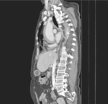
A 69-year-old woman presented emergently with acute aortic syndrome. Five years earlier, due to esophageal perforation from fish-bone ingestion, she had undergone esophageal reconstruction with a free jejunal flap and substernal colon interposition. The patient had a long history of hypertension, without regular medical control. She was hemodynamically stable with visible surgical scars and a bulging mass at the base of the neck (Fig. 1). Computed tomography (CT) of the chest revealed the colon substitute in the substernal area and an acute intramural hematoma that involved the ascending and descending aorta (Fig. 2). The maximal aortic diameter was 45 mm, and the hematoma was 10.5 mm thick. A sagittal CT view showed that the colon substitute was between the sternum and the intramural hematoma (Fig. 3).
Fig. 1 Photograph shows clinical appearance of the huge bulging colon in the patient's lower neck. Multiple scars from previous operations are visible.
Fig. 2 Axial computed tomogram shows an acute intramural hematoma involving the ascending and descending aorta, along with colon substitute in the substernal area.
Fig. 3 Computed tomography (sagittal view) shows the colon substitute between the sternum and the intramural hematoma.
Because of the possible difficulty of repeat sternotomy, and controversy surrounding the treatment of type A intramural hematoma, the patient was treated conservatively. She was closely monitored in intensive care on β-blocker therapy for control of heart rate and blood pressure. She was discharged on the 7th hospital day without any complication. Three months later, CT showed complete resolution of the intramural hematoma and no evidence of aortic dissection. A CT scan at 13 months showed no pathologic condition (Fig. 4).
Fig. 4 Follow-up computed tomogram at 13 months shows complete resolution of the intramural hematoma.
Comment
The optimal initial treatment for a type A intramural hematoma can be individualized. Emergent surgery is recommended for patients with cardiac tamponade, myocardial ischemia, or aortic regurgitation. If patients do not have complications, supportive medical therapy with frequent follow-up imaging is a reasonable option.1 Furthermore, on the basis of previously reported data, patients with an ascending aortic diameter of more than 50 mm or a subadventitial hematoma more than 12 mm thick should be considered as candidates for early surgery.2
Footnotes
Address for reprints: Chiung-Lun Kao, MD, Division of Thoracic & Cardiovascular Surgery, Chang Gung Memorial Hospital at Chiayi, No. 6, Sec. West, Chia Pu Rd., Putzu City, Chiayi Hsien, Taiwan 613, ROC
E-mail: sa11421@adm.cgmh.org.tw
References
- 1.Kitai T, Kaji S, Yamamuro A, Tani T, Tamita K, Kinoshita M, et al. Clinical outcomes of medical therapy and timely operation in initially diagnosed type A aortic intramural hematoma: a 20-year experience. Circulation 2009;120(11 Suppl):S292–8. [DOI] [PubMed]
- 2.Kan CB, Chang RY, Chang JP. Optimal initial treatment and clinical outcome of type A aortic intramural hematoma: a clinical review. Eur J Cardiothorac Surg 2008;33(6):1002–6. [DOI] [PubMed]


