Abstract
The clostridial botulinum neurotoxins (BoNTs) are the most potent protein toxins known. The carboxyl-terminal fragment of the toxin heavy chain (Hc) has been intensively investigated as a BoNT vaccine immunogen. We sought to determine whether targeting Hc to antigen-presenting cells (APCs) could accelerate the immune responses to vaccination with BoNT serotype A (BoNT/A) Hc. To test this hypothesis, we targeted Hc to the Fc receptors for IgG (FcγRs) expressed by dendritic cells (DCs) and other APCs. Hc was expressed as a fusion protein with a recombinant ligand for human FcγRs (R4) to produce HcR4 or a similar ligand for murine FcγRs to produce HcmR4. HcR4, HcmR4, and Hc were produced as secreted proteins using baculovirus-mediated expression in SF9 insect cells. In vitro receptor binding assays showed that HcR4 effectively targets Hc to all classes of FcγRs. APCs loaded with HcR4 or HcmR4 are substantially more effective at stimulating Hc-reactive T cells than APCs loaded with nontargeted Hc. Mice immunized with a single dose of HcmR4 or HcR4 had earlier and markedly higher Hc-reactive antibody titers than mice immunized with nontargeted Hc. These results extend to BoNT neutralizing antibody titers, which are substantially higher in mice immunized with HcmR4 than in mice immunized with Hc. Our results demonstrate that targeting Hc to FcγRs augments the pace and magnitude of immune responses to Hc.
INTRODUCTION
The clostridial botulinum neurotoxins (BoNTs) comprise a group of seven antigenically distinct proteins (serotypes A to G). BoNTs are the most potent protein toxins known. Human botulism is primarily caused by serotypes A, B, and E and typically occurs through ingestion of the toxin in contaminated food, though wound botulism and infant botulism (colonizing infection in neonates) also occur (10). Due to their lethality and potential for misuse, BoNTs are classified as category A biothreats by the Centers for Disease Control and Prevention (2).
BoNT is expressed as a single-chain, 150-kDa polypeptide that is subsequently cleaved, resulting in a 50-kDa light chain linked by a disulfide bond to a 100-kDa heavy chain. BoNT activities map to discrete regions within the chains: endoprotease activity resides within the light chain, while the translocation and receptor binding domains are located in the heavy chain. The heavy chain can be structurally subdivided into an amino-terminal fragment (HN) containing the translocation domain and a carboxyl-terminal fragment (Hc) containing the receptor binding domain (41, 42).
At present, protection against BoNT intoxication is provided by a formalin-inactivated pentavalent toxoid vaccine against serotypes A to E. The toxoid vaccine has several disadvantages: the toxoid preparations are crude and dangerous to produce, and multiple boosters are needed to achieve protective immunity (11). Recently, new strategies for BoNT vaccine development have emerged, with emphasis given to the use of recombinant Hc as the immunogen (43).
An early and rate-limiting step in the immune response to vaccination is the uptake, processing, and presentation of antigen by dendritic cells (DCs). DCs are professional antigen-presenting cells (APCs) that acquire antigen in peripheral sites, traffic to lymphoid tissues, and efficiently stimulate B and T cells. By improving the uptake of vaccine proteins by DCs or other APCs, more rapid and robust responses to vaccination can ensue. Targeting antigen to DC surface receptors has emerged as an effective means to load DCs with antigen in vivo (30, 48). The Fc receptors for IgG (FcγRs), expressed on DCs and APCs, bind and internalize antigen-IgG immune complexes via endocytic and phagocytic routes, resulting in the accumulation of exogenous antigen within DCs. Antigen-loaded DCs efficiently degrade antigenic proteins into peptides which, once loaded onto major histocompatibility complex (MHC) class II molecules, can be presented to CD4+ T cells. Additionally, antigen uptake through FcγRs by immature DCs can facilitate “cross-presentation” of exogenously derived antigen onto MHC class I molecules, which can be presented to naive CD8+ T cells (16, 36). These features of FcγRs make them attractive targets for in vivo delivery of antigens to DCs.
Humans express three classes of FcγR (31, 37). FcγRI (CD64) binds monomeric IgG and immune complexes with high affinity. FcγRIIa/b (CD32a/b) and FcγRIIIa/b (CD16a/b), the low-affinity receptors for Fc, bind monomeric IgG poorly but bind IgG immune complexes avidly. Ligation of FcγRI, FcγRIIa, or FcγRIIIa initiates cellular activation, whereas FcγRIIb delivers an inhibitory signal (18). FcγRs are transmembrane receptors, with the exception of FcγRIIIb, which is attached to the membrane with a glycophosphatidyl inositol link. Both high- and low-affinity FcγRs contribute to antigen uptake and processing by APCs (1, 51). Mouse orthologs exist for the human FcγRs, with the exceptions of FcγRIIa and FcγRIIIb.
Previously, we developed a novel recombinant ligand for FcγRs (R4) (see Fig. 2A) that targets antigen to FcγRs expressed on DCs in vivo and results in greatly enhanced humoral immune responses to the antigens delivered (19, 49). In the present study, we sought to determine whether in vivo DC loading could improve immune responses to BoNT serotype A (BoNT/A) Hc. To test this hypothesis, we used the R4 ligand to target BoNT/A Hc to FcγRs in vivo. We characterized the early immune responses to single low doses of antigen. Our results demonstrate that targeting Hc to FcγRs leads to earlier and more robust antibody responses than nontargeted Hc.
Fig. 2.
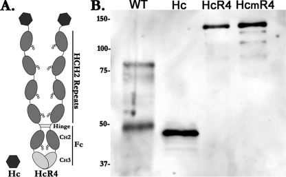
Structure and expression of recombinant Hc antigens. (A) Schematic of recombinant Hc and HcR4. HcR4 consists of Hc expressed as a fusion protein with R4, a tandem repeat composed of 4 copies of the hinge and CH2 sequences (HCH2) from IgG fused to an Fc region of IgG. The HCH2 sequence contains the region of IgG that binds to FcγRs. (B) Western blot analysis of Hc antigens. Proteins were separated on SDS-PAGE gels under reducing conditions and transferred to nitrocellulose membranes. Hc antigens were detected by using an anti-BoNT/A1 polyclonal antibody. Lane 1, wild-type BoNT/A1 Hc (WT; 200 ng); lane 2, recombinant Hc (50 ng); lane 3, HcR4 (150 ng); lane 4, HcmR4 (150 ng). Positions of molecular weight markers, in thousands, are shown to the left.
MATERIALS AND METHODS
Construction and expression of BoNT/A Hc, HcR4, and HcmR4.
A synthetic Hc gene, encoding V857 to L1296 of BoNT/A1, was assembled from fragments synthesized de novo. Codon usage in the synthetic BoNT/A Hc gene was optimized for expression in the Spodoptera frugiperda (SF) insect cell line using UPGENE codon optimization software (12). Naturally occurring restriction sites were removed, and a 5′ XhoI, a 3′ EcoRI, and an internal NdeI site were introduced into the sequence.
R4 is an IgG-like fusion protein that contains a tandem repeat composed of 4 copies of the hinge and CH2 (HCH2) regions of IgG (19, 49). The HCH2 region of IgG encompasses those sequences that bind FcγRs. To express Hc as a fusion protein with R4, the Hc gene fragment was subcloned into the XhoI and EcoRI sites of the pFastBac1 baculovirus expression vector (pFB) (Invitrogen) that had been previously modified with the introduction of R4 coding sequences (19). The Hc gene fragment was also subcloned into the XhoI and EcoRI sites of the pFB-His expression vector to express the Hc gene with a 6×His tag as previously described (19). To direct secretion of recombinant proteins into the medium, the constructs were preceded in-frame by the IgG heavy chain leader sequence inserted into pR4-FB and pFB-His.
We derived a murine R4 ligand (mR4) based on IgG2a sequences, the murine homolog of human IgG1. mR4 was constructed as previously described (49) and inserted into the EcoRI and SalI sites of pFB to produce pmR4-FB. The IgG leader sequence and Hc gene fragment were transferred as a BamHI-EcoRI fragment into pmR4-FB to produce the HcmR4 expression vector.
Recombinant baculovirus was used to infect SF9 cells, and recombinant proteins were purified directly from the growth medium, as previously described (19). Eluted proteins were dialyzed extensively against endotoxin-free phosphate-buffered saline (PBS), pH 7.0, aliquoted, and stored at −70°C until used.
Western blot analysis.
Protein samples were denatured by boiling in a loading buffer containing 2-mercaptoethanol as a reducing agent, resolved on 7% SDS–polyacrylamide gels, and transferred to nitrocellulose membranes. Membranes were incubated with rabbit anti-BoNT/A polyclonal antibody (Metabiologics) at a 1:8,000 dilution in 0.2% nonfat milk in Tris-buffered saline (TBS), pH 7.4. Secondary detection was with horseradish peroxidase (HRPO)-conjugated goat anti-rabbit polyclonal antibody (Bethyl) at a 1:10,000 dilution in 0.2% nonfat milk in TBS. Blots were incubated with a chemiluminescent HRPO substrate (Pierce), and images captured using a ChemiGenius2 imager (Syngene).
FcγR binding assay.
The extracellular ligand-binding domains of human FcγRs were expressed in HEK293 cells and purified as His-tagged recombinant proteins. Recombinant low-affinity FcγRs were diluted to 4 μg/ml in Dulbecco's PBS, pH 7.6, and coated onto 96-well enzyme-linked immunosorbent assay (ELISA) plates at 100 μl/well. Recombinant FcγRI was diluted to 2 μg/ml in PBS and coated onto ELISA plates at 100 μl/well. Plates were incubated overnight at 4°C. Wells were washed once with PBS plus 0.05% Tween 20 (PBST) and blocked by adding 200 μl of 1% nonfat milk in PBS. Plates were washed 4 times with PBST, human IgG (Sigma) or HcR4 was diluted in 0.25% nonfat milk in PBS at the concentrations indicated in the legend to Fig. 3, and 0.1-ml amounts were added to duplicate wells and incubated for 3 h. The plates were washed 4 times with PBST, and bound ligands were detected by the addition of protein G conjugated to HRPO. Protein G binds the CH3-CH2 interface of IgG and, thus, binds equivalently to both IgG and HcR4. Wells were washed with PBST, and 0.2 ml of o-phenylenediamine (1 mg/ml) (Sigma) with 1 μl/ml of 30% H202 in citrate buffer (0.1 M, pH 4.5) was added to each well and incubated for 15 min. Data were acquired on a ThermoMax plate reader using the dual-wavelength endpoint (450 nm/650 nm) method and expressed as the optical density at 450 nm (OD 450) after correction for blank absorbance.
Mice.
Female SJL and BALB/c mice, aged 6 to 7 weeks (Charles River), were acclimated for 1 week prior to immunization. Animal care and experimental procedures were approved by the animal care and use committee at the University of Chicago and were performed in accordance with NIH guidelines.
Immunizations.
Oil-water-monophosphoryl lipid A immunization was performed as follows. One milligram of monophosphoryl lipid A (Sigma) was solubilized in 40 μl of squalene (Sigma) with warming. One milliliter of a 0.4% solution of Tween 80 (Sigma) in PBS, pH 7.4, was added, and the mixture was vortexed to produce an emulsion. Hc, HcmR4, or HcR4 was diluted in PBS, pH 7.4, and mixed 1:1 with the emulsion. Mice were immunized subcutaneously (s.c.) with equal molar amounts of Hc antigen or, in some experiments, an equal mass of Hc, HcmR4, or HcR4 as indicated in the figure legends. Mice were bled from the saphenous vein on days 7 and 14 after immunization to obtain serum. For nasal immunization, 25 μg of HcR4 in a total volume of 10 μl of PBS was instilled into each nostril on days 0, 7, and 14. Serum was obtained on day 21. The 10-μl volume used to deliver HcR4 is within the accepted range for distribution to the upper airways in mice (44).
Mouse serum IgG titers (ELISA).
IgG titers were determined using ELISA as described previously (19). Recombinant Hc was used as the capture antigen at 3 μg/ml. Data were acquired on a ThermoMax plate reader as described above. Serum dilutions were considered positive when their mean OD values exceeded twice the value obtained from wells containing nonimmune sera.
Antigen presentation assay.
The antigen presentation assay was performed as described previously (19). Briefly, SJL mice were immunized with 1 μg of Hc, and draining lymph nodes were harvested 14 days later. T cells were isolated from the draining lymph nodes. APCs were isolated from the spleens of naive mice, and their proliferation blocked with mitomycin C. T cells and APC were cocultured in the presence of 1.2 × 10⁻⁸ M Hc antigens. After 72 h of culture, wells were pulsed with 1 μCi of [3H]thymidine for 8 h, at which time cells were harvested and thymidine incorporation determined using a scintillation counter.
Botulinum neurotoxin.
Pure botulinum neurotoxin type A1 was purified from C. botulinum strain Hall A hyper as described previously (28). The specific activity was determined via a mouse bioassay (17, 40) to be 7.8 pg/mouse 50% lethal dose (LD50). Toxin was stored in 40% glycerol, 15 mM sodium phosphate, 90 mM NaCl at −20°C.
RSC neutralization assay.
Primary rat spinal cord cells were prepared as described previously (34) and seeded onto collagen-coated 96-well plates (BD Biosciences) at a density of 75,000 cells/well. The BoNT/A1 neutralization assay was performed essentially as described previously (34). Briefly, 0.25 to 8 μl of mouse serum was preincubated with 0.5 units of BoNT/A1 (approximately 0.5 pM) in a total volume of 50 μl of culture medium (Neurobasal medium supplemented with B27, Glutamax, and penicillin-streptomycin, all from Invitrogen) at 37°C for 1 h. Matured primary rat spinal cord cells were exposed to the toxin-antibody mixtures for 48 h, and cell lysates were analyzed by Western blotting as described previously (34), except that an anti-SNAP-25 antibody produced in rabbit (kindly provided by Reinhard Jahn) and a secondary anti-rabbit alkaline phosphatase-conjugated antibody (KPL) were used. The extent of toxin neutralization was determined by the analysis of SNAP-25 cleavage using quantitative densitometry of Western blots and expressed as percent protection. All serum samples were tested in triplicate.
Select agent registration.
Work involving select agents was performed in E. A. Johnson's laboratories. E. A. Johnson's laboratories and personnel at UW-Madison are registered for research involving botulinum neurotoxins. The University of Wisconsin-Madison is registered with the CDC Select Agent program, registration number C20101208-1150.
Statistical analyses.
Results are expressed throughout as means ± standard errors of the means (SEM). The statistical significance of the differences between the means of treatment groups was measured using a paired Student t test on log-transformed data. In reporting the data, the following notation is used: *, P < 0.05; **; P < 0.01; and ***, P < 0.005.
RESULTS
Design and expression of Hc, HcR4, and HcmR4.
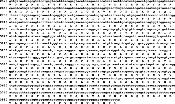
Codon optimization was used to design a gene for high-level expression of recombinant BoNT/A Hc (857V to 1296L) in SF9 cells (Fig. 1). The optimization resulted in the reduction or elimination of rare codons, as well as balanced codon usage. Table 1 compares the codon usage for the SF9-optimized gene to the codon usage for an optimized gene expressed in yeast (5, 8) and to wild-type C. botulinum.
Fig. 1.
Nucleotide sequence of the synthetic gene for BoNT/A Hc. The synthetic gene encoding V857 to L1296 of BoNT/A1 was designed for optimal expression in insect cells. For reference, the nucleic acid sequence is numbered relative to the sequence deposited in GenBank under accession number X52066. The amino acid translation is shown below the nucleotide sequence.
Table 1.
Codon usage is compared for BoNT/A Hc gene fragments
| Amino acid | Codon | No. of times codon occursa |
||
|---|---|---|---|---|
| WT | Y | SF | ||
| Ala | GCG | 0 | 1 | 0 |
| Ala | GCA | 5 | 0 | 3 |
| Ala | GCT | 5 | 9 | 3 |
| Ala | GCC | 0 | 0 | 4 |
| Cys | TGT | 2 | 1 | 1 |
| Cys | TGC | 2 | 3 | 3 |
| Asp | GAT | 22 | 5 | 7 |
| Asp | GAC | 2 | 19 | 17 |
| Glu | GAG | 1 | 1 | 12 |
| Glu | GAA | 17 | 17 | 6 |
| Phe | TTT | 17 | 0 | 6 |
| Phe | TTC | 0 | 17 | 11 |
| Gly | GGG | 3 | 0 | 0 |
| Gly | GGA | 8 | 0 | 4 |
| Gly | GGT | 10 | 22 | 11 |
| Gly | GGC | 2 | 1 | 8 |
| His | CAT | 4 | 0 | 1 |
| His | CAC | 0 | 4 | 3 |
| Ile | ATA | 24 | 0 | 6 |
| Ile | ATT | 22 | 0 | 15 |
| Ile | ATC | 3 | 49 | 28 |
| Lys | AAG | 4 | 8 | 23 |
| Lys | AAA | 29 | 25 | 10 |
| Leu | TTG | 3 | 1 | 5 |
| Leu | TTA | 23 | 0 | 0 |
| Leu | CTG | 1 | 33 | 17 |
| Leu | CTA | 4 | 0 | 0 |
| Leu | CTT | 3 | 0 | 3 |
| Leu | CTC | 0 | 0 | 9 |
| Met | ATG | 9 | 9 | 9 |
| Asn | AAT | 53 | 25 | 19 |
| Asn | AAC | 4 | 32 | 38 |
| Pro | CCG | 0 | 9 | 0 |
| Pro | CCA | 5 | 0 | 3 |
| Pro | CCT | 4 | 0 | 2 |
| Pro | CCC | 0 | 0 | 4 |
| Gln | CAG | 4 | 15 | 10 |
| Gln | CAA | 11 | 0 | 5 |
| Arg | AGG | 5 | 0 | 5 |
| Arg | AGA | 14 | 0 | 2 |
| Arg | CGG | 0 | 0 | 0 |
| Arg | CGA | 0 | 0 | 0 |
| Arg | CGT | 1 | 13 | 6 |
| Arg | CGC | 0 | 7 | 7 |
| Ser | AGT | 15 | 0 | 3 |
| Ser | AGC | 2 | 0 | 9 |
| Ser | TCG | 0 | 0 | 0 |
| Ser | TCA | 11 | 0 | 5 |
| Ser | TCT | 6 | 20 | 6 |
| Ser | TCC | 0 | 14 | 11 |
| Thr | ACG | 0 | 0 | 1 |
| Thr | ACA | 6 | 0 | 4 |
| Thr | ACT | 10 | 7 | 4 |
| Thr | ACC | 0 | 9 | 7 |
| Val | GTG | 0 | 0 | 12 |
| Val | GTA | 19 | 9 | 3 |
| Val | GTT | 3 | 13 | 2 |
| Val | GTC | 1 | 1 | 6 |
| Trp | TGG | 9 | 9 | 9 |
| Tyr | TAT | 26 | 0 | 5 |
| Tyr | TAC | 2 | 28 | 23 |
| End | TGA | 0 | 0 | 0 |
| End | TAG | 0 | 0 | 0 |
| End | TAA | 0 | 0 | 0 |
Hc was expressed with a 6×His tag or as a fusion protein with the R4 and mR4 ligands to produce Hc, HcR4, and HcmR4, respectively. Hc, HcR4, and HcmR4 were produced as soluble secreted proteins using baculovirus-mediated expression in SF9 insect cells. Western blot analyses show that Hc, HcR4, HcmR4, and wild-type Hc from C. botulinum are recognized by a specific anti-BoNT/A antibody (Fig. 2B). As shown in Fig. 2B, 200 ng of wild-type Hc (Metabiologics) resulted in fainter bands on Western blots than did 50 ng of recombinant Hc. We attribute the lower intensity of wild-type Hc to reduced purity, partial cleavage (note a faint, higher band which is probably the uncleaved heavy chain), and/or the effects of the urea in the wild-type preparation on sample electrophoresis. The recombinant HcR4 ligands, schematically depicted in Fig. 2A, are IgG-like homodimers held together by disulfide bonds. Reducing SDS-PAGE disrupts the disulfide bonds, and as a result, the observed molecular mass of 140 kDa for HcmR4 and HcR4 using Western blotting is half their actual size. HcR4 and HcmR4 have predicted molecular masses of 278 kDa and deliver 2 copies of Hc per molecule, whereas Hc has a molecular mass of 45 kDa. Therefore, 1 μg of HcR4 or HcmR4 contains/delivers 0.32 μg of Hc antigen.
HcR4 binding to FcγRs.
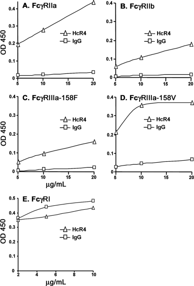
HcR4 binding to low-affinity FcγRs was analyzed using in vitro receptor binding assays. HcR4 has markedly better binding to low-affinity FcγRs than IgG at all concentrations tested (Fig. 3). In humans, FcγRIIIa has two alleles. The FcγRIIIa-158V allele has higher affinity for both IgG and IgG immune complexes than the FcγRIIIa-158F allele (22, 50). HcR4 binds robustly to FcγRIIIa-158V, with evidence of saturation-like binding above 10 μg/ml (Fig. 3D). Saturation binding may result from a higher affinity specific to FcγRIIIa-158V for HcR4 relative to the other low-affinity FcγRs. HcR4 also has 10-fold-improved binding over that of monomeric IgG to the FcγRIIIa-158F allele (Fig. 3C). Thus, HcR4 is competent to deliver Hc antigen to low-affinity FcγRs, and binding is greatly enhanced compared to that of IgG. We also employed a receptor binding assay to establish that HcR4 binds to FcγRI, the high-affinity FcγR (Fig. 3E).
Fig. 3.
HcR4 targets Hc to FcγRs. Recombinant ligand-binding domains from FcγRs were immobilized on ELISA plates, and binding of HcR4 or monomeric human IgG was assessed at 5, 10, and 20 μg/ml for low-affinity FcγRs and at 2, 5, and 10 μg/ml for the high-affinity receptor, FcγRI. FcγRIIa (A); FcγRIIb (B); FcγRIIIa-158F (C); FcγRIIIa-158V (D); FcγRI (E). HcR4 has markedly better binding to low-affinity FcγRs than IgG at all concentrations tested. The data presented are from a representative assay of three performed.
APCs process and present Hc antigens more effectively when delivered as HcR4.
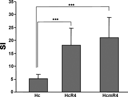
We used an in vitro T cell proliferation assay (19) to determine whether R4-mediated targeting of Hc leads to more efficient presentation of Hc antigens by APCs than nontargeted Hc. In this assay, antigens that are taken up and processed by APCs stimulate antigen-reactive T cells to proliferate, and thus, T cell proliferation provides a measure of antigen uptake efficiency. Proliferation-arrested APCs were loaded with equal concentrations of Hc, HcR4, and HcmR4 in the presence of Hc-reactive T cells, and proliferation was measured after 3 days of coculture. Loading APCs with HcR4 or HcmR4 resulted in 3.5-fold- and 3.8-fold-higher T cell proliferation, respectively, than was seen when APCs were loaded with Hc (Fig. 4). Therefore, both HcR4 and HcmR4 are highly effective at directing antigen entry into APCs.
Fig. 4.
Targeting Hc antigens to FcγRs results in highly effective antigen entry into APCs. Hc-specific T cell proliferation was used to compare efficacy of antigen entry into APCs. T cells were isolated from the lymph nodes of Hc-sensitized mice. Splenocytes from naïve mice were isolated as a source of APC and treated with mitomycin C to arrest proliferation. T cells and APCs were cultured together in the presence of 1.2 × 10⁻⁸ M of Hc, HcR4, or HcmR4 in 96-well plates. Proliferation was determined after 72 h of culture. The stimulation index (SI) is the fold increase over the results for unstimulated control cultures. Results show the means ± SEM for triplicate cultures. ***, P < 0.005.
Immunization with HcmR4 or HcR4 results in earlier and higher antibody titers than immunization with Hc.
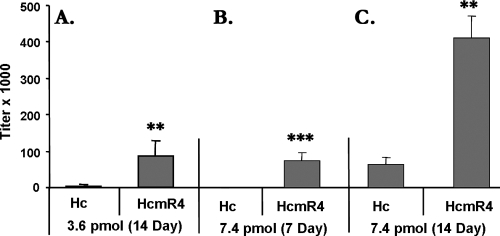
To determine whether HcmR4 can improve the magnitude of the antibody response, BALB/c mice were immunized s.c. with a single 3.6-pmol dose of Hc antigen (165 ng of Hc or 500 ng of HcmR4). Serum was collected on day 14 after immunization, and Hc-reactive antibody titers were determined using ELISA. Mice immunized with HcmR4 developed 48-fold-higher anti-Hc titers than mice receiving nontargeted Hc (Fig. 5A).
Fig. 5.
Antibody responses of BALB/c mice after a single immunization with Hc or HcmR4. Mice (n = 5 per group) were immunized with Hc or HcmR4 at the indicated dose of Hc antigen. Serum was collected 7 or 14 days after immunization, and antibody titers were determined by ELISA. (A) Day-14 serum titers from mice immunized with 3.6 pmol of Hc antigen. (B and C) Serum titers 7 and 14 days after immunization with 7.4 pmol of Hc antigen. Results show means ± SEM. **, P < 0.01; ***, P < 0.005.
In order to evaluate the rapidity of the antibody response, mice were immunized with a 2-fold-higher dose of antigen (7.4 pmol), and serum titers were determined at 7 and 14 days after immunization. At 7 days, HcmR4-immunized mice had 74-fold-higher Hc-reactive titers than mice given nontargeted Hc (Fig. 5B). At 14 days, mice given HcmR4 had 6.5-fold-higher anti-Hc titers than mice receiving Hc (Fig. 5C). These results indicate that targeting Hc to FcγRs accelerates the immune response. In addition, while doubling the antigen load lessened the relative magnitude of the difference between day 14 responses, it did not overcome the effects of targeting antigen to APCs.
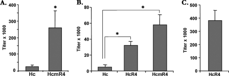
Due to the higher molecular weight of HcR4 and HcmR4 relative to that of Hc, our equimolar dosing studies required the administration of a greater mass of HcR4 and HcmR4 than of Hc. Thus, the improved results observed with R4 ligands could potentially occur as a consequence of the larger mass of immunogen rather than the effects of antigen targeting. To distinguish between these possibilities, we next examined the immune responses in mice immunized with equal masses of Hc, HcR4, and HcmR4. For these studies, we used SJL mice, a strain in which the humoral response to BoNT antigens has been studied extensively (32). Mice were immunized with 1 μg of either Hc or HcmR4, and titers were determined in serum collected on day 7. Immunization with HcmR4 resulted in 10-fold higher anti-Hc titers than those seen in mice receiving Hc alone (Fig. 6A). The effectiveness of R4 targeting in both the SJL and BALB/c mouse strains indicates that the effects seen are not dependent on the genetic background of a single mouse strain. To determine if HcR4, the human R4 ligand, could also augment responses to Hc in vivo, mice were immunized with 0.5 μg of Hc, HcR4, or HcmR4 and serum titers were determined on day 14. Mice receiving HcR4 or HcmR4 had 6-fold- and 11-fold-higher anti-Hc titers, respectively, than mice given nontargeted Hc (Fig. 6B). The results indicate that human HcR4 ligand also augments antibody responses. These differences are notable given that HcR4 and HcmR4 deliver only one-third the amount of Hc antigen as an equal mass of recombinant Hc.
Fig. 6.
Antibody responses of SJL mice. Mice (n = 5 per group) were immunized with Hc, HcmR4, or HcR4. Hc-reactive serum antibody titers were determined by ELISA. (A) Serum titers 7 days after a single s.c. immunization with 1 μg of Hc or HcmR4. (B) Serum titers 14 days after sc immunization with 500 ng of Hc, HcR4, or HcmR4. (C) Antibody responses to HcR4 given nasally. Mice (n = 5) were given three doses of 25 μg of HcR4 in 10 μl of PBS instilled into each nostril on days 0, 7, and 14. Serum was collected on day 21, and Hc-reactive antibody titers were determined. Results show means ± SEM. *, P < 0.05.
Nasal vaccination is an effective route for HcR4 immunization.
Studies from the Simpson laboratory indicate that Hc can transcytose the mucosal epithelium and evoke systemic immune responses when administered nasally (38). Fusion to R4 may potentially alter this function of Hc. To test this hypothesis, we nasally vaccinated SJL mice, following the multidose protocol established by Simpson's group with the exception that we used a three-dose regimen rather than the four doses used in their protocol. Blood was sampled 1 week after the last dose, and the Hc-reactive titers were determined. HcR4 evoked robust Hc-reactive antibody responses in serum, indicating that HcR4 is effective at inducing Hc-specific immune responses via the nasal route (Fig. 6C).
Quantitative assessment of neutralizing antibodies.
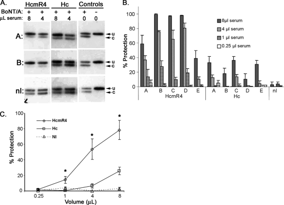
To determine whether immunization with HcmR4 induces neutralizing antibodies to Hc, we used the rat spinal cord cell (RSC) assay (34, 35). The RSC assay has been validated against the mouse lethality assay for assessing BoNT/A neutralizing antibodies in sera (34). The read-out of the RSC assay is SNAP-25 cleavage, which occurs only after all previous steps in the intoxication process have been completed. Analysis of the extent of SNAP-25 cleavage provides a quantitative measure of the level of protection afforded by neutralizing antibodies present in serum. We analyzed the sera from BALB/c mice immunized with 7.4 pmol of Hc or HcmR4 that had been previously characterized for total anti-Hc titers (Fig. 5C). Mice immunized with HcmR4 had higher levels of neutralizing antibodies at day 14 than mice immunized with nontargeted Hc (Fig. 7A). The level of toxin neutralization was determined by quantitative densitometric analysis of SNAP-25 cleavage and expressed as percent protection (Fig. 7B). The results show that immunization with HcmR4 results in higher levels of neutralization than immunization with Hc at all volumes of serum tested, with significant differences (P < 0.05) occurring with the 8-, 4-, and 1-μl volumes (Fig. 7C). Overall, serum from HcmR4-immunized mice had a 5.1-fold increase in the level of neutralization in comparison to the results with nontargeted Hc.
Fig. 7.
Neutralizing antibodies to BoNT/A1 were quantitated using the RSC serum neutralization assay. Primary rat spinal cord cells were exposed to 0.5 units (0.5 pM) of BoNT/A1 preincubated with the indicated mouse serum. Cell lysates were analyzed by Western blotting using an anti-SNAP-25 antibody that recognizes both BoNT/A1-cleaved (c) and uncleaved (u) SNAP-25. Uncleaved SNAP-25 is indicative of toxin neutralization. BALB/c mice were immunized with 7.4 pmol of HcmR4 or Hc and serum collected on day 14. (A) Sera were analyzed for neutralizing antibodies. As a control, sera from nonimmunized BALB/c mice (nI) were also analyzed. Data presented are representative of three assays performed. (B) The level of toxin neutralization was determined by quantitative analysis of SNAP-25 cleavage by Western blot densitometry and expressed as percent protection. Shown are the means ± SD for three independent analyses for each serum sample analyzed and for all volumes analyzed. (C) Protection is plotted against serum volume to reveal the extent of protection provided by immunization with HcmR4 or Hc. Results show means ± SEM for each volume of serum analyzed. *, P < 0.05.
DISCUSSION
The presence of protective epitopes within BoNT/A Hc is well established, but the results from vaccinations with Hc are frequently suboptimal. Single immunizations with Hc typically result in limited protection (5, 52), whereas the doses required to achieve full protection can be massive (24). Nonetheless, the foregoing suggests that improvements in vaccine methodology might permit the successful development of single-dose Hc-based vaccines.
Targeting antigens to DCs has been shown to be an effective means for improving vaccine efficacy. DCs have been referred to as “nature's adjuvant,” given their ability to potently stimulate B and T cells (46). A critical early step in the immune response is the uptake and processing of antigen by DCs, and strategies that target antigen to DCs can improve uptake and accelerate immune responses. DCs express a variety of receptors that have been investigated as targets for antigen delivery, including DEC-205, DC-sign, CD40, and the FcγRs (3, 23, 45, 48). Several features of FcγRs make them attractive for directing antigen into DCs in vivo: (i) multiple classes of FcγRs are expressed on immature DCs; (ii) FcγRs bind IgG immune complexes avidly; (iii) antigen uptake via FcγRs promotes efficient processing of antigenic proteins and presentation of antigen-derived peptides in the context of MHC class I and class II molecules; and (iv) FcγR ligation by immune complexes induces DC maturation.
Various strategies for targeting antigen to FcγRs have been pursued. Using recombinant techniques, peptide epitopes have been introduced into the complementarity-determining region loops of IgG to produce “antigenized antibodies” (4, 53). Alternatively, antigen has been attached to monoclonal antibodies and to Fab fragments specific for individual classes of FcγR (15, 20). The R4 ligand used in these studies has several useful features for the delivery of antigen to FcγRs, including the ability to target both low- and high-affinity FcγRs and its expression as a single recombinant protein of relatively small size (49). An additional consideration relates to the number of Fc regions available to bind FcγRs, or Fc valency, of R4. The Fc valency of immune complexes determines the extent of their interactions with low-affinity FcγRs. Low valencies favor interaction with activatory FcγRIII (21, 29, 47) whereas higher Fc valencies have been implicated as triggers for inhibitory FcγRIIb signaling (1). Therefore, some of the enhancing effects seen with R4, which has 10 potential FcγR binding sites, may reflect a more favorable engagement of activatory rather than inhibitory FcγRs.
By expressing BoNT/A Hc as a fusion protein with R4, we reasoned that the Hc antigen could be effectively targeted to DCs and to APCs. We used in vitro techniques to establish that R4 indeed targets Hc to FcγRs and leads to enhanced antigen presentation. Binding assays established that HcR4 engaged the low-affinity receptors with markedly stronger binding than was seen with monomeric IgG. In particular, HcR4 binds avidly to both FcγRIIIa allotypes (158F and 158V). The FcγRIIIa-158F allele is the more commonly expressed FcγRIIIa allotype. The FcγRIIIa-158F receptor binds IgG immune complexes with lower affinity than the 158V allele and correlates with poor clinical responses to monoclonal antibody therapy for several types of cancer (6, 7). Our finding of improved binding of R4 to the lower-affinity 158F allele may suggest a possible therapeutic relevance for R4 in addition to augmenting antigen acquisition. A second line of evidence is provided by the results from an antigen presentation assay. We found that Hc-reactive T cells stimulated with APCs loaded with HcR4 or HcmR4 had markedly higher proliferative responses than when APCs were loaded with Hc. As the antigens were added directly to APCs, these results support the argument that the R4 ligand alone confers the increased efficacy in antigen targeting. Taken together, these findings show that HcR4 is a potent ligand for all FcγRs and enhances antigen presentation, features shared with small immune complexes.
Immune complexes can stimulate rapid and efficient immune responses, with significant antigen-specific antibody responses occurring, in some instances, within 4 days of immunization (13). Vaccination with HcR4 or HcmR4 leads to early and large immune responses against Hc, with effects evident as early as 7 days after immunization, at which time responses to nontargeted Hc were low or absent. Vaccination with either equal moles or equal masses of the Hc immunogens leads to similar results, providing support for the hypothesis that it is the targeting of Hc to DCs that improves responses. The magnitude of the responses to HcR4 and HcmR4 is in line with our previous findings using R4 to augment immune responses to a model antigen from human serum albumin (19).
Hc drives transcytosis across mucosal epithelial cells, providing a means for BoNT transit across mucosal surfaces (9, 26). This feature of Hc has been exploited to use it as a nasal vaccine (33, 38), and we sought to determine whether HcR4 retained this capacity. We found that nasal vaccination with HcR4 leads to robust systemic Hc-reactive antibody responses and conclude that fusion to R4, at a minimum, does not inhibit Hc from inducing mucosal responses. These results further suggest that Hc may transport R4 as “cargo” across the mucosal interface, as has been observed for other protein domains fused to Hc (27). Future studies will be needed to determine whether R4 can augment responses to nasal vaccination by targeting Hc to FcγRs expressed on DCs and APCs located in the nasal mucosa (14).
During the early phase of the primary immune response, antibodies directed against appropriate neutralizing epitopes may lack sufficient avidity to neutralize BoNT. We used the RSC assay to examine the levels of neutralization in sera from mice collected 14 days after immunization with HcmR4 or Hc. Immunization with HcmR4 resulted in a 5.1-fold-higher level of neutralization than was seen in mice immunized with Hc. This increase mirrors the 6.5-fold increase in total Hc-reactive antibody titers observed at this time point. These results are consistent with those of a number of studies that find a correlation between neutralization and total serum ELISA titers (5, 25). It will be of interest to determine whether neutralizing antibodies to BoNT can be induced at earlier time points, yet their presence within 2 weeks of immunization with a single low dose of HcR4 strongly suggests that targeting Hc to FcγRs accelerates the induction of neutralizing antibodies.
The present study provides evidence that targeting Hc antigens to FcγRs in vivo accelerates the critical early steps of antigen acquisition and leads to rapid and robust immune responses directed against BoNT. We employed Hc from BoNT/A, which is among the more immunogenic of the BoNT serotypes (39). We posit that Hc targeting to FcγRs will also improve immune responses to less-immunogenic BoNT serotypes, such as BoNT/B, and in this way, may facilitate the development of multiserotype BoNT vaccines. These studies also provide additional support for the utility of using R4 to target immunogens to FcγRs. R4 has the potential to reduce the amount of antigen needed to vaccinate an individual while enhancing humoral responses against the targeted antigen.
ACKNOWLEDGMENTS
We thank Irshad Shakir and Matthew Gunkel for excellent technical assistance. The anti-SNAP-25 antibody was generously provided by Reinhard Jahn, Department of Neurobiology, Max-Planck Institut Göttingen.
These studies were supported by Public Health Service grant R21AI058003 from NIH/NIAID (D.M.W.) and by a generous gift from Jack Schaps.
Footnotes
Published ahead of print on 16 May 2011.
REFERENCES
- 1. Akiyama K., et al. 2003. Targeting apoptotic tumor cells to Fc gamma R provides efficient and versatile vaccination against tumors by dendritic cells. J. Immunol. 170:1641–1648 [DOI] [PubMed] [Google Scholar]
- 2. Arnon S. S., et al. 2001. Botulinum toxin as a biological weapon: medical and public health management. JAMA 285:1059–1070 [DOI] [PubMed] [Google Scholar]
- 3. Birkholz K., et al. 2010. Targeting of DEC-205 on human dendritic cells results in efficient MHC class II-restricted antigen presentation. Blood 116:2277–2285 [DOI] [PubMed] [Google Scholar]
- 4. Brumeanu T. D., et al. 1996. Engineering of doubly antigenized immunoglobulins expressing T and B viral epitopes. Immunotechnology 2:85–95 [DOI] [PubMed] [Google Scholar]
- 5. Byrne M. P., Smith T. J., Montgomery V. A., Smith L. A. 1998. Purification, potency, and efficacy of the botulinum neurotoxin type A binding domain from Pichia pastoris as a recombinant vaccine candidate. Infect. Immun. 66:4817–4822 [DOI] [PMC free article] [PubMed] [Google Scholar]
- 6. Cartron G. 2009. FCGR3A polymorphism story: a new piece of the puzzle. Leuk. Lymphoma 50:1401–1402 [DOI] [PubMed] [Google Scholar]
- 7. Cartron G., et al. 2002. Therapeutic activity of humanized anti-CD20 monoclonal antibody and polymorphism in IgG Fc receptor FcgammaRIIIa gene. Blood 99:754–758 [DOI] [PubMed] [Google Scholar]
- 8. Clayton M. A., Clayton J. M., Brown D. R., Middlebrook J. L. 1995. Protective vaccination with a recombinant fragment of Clostridium botulinum neurotoxin serotype A expressed from a synthetic gene in Escherichia coli. Infect. Immun. 63:2738–2742 [DOI] [PMC free article] [PubMed] [Google Scholar]
- 9. Couesnon A., Pereira Y., Popoff M. R. 2008. Receptor-mediated transcytosis of botulinum neurotoxin A through intestinal cell monolayers. Cell. Microbiol. 10:375–387 [DOI] [PubMed] [Google Scholar]
- 10. Dembek Z. F., Smith L. A., Rusnak J. M. 2007. Botulism: cause, effects, diagnosis, clinical and laboratory identification, and treatment modalities. Disaster Med. Public Health Prep. 1:122–134 [DOI] [PubMed] [Google Scholar]
- 11. Fiock M. A., Cardella M. A., Gearinger N. F. 1963. Studies on immunity to toxins of Clostridium botulinum. IX. Immunologic response of man to purified pentavalent ABCDE botulinum toxiod. J. Immunol. 90:697–702 [PubMed] [Google Scholar]
- 12. Gao W., Rzewski A., Sun H., Robbins P. D., Gambotto A. 2004. UpGene: application of a web-based DNA codon optimization algorithm. Biotechnol. Prog. 20:443–448 [DOI] [PubMed] [Google Scholar]
- 13. Goins C. L., Chappell C. P., Shashidharamurthy R., Selvaraj P., Jacob J. 2010. Immune complex-mediated enhancement of secondary antibody responses. J. Immunol. 184:6293–6298 [DOI] [PubMed] [Google Scholar]
- 14. Gosselin E. J., Bitsaktsis C., Li Y., Iglesias B. V. 2009. Fc receptor-targeted mucosal vaccination as a novel strategy for the generation of enhanced immunity against mucosal and non-mucosal pathogens. Arch. Immunol. Ther. Exp. (Warsz.) 57:311–323 [DOI] [PubMed] [Google Scholar]
- 15. Graziano R. F., et al. 1997. Targeting tumor cell destruction with CD64-directed bispecific fusion proteins. Cancer Immunol. Immunother 45:124–127 [DOI] [PMC free article] [PubMed] [Google Scholar]
- 16. Guermonprez P., Valladeau J., Zitvogel L., Thery C., Amigorena S. 2002. Antigen presentation and T cell stimulation by dendritic cells. Annu. Rev. Immunol. 20:621–667 [DOI] [PubMed] [Google Scholar]
- 17. Hatheway C. L. 1988. Botulism, p. 111–133.In Balows A. Laboratory diagnosis of infectious diseases: principles and practice. Springer-Verlag, New York, NY [Google Scholar]
- 18. Isakov N. 1997. ITIMs and ITAMs. The yin and yang of antigen and Fc receptor-linked signaling machinery. Immunol. Res. 16:85–100 [DOI] [PubMed] [Google Scholar]
- 19. Jensen M. A., Arnason B. G., White D. M. 2007. A novel Fc gamma receptor ligand augments humoral responses by targeting antigen to Fc gamma receptors. Eur. J. Immunol. 37:1139–1148 [DOI] [PubMed] [Google Scholar]
- 20. Keler T., et al. 2000. Targeting weak antigens to CD64 elicits potent humoral responses in human CD64 transgenic mice. J. Immunol. 165:6738–6742 [DOI] [PubMed] [Google Scholar]
- 21. Klaassen R. J., Goldschmeding R., Tetteroo P. A., Von dem Borne A. E. 1988. The Fc valency of an immune complex is the decisive factor for binding to low-affinity Fc gamma receptors. Eur. J. Immunol. 18:1373–1377 [DOI] [PubMed] [Google Scholar]
- 22. Koene H. R., et al. 1997. Fc gammaRIIIa-158V/F polymorphism influences the binding of IgG by natural killer cell Fc gammaRIIIa, independently of the Fc gammaRIIIa-48L/R/H phenotype. Blood 90:1109–1114 [PubMed] [Google Scholar]
- 23. Kretz-Rommel A., et al. 2007. In vivo targeting of antigens to human dendritic cells through DC-SIGN elicits stimulatory immune responses and inhibits tumor growth in grafted mouse models. J. Immunother. 30:715–726 [DOI] [PubMed] [Google Scholar]
- 24. Lee J. C., et al. 2007. C terminal half fragment (50 kDa) of heavy chain components of Clostridium botulinum type C and D neurotoxins can be used as an effective vaccine. Microbiol. Immunol. 51:445–455 [DOI] [PubMed] [Google Scholar]
- 25. Lee J. S., et al. 2001. Candidate vaccine against botulinum neurotoxin serotype A derived from a Venezuelan equine encephalitis virus vector system. Infect. Immun. 69:5709–5715 [DOI] [PMC free article] [PubMed] [Google Scholar]
- 26. Maksymowych A. B., Simpson L. L. 1998. Binding and transcytosis of botulinum neurotoxin by polarized human colon carcinoma cells. J. Biol. Chem. 273:21950–21957 [DOI] [PubMed] [Google Scholar]
- 27. Maksymowych A. B., Simpson L. L. 2004. Structural features of the botulinum neurotoxin molecule that govern binding and transcytosis across polarized human intestinal epithelial cells. J. Pharmacol. Exp. Ther. 310:633–641 [DOI] [PubMed] [Google Scholar]
- 28. Malizio C. J., Goodnough M. C., Johnson E. A. 2000. Purification of Clostridium botulinum type A neurotoxin. Methods Mol. Biol. 145:27–39 [DOI] [PubMed] [Google Scholar]
- 29. Manca F., Fenoglio D., Li Pira G., Kunkl A., Celada F. 1991. Effect of antigen/antibody ratio on macrophage uptake, processing, and presentation to T cells of antigen complexed with polyclonal antibodies. J. Exp. Med. 173:37–48 [DOI] [PMC free article] [PubMed] [Google Scholar]
- 30. Nchinda G., et al. 2008. The efficacy of DNA vaccination is enhanced in mice by targeting the encoded protein to dendritic cells. J. Clin. Invest. 118:1427–1436 [DOI] [PMC free article] [PubMed] [Google Scholar]
- 31. Nimmerjahn F., Ravetch J. V. 2006. Fcγ receptors: old friends and new family members. Immunity 24:19–28 [DOI] [PubMed] [Google Scholar]
- 32. Oshima M., Hayakari M., Middlebrook J. L., Atassi M. Z. 1997. Immune recognition of botulinum neurotoxin type A: regions recognized by T cells and antibodies against the protective H(C) fragment (residues 855-1296) of the toxin. Mol. Immunol. 34:1031–1040 [DOI] [PubMed] [Google Scholar]
- 33. Park J. B., Simpson L. L. 2003. Inhalational poisoning by botulinum toxin and inhalation vaccination with its heavy-chain component. Infect. Immun. 71:1147–1154 [DOI] [PMC free article] [PubMed] [Google Scholar]
- 34. Pellett S., Tepp W. H., Clancy C. M., Borodic G. E., Johnson E. A. 2007. A neuronal cell-based botulinum neurotoxin assay for highly sensitive and specific detection of neutralizing serum antibodies. FEBS Lett. 581:4803–4808 [DOI] [PMC free article] [PubMed] [Google Scholar]
- 35. Pellett S., Tepp W. H., Toth S. I., Johnson E. A. 2010. Comparison of the primary rat spinal cord cell (RSC) assay and the mouse bioassay for botulinum neurotoxin type A potency determination. Sensitive cell-based assay for the quantitative detection of botulinum neurotoxin type A. J. Pharmacol. Toxicol. Methods 60:304–310 [DOI] [PubMed] [Google Scholar]
- 36. Rafiq K., Bergtold A., Clynes R. 2002. Immune complex-mediated antigen presentation induces tumor immunity. J. Clin. Invest. 110:71–79 [DOI] [PMC free article] [PubMed] [Google Scholar]
- 37. Raghavan M., Bjorkman P. J. 1996. Fc receptors and their interactions with immunoglobulins. Annu. Rev. Cell Dev. Biol. 12:181–220 [DOI] [PubMed] [Google Scholar]
- 38. Ravichandran E., et al. 2007. Trivalent vaccine against botulinum toxin serotypes A, B, and E that can be administered by the mucosal route. Infect. Immun. 75:3043–3054 [DOI] [PMC free article] [PubMed] [Google Scholar]
- 39. Rusnak J. M., Smith L. A. 2009. Botulinum neurotoxin vaccines: past history and recent developments. Hum. Vaccine 5:794–805 [DOI] [PubMed] [Google Scholar]
- 40. Schantz E. J., Kautter D. A. 1978. Standardized assay for Clostridium botulinum toxins. J. Assoc. Off. Anal. Chem. 61:96–99 [Google Scholar]
- 41. Simpson L. L. 2004. Identification of the major steps in botulinum toxin action. Annu. Rev. Pharmacol. Toxicol. 44:167–193 [DOI] [PubMed] [Google Scholar]
- 42. Singh B. R. 2000. Intimate details of the most poisonous poison. Nat. Struct. Biol. 7:617–619 [DOI] [PubMed] [Google Scholar]
- 43. Smith L. A. 2009. Botulism and vaccines for its prevention. Vaccine 27(Suppl. 4):D33–D39 [DOI] [PubMed] [Google Scholar]
- 44. Southam D. S., Dolovich M., O'Byrne P. M., Inman M. D. 2002. Distribution of intranasal instillations in mice: effects of volume, time, body position, and anesthesia. Am. J. Physiol. Lung Cell. Mol. Physiol. 282:L833–L839 [DOI] [PubMed] [Google Scholar]
- 45. Steinman R. M., Banchereau J. 2007. Taking dendritic cells into medicine. Nature 449:419–426 [DOI] [PubMed] [Google Scholar]
- 46. Steinman R. M., Pope M. 2002. Exploiting dendritic cells to improve vaccine efficacy. J. Clin. Invest. 109:1519–1526 [DOI] [PMC free article] [PubMed] [Google Scholar]
- 47. Strohmeier G. R., et al. 1995. Role of the Fc gamma R subclasses Fc gamma RII and Fc gamma RIII in the activation of human neutrophils by low and high valency immune complexes. J. Leukoc. Biol. 58:415–422 [DOI] [PubMed] [Google Scholar]
- 48. Tacken P. J., de Vries I. J., Torensma R., Figdor C. G. 2007. Dendritic-cell immunotherapy: from ex vivo loading to in vivo targeting. Nat. Rev. Immunol. 7:790–802 [DOI] [PubMed] [Google Scholar]
- 49. White D. M., Jensen M. A., Shi X., Qu Z., Arnason B. G. 2001. Design and expression of polymeric immunoglobulin fusion proteins: a strategy for targeting low-affinity Fcgamma receptors. Protein Expr. Purif. 21:446–455 [DOI] [PubMed] [Google Scholar]
- 50. Wu J., et al. 1997. A novel polymorphism of FcgammaRIIIa (CD16) alters receptor function and predisposes to autoimmune disease. J. Clin. Invest. 100:1059–1070 [DOI] [PMC free article] [PubMed] [Google Scholar]
- 51. Yada A., et al. 2003. Accelerated antigen presentation and elicitation of humoral response in vivo by FcγRIIB- and FcγRI/III-mediated immune complex uptake. Cell Immunol. 225:21–32 [DOI] [PubMed] [Google Scholar]
- 52. Yu Y. Z., et al. 2009. Protection with a recombinant Hc of Clostridium botulinum neurotoxin serotype A from Escherichia coli as an effective subunit vaccine. Immunopharmacol. Immunotoxicol. 31:261–266 [DOI] [PubMed] [Google Scholar]
- 53. Zaghouani H., et al. 1993. Presentation of a viral T cell epitope expressed in the CDR3 region of a self immunoglobulin molecule. Science 259:224–227 [DOI] [PubMed] [Google Scholar]


