

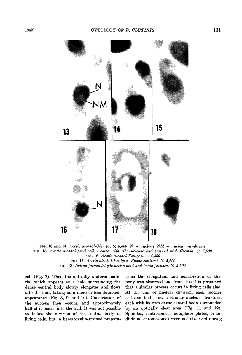
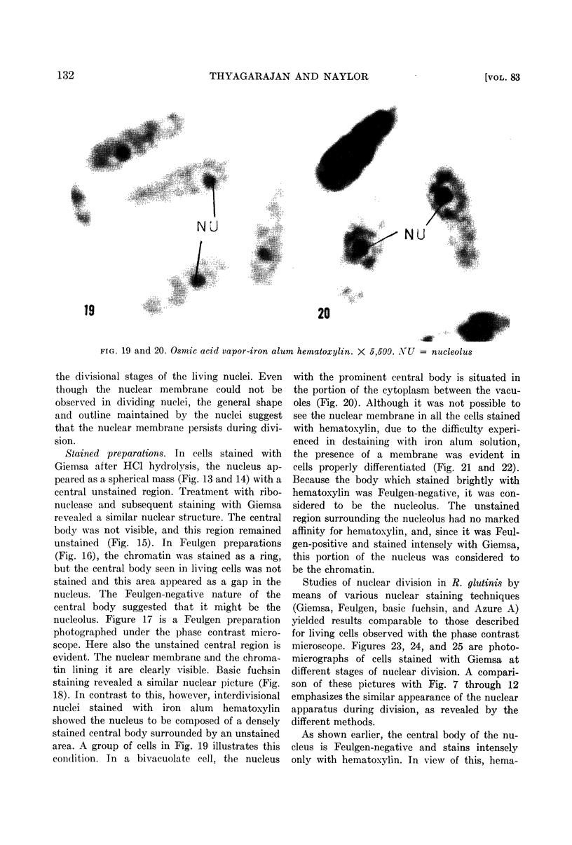
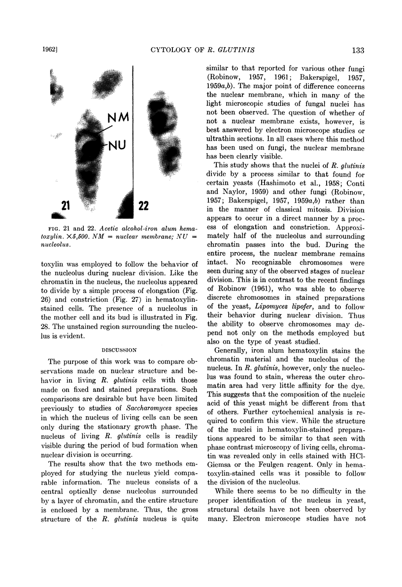
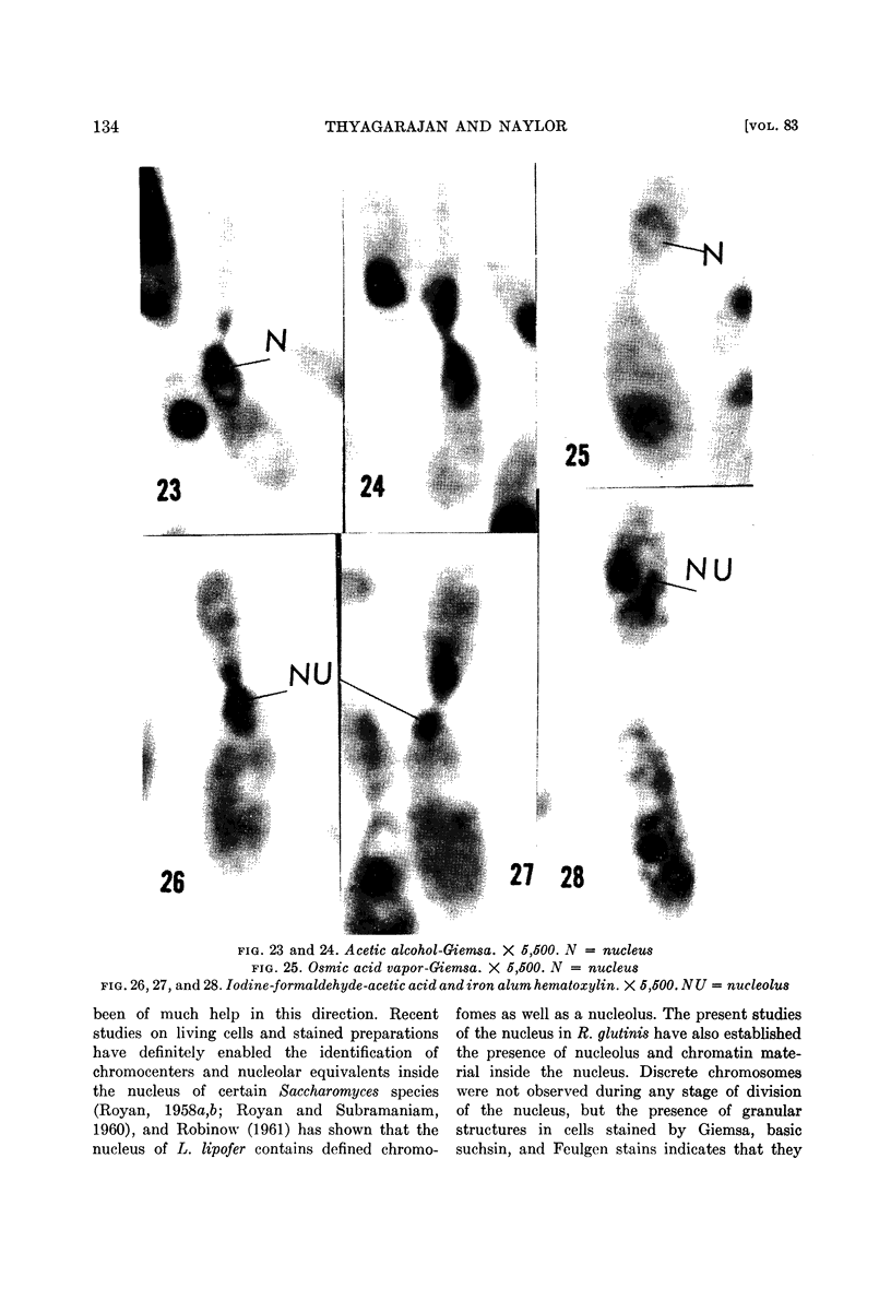
Abstract
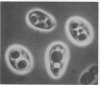
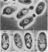
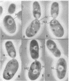
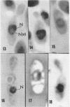
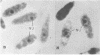
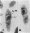
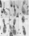
Thyagarajan, T. R. (Cornell University, Ithaca, N. Y.) and H. B. Naylor. Cytology of Rhodotorula glutinis. J. Bacteriol. 83:127–136. 1962.—The structure and manner of division of nuclei in actively dividing cells of Rhodotorula glutinis were studied with the phase contrast microscope. The nucleus consists of a dense central body, surrounded by a shell of optically uniform material of low density. The entire structure is enclosed within a nuclear membrane. Various fixation and staining techniques were employed to confirm the observations made from living cells. Since the dense central body is Feulgen-negative and is readily stained by iron alum hematoxylin, it is identified as the nucleolus. The material surrounding the nucleolus has no marked affinity for hematoxylin but is Feulgen-positive and stains intensely with Giemsa and basic fuchsin. The nucleus appears to divide by a process of elongation and constriction during which roughly half of the nucleolus, along with the surrounding chromatin, passes into the bud. The nuclear membrane was found to persist during all stages of division. Vacuoles were seldom observed in actively dividing cells. The nucleus of R. glutinis is similar in structure to the nuclei of higher organisms, but its behavior during division is quite different.
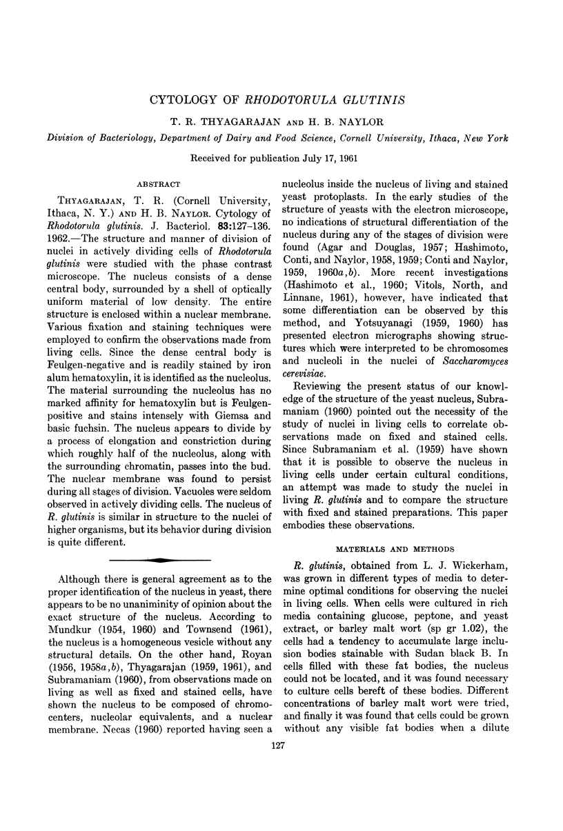
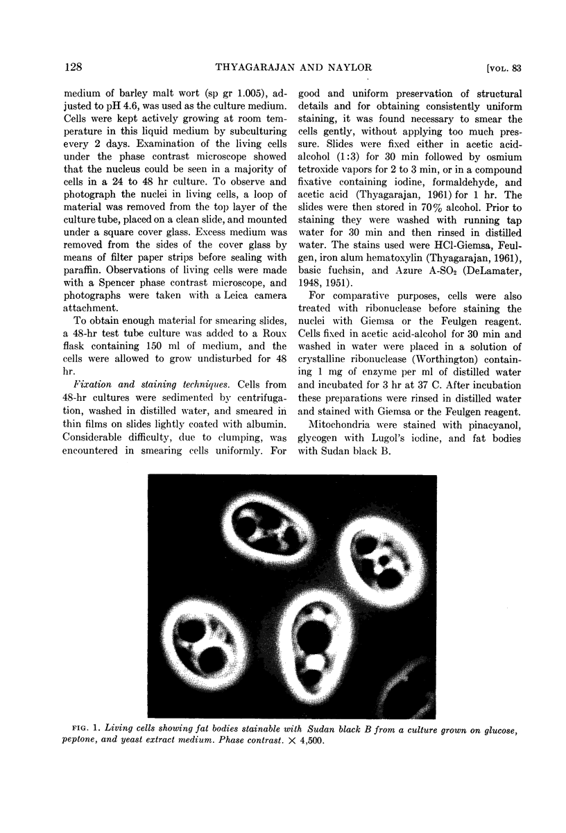
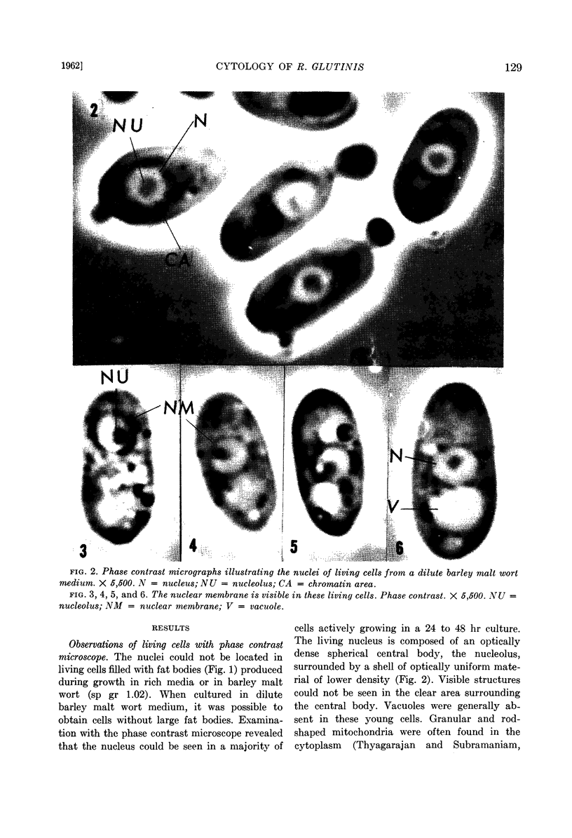
Full text
PDF
Images in this article
Selected References
These references are in PubMed. This may not be the complete list of references from this article.
- AGAR H. D., DOUGLAS H. C. Studies on the cytological structure of yeast: electron microscopy of thin sections. J Bacteriol. 1957 Mar;73(3):365–375. doi: 10.1128/jb.73.3.365-375.1957. [DOI] [PMC free article] [PubMed] [Google Scholar]
- BAKERSPIGEL A. The structure and manner of division of the nuclei in the vegetative mycelium of Gelasinospora tetrasperma Dowd. Can J Microbiol. 1959 Apr;5(2):125–130. doi: 10.1139/m59-016. [DOI] [PubMed] [Google Scholar]
- BAKERSPIGEL A. The structure and mode of division of the nuclei in the yeast cells and mycelium of Blastomyces dermatitidis. Can J Microbiol. 1957 Oct;3(6):923–936. doi: 10.1139/m57-102. [DOI] [PubMed] [Google Scholar]
- CONTI S. F., NAYLOR H. B. Electron microscopy of ultrathin sections of Schizosaccharomyces octosporus. I. Cell division. J Bacteriol. 1959 Dec;78:868–877. doi: 10.1128/jb.78.6.868-877.1959. [DOI] [PMC free article] [PubMed] [Google Scholar]
- CONTI S. F., NAYLOR H. B. Electron microscopy of ultrathin sections of Schizosaccharomyces octosporus. II. Morphological and cytological changes preceding ascospore formation. J Bacteriol. 1960 Mar;79:331–340. doi: 10.1128/jb.79.3.331-340.1960. [DOI] [PMC free article] [PubMed] [Google Scholar]
- CONTI S. F., NAYLOR H. B. Electron microscopy of ultrathin sections of Schizosaccharomyces octosporus. III. Ascosporogenesis, ascospore structure, and germination. J Bacteriol. 1960 Mar;79:417–425. doi: 10.1128/jb.79.3.417-425.1960. [DOI] [PMC free article] [PubMed] [Google Scholar]
- DeLAMATER E. D. A staining and dehydrating procedure for the handling of microorganisms. Stain Technol. 1951 Jul;26(3):199–204. doi: 10.3109/10520295109113208. [DOI] [PubMed] [Google Scholar]
- HASHIMOTO T., CONTI S. F., NAYLOR H. B. Fine structure of microorganisms. III. Electron microscopy of resting and germinating ascospores of Saccharomyces cerevisiae. J Bacteriol. 1958 Oct;76(4):406–416. doi: 10.1128/jb.76.4.406-416.1958. [DOI] [PMC free article] [PubMed] [Google Scholar]
- HASHIMOTO T., CONTI S. F., NAYLOR H. B. Studies of the fine structure of microorganisms. IV. Observations on budding Saccharomyces cerevisiae by light and electron microscopy. J Bacteriol. 1959 Mar;77(3):344–354. doi: 10.1128/jb.77.3.344-354.1959. [DOI] [PMC free article] [PubMed] [Google Scholar]
- HASHIMOTO T., GERHARDT P., CONTI S. F., NAYLOR H. B. Studies on the fine structure of microorganisms. V. Morphogenesis of nuclear and membrane structures during ascospore formation in yeast. J Biophys Biochem Cytol. 1960 Apr;7:305–310. doi: 10.1083/jcb.7.2.305. [DOI] [PMC free article] [PubMed] [Google Scholar]
- LINDEGREN C. C., WILLIAMS M. A., MCCLARY D. O. The distribution of chromatin in budding yeast cells. Antonie Van Leeuwenhoek. 1956;22(1):1–20. doi: 10.1007/BF02538308. [DOI] [PubMed] [Google Scholar]
- MUNDKUR B. D. The nucleus of Saccharomyces; a cytological study of a frozen-dried polyploid series. J Bacteriol. 1954 Nov;68(5):514–529. doi: 10.1128/jb.68.5.514-529.1954. [DOI] [PMC free article] [PubMed] [Google Scholar]
- MUNDKUR B. Submicroscopic morphology of frozen-dried yeast. Exp Cell Res. 1960 Oct;21:201–205. doi: 10.1016/0014-4827(60)90361-x. [DOI] [PubMed] [Google Scholar]
- ROBINOW C. F. Mitosis in the yeast Lipomyces lipofer. J Biophys Biochem Cytol. 1961 Apr;9:879–892. doi: 10.1083/jcb.9.4.879. [DOI] [PMC free article] [PubMed] [Google Scholar]
- ROBINOW C. F. The structure and behavior of the nuclei in spores and growing hyphae of Mucorales. I. Mucor hiemalis and Mucor fragilis. Can J Microbiol. 1957 Aug;3(5):771–789. doi: 10.1139/m57-087. [DOI] [PubMed] [Google Scholar]
- TOWNSEND G. F. Demonstrating the nuclear membrane of the macro yeast cell with KMnO4 staining. Stain Technol. 1961 Jan;36:40–41. doi: 10.3109/10520296109113238. [DOI] [PubMed] [Google Scholar]
- VITOLS E., NORTH R. J., LINNANE A. W. Studies on the oxidative metabolism of Saccharomyces cerevisiae. I. Observations on the fine structure of the yeast cell. J Biophys Biochem Cytol. 1961 Mar;9:689–699. doi: 10.1083/jcb.9.3.689. [DOI] [PMC free article] [PubMed] [Google Scholar]
- YOTSUYANAGI Y. Etude au microscope électronique des coupes ultra-fines de la levure. C R Hebd Seances Acad Sci. 1959 Jan 12;248(2):274–277. [PubMed] [Google Scholar]
- YOTSUYANAGI Y. [Electron-microscopic demonstration of chromosomes in yeasts by means of specific staining]. C R Hebd Seances Acad Sci. 1960 Feb 22;250:1522–1524. [PubMed] [Google Scholar]