Abstract
Background
Erythropoietin (Epo) administration has been reported to have tumor-promoting effects in anemic cancer patients. We investigated the prognostic impact of endogenous Epo in patients with pancreatic ductal adenocarcinoma (PDAC).
Methodology
The clinico-pathological relevance of hemoglobin (Hb, n = 150), serum Epo (sEpo, n = 87) and tissue expression of Epo/Epo receptor (EpoR, n = 104) was analyzed in patients with PDAC. Epo/EpoR expression, signaling, growth, invasion and chemoresistance were studied in Epo-exposed PDAC cell lines.
Results
Compared to donors, median preoperative Hb levels were reduced by 15% in both chronic pancreatitis (CP, p<0.05) and PDAC (p<0.001), reaching anemic grade in one third of patients. While inversely correlating to Hb (r = −0.46), 95% of sEPO values lay within the normal range. The individual levels of compensation were adequate in CP (observed to predicted ratio, O/P = 0.99) but not in PDAC (O/P = 0.85). Strikingly, lower sEPO values yielding inadequate Epo responses were prominent in non-metastatic M0-patients, whereas these parameters were restored in metastatic M1-group (8 vs. 13 mU/mL; O/P = 0.82 vs. 0.96; p<0.01)—although Hb levels and the prevalence of anemia were comparable. Higher sEpo values (upper quartile ≥16 mU/ml) were not significantly different in M0 (20%) and M1 (30%) groups, but were an independent prognostic factor for shorter survival (HR 2.20, 10 vs. 17 months, p<0.05). The pattern of Epo expression in pancreas and liver suggested ectopic release of Epo by capillaries/vasa vasorum and hepatocytes, regulated by but not emanating from tumor cells. Epo could initiate PI3K/Akt signaling via EpoR in PDAC cells but failed to alter their functions, probably due to co-expression of the soluble EpoR isoform, known to antagonize Epo.
Conclusion/Significance
Higher sEPO levels counteract anemia but worsen outcome in PDAC patients. Further trials are required to clarify how overcoming a sEPO threshold ≥16 mU/ml by endogenous or exogenous means may predispose to or promote metastatic progression.
Introduction
Anemia in cancer patients is a common symptom caused either by the tumor itself or by cytotoxic treatment [1]. In response to decreased hemoglobin (Hb) levels, the erythropoiesis-stimulating agent erythropoietin (Epo) is produced in the kidney and subsequently triggers erythropoiesis in the bone marrow and the release of erythrocytes into the blood circulation, thus restoring the Hb level. In cancer, this feedback mechanism seems to be frequently disrupted, yielding an inadequate Epo response [2], [3]. Administration of exogenous (recombinant human) rhEpo has been approved for the correction of chemotherapy-induced anemia in patients with non-hematopoietic malignancies, leading to a reduction in blood transfusion requirements and an improvement in quality of life [4]–[6]. According to current guidelines, rhEpo can be administered to patients when dosed to a target Hb level of less than 12 g/dl [7], [8]. A meta-analysis revealed no negative effect on tumor progression if rhEpo was used in accordance with those guidelines [9]. Still, several clinical trials have shown a higher risk of thrombovascular events, decreased survival, and worse tumor control, calling into question the safety and benefit of rhEpo treatment in patients with solid tumors [8]–[10]. As summarized in a recent report, only one out of 19 clinical trials showed a positive impact of rhEpo on overall survival (hazard ratio 1.3), whereas 10 studies did not demonstrate any effect and 8 trials demonstrated worse survival for the Epo arm [11].
Those data also turned attention to the role of endogenous Epo in carcinogenesis [12]–[16]. Several studies have attempted to explore direct effects of Epo on tumor cells and the possible mechanisms for Epo-mediated tumor progression, but the data are still controversial. Epo is a 30.4-kDa protein whose binding to the transmembrane Epo receptor (EpoR) initiates signaling through several transduction cascades: Jak2/STAT5, PI3K/Akt, ERK1/2, phospholipase C and D, and NF-κB [17]–[20]. These pathways appear to transduce Epo/EpoR signals not only in erythroid precursors but also in malignant cells [12], [14], [21], [22]. The published data reporting an Epo-mediated impact on signaling, proliferation, survival or invasion varied greatly with different tumor cells and respective experimental conditions. The controversy over the functionality of EpoR in malignant cells was heightened by the discovery that cells may express multiple EpoR isoforms, with only a very small fraction being at the cell surface [23]–[25]. Importantly, the finding that the widely used anti-EpoR antibody C-20 cross-reacts with heat-shock protein 70 (HSP70) called into question C-20-based findings of EpoR in tumor cells and tissues [26], [27].
Pancreatic ductal adenocarcinoma (PDAC) is one of the most aggressive and deadliest malignancies, and also requires the highest rate of transfusions among cancer patients undergoing cytotoxic therapies [28]–[30]. Chemotherapy is standard in the adjuvant and palliative settings, and aggravates anemia. Considering the reported negative effects of rhEpo treatment, the use of rhEpo to correct anemia in PDAC patients should be carefully assessed. In the present study, we hypothesized that the level of endogenous Epo might be a risk factor for PDAC progression in both anemic and non-anemic patients, and therefore investigated whether and how the individual Epo response can determine the degree of cancer aggressiveness in PDAC patients. The expression of Epo/EpoR in blood and tissue samples was analyzed in the context of clinico-pathological parameters in donors, chronic pancreatitis (CP) patients, and PDAC patients. The possibility of direct pro-malignant effects and increased chemoresistance was assessed in PDAC cells exposed to rhEpo.
Materials and Methods
Patients and specimens
The study was conducted in accordance with the Helsinki Declaration; specimen collection was approved by the ethical committee of the University of Heidelberg (votes 301/2001 and 159/2002) and written informed consent was obtained from the patients. The study was performed with tissue and blood samples obtained from patients admitted to the Department of General, Visceral and Transplantation Surgery, University Hospital Heidelberg [30], between February 2002 and February 2005 for surgical treatment of PDAC (n = 150) or chronic pancreatitis (CP, n = 42). Diagnoses were established by a pathologist according to the World Health Organization (WHO) classification. Clinical stages are given as defined by the Union for International Cancer Control (AJCC/UICC; 7th Edition, 2009). The normal pancreatic specimens (n = 29) were collected during donor organ procurement if no suitable recipient had been found for the donor's pancreas through the Eurotransplant program. The general characteristics of the patients are given in Table 1: the blood specimens were processed to measure levels of Hb and Epo, whereas tissues were used for analyses of Epo/EpoR mRNA and protein expression. The exact number of analyzed specimens in each study group is also shown in the figure legends.
Table 1. Patients information.
| A. General characteristics of the groups | ||||||||
| Groups | No. of patients | Age [years]Median (IQR) | Hb [g/dl]Median (IQR) | Serum Epo [mU/ml]Median (IQR) | O/P ratioMean±SD | Epo mRNA [copies/10 kCPB]Median (IQR) | EpoR mRNA [copies/10 kCPB]Median (IQR) | Survival [months]Median |
| Donor | 38 | 50.5 (34.0–59.8) | 15.1 (13.9–16.3) n = 9 | 12.7 (8.7–13.3) n = 9 | 1.01±0.12 | 20 (13–25) n = 29 | 174 (124–281) n = 29 | |
| Male | 25 | 46.0 (30.5–60.5) | 15.9 (14.9–16.3) n = 6 | 12.9 (11.3–13.7) n = 6 | 1.06±0.10 | 16 (10–25) n = 19 | 148 (104–255) n = 19) | |
| Female | 13 | 52.0 (43.5–60.5) | 13.8 (13.5–13.9) n = 3 | 9.2 (8.1–12.7) n = 3 | 0.90±0.08 | 22 (15–26) n = 10 | 240 (152–544) n = 10) | |
| CP | 42 | 47.0 (38.0–54.0) | 12.9 (12.0–14.2) n = 42 | 12.0 (8.9–18.8) n = 13 | 0.99±0.23 | 7 (2–13) n = 29 | 351 (219–533) n = 29) | |
| Male | 31 | 50.0 (37.0–56.0) | 13.6 (12.3–14.5) n = 31 | 11.3 (8.0–15.1) n = 10 | 0.92±0.17 | 9 (2–16) n = 21 | 355 (219–533) n = 21) | |
| Female | 11 | 44.0 (40.0–48.0) | 11.8 (10.8–12.9) n = 11 | 18.2 (13.7–74.4) n = 3 | 1.25±0.25 | 5 (2–9) n = 8) | 322 (218–538) n = 8) | |
| PDAC | 150 | 65.0 (56.0–70.3) | 13.0 (11.9–13.9) n = 150 | 9.8 (5.6–15.2) n = 87 | 0.85+0.24 | 1 (0–2) n = 104 | 176 (94–302) n = 104) | 18.0 |
| Male | 80 | 63.5 (56.0–70.8) | 13.7 (12.0–14.5) n = 80 | 8.5 (5.3–15.5) n = 42 | 0.87±0.25 | 0 (0–4) n = 60 | 149 (95–281) n = 60) | 17.0 |
| Female | 70 | 65.0 (57.8–70.3) | 12.6 (11.8–13.5) n = 70 | 10.0 (6.0–15.3) n = 45 | 0.84±0.24 | 1 (0–2) n = 44 | 183 (94–418) n = 44) | 18.5 |
Hematological analyses
Hb values were determined in pre-operative blood samples of PDAC (n = 150) and CP (n = 42) patients and used to diagnose anemia as defined by the European Organization for the Research and Treatment of Cancer (EORTC) at Hb levels ≤12 g/dl. Epo levels in sera of PDAC (n = 87) and CP (n = 13) sub-cohorts as well as healthy volunteers (n = 9) were analyzed using the IMMULITE® detection system and Epo kit according to the manufacturer's instructions (DPC Biermann GmbH, Bad Nauheim, Germany).
The adequacy of Epo response to a given degree of anemia was defined by a standard method calculating observed to predicted sEPO ratio (O/P) [3], [31]–[34]. The regression equation log(Epo) = 1.666−(0.04147×Hb) was established with sEpo and Hb values from 22 non-cancerous reference subjects (donors and CP patients) and then used to predict Hb-adequate Epo values in the individuals with PDAC. The mean O/P ratio in reference subjects was 1.00±0.19 (95% confidence interval: 0.92–1.09).
Cell cultures and Epo treatment
The authenticity of established pancreatic cell lines AsPC-1, BxPC3, Colo357, Capan-1, MiaPaCa-2, PANC-1, Su8686, and T3M4 was certified by DSMZ (German Collection of Microorganisms and Cell Cultures, GmbH, Braunschweig, Germany). The cells were cultured under hypoxic (0.75% O2+10% CO2; in a chamber produced by Billups-Rothenberg, Del Mar, CA) or normoxic (21% O2+5% CO2) conditions in RPMI-1640 (PAA, Cölbe, Germany) supplemented with 0–10% of fetal bovine serum (FBS, PAN Biotech, Aidenbach, Germany). The IL-3-dependent pro-B cell line Ba/F3 or the fibroblast cell line NIH/3T3 were stably transduced with either pMOWS-hEpoR or the empty vector (mock-transduction) and served as a positive or negative control, respectively. Ba/F3 cells were maintained with 10% of WEHI-3B supernatant as a source of IL-3. [20].
The cells were exposed to rhEpo (Erypo®, Ortho Biotech, Janssen-Cilag GmbH, Neuss, Germany) at final concentrations of 1–50 U/ml. Depending on the type of experiment, the cells were harvested at different time points, as indicated in the main text. The signal transduction studies included pretreatment of cells with the PI3K-inhibitor LY2940002 (Sigma, Deisenhofen, Germany) at a final concentration of 30 µM added 30 min prior to the experiments or addition of the monoclonal anti-EpoR antibody (MAB307, Table 3) at 30 µg/ml as previously described to antagonize binding of Epo to the EpoR [16], [20].
Table 3. Primary antibodies.
| Antigen | Antibody type | Application* and Dilution | Manufacturer |
| hEpoR (C-20, cytoplasmic domain) | Rabbit polyclonal | IB, 1∶2000–1∶5000 | Santa Cruz Biotechnologies, Heidelberg, Germany |
| hEpoR (M-20, cytoplasmic domain) | Rabbit polyclonal | IB, 1∶500; IP, 1∶250 | Santa Cruz Biotechnologies, Heidelberg, Germany |
| hEpoR (clone 38409, extracellular domain) | Mouse monoclonal | FC, 10 µL/2×105 cells | R&D Systems, Minneapolis, MN, USA |
| Mouse IgG2b Isotype control | Mouse monoclonal | FC, 20 µL/2×105 cells | R&D Systems, Minneapolis, MN, USA |
| hEpoR (clone MAB307, extracellular domain) | Mouse monoclonal | BA, 30 µg/µl | R&D Systems, Minneapolis, MN, USA |
| hEpoR (3D10, extracellular domain) | Mouse monoclonal | IF, 1∶25; IB, 1∶500; IHC, 1∶100; IP, 3 µL | Sigma, Deisenhofen, Germany |
| Epo (n-19) | Goat polyclonal | IHC 1∶50 | Santa Cruz Biotechnologies, Heidelberg, Germany |
| Akt | Rabbit polyclonal | IB, 1∶1000 | Cell Signaling Technology, Inc., Danvers, MA, USA |
| Phospho-Akt (ser473) | Rabbit polyclonal | IB, 1∶1000 | Cell Signaling Technology, Inc., Danvers, MA,USA |
| STAT5 (C-17) | Rabbit polyclonal | IP, 5 µL; IB, 1∶1000 | Santa Cruz Biotechnologies, Heidelberg, Germany |
| Phospho-STAT5 (Tyr 694) | Rabbit polyclonal | IB, 1∶1000 | Cell Signaling Technology, Inc., Danvers, MA,USA |
*BA, blocking antibody; IB, immunoblot; IF, immunofluorescence; IHC, immunohistochemistry; IP, immunoprecipitation; FC, flow cytometry.
Analysis of Epo and EpoR mRNA expression
mRNA was isolated from tissues and cells, converted to cDNA, and amplified by PCR (real-time quantitative qRT-PCR) using kits and automated equipment from Roche Applied Sciences (RAS, Mannheim, Germany) as described previously [35]. The number of Epo and EpoR transcripts in the samples measured in the LightCycler® (RAS) with commercially available kits (Search-LC, Heidelberg, Germany) was normalized to the expression of the housekeeping gene cyclophilin B. The final data are presented as number of Epo or EpoR transcripts per 10,000 CPB copies (10 kCPB).
To check the expression of EpoR isoforms, PCR was performed using primers previously published by Arcasoy et al., as listed in Table 2 [24]. PCR products were separated by agarose gel electrophoresis and visualized by staining the gel with ethidium bromide.
Table 2. Overview of EpoR RT-PCR primers [24].
| Primer | EpoR gene location | Sequence | Product size (bp) | Isoform |
| EpoR_FL1 | Exon 8 sense | GCTCCCAGCTCTTGCGTCCA | 316 | Full length EpoR |
| EpoR_FL2 | Non-coding antisense | TCGCCATCCCTGTTCCATAA | ||
| EpoR_FL3 | Exon 7 sense | ATCCCGAGCCCAGAGAGCGAGT | 137 | Full length EpoR |
| EpoR_FL4 | Exon 8 antisense | AGGGAAGCAGGTGGGTCCTCCGTG | ||
| EpoR_S5 | Intron 5 sense | GGAGCCAGGGCGAATCACGG | 204 | Sol-EpoR (Intron 5 unspliced) |
| EpoR_S6 | Exon 7 antisense | GCCTTCAAACTCGCTCTCTG |
Testing of anti-EpoR antibodies
Recent studies revealed a lack of specificity for C-20 and other anti-EpoR antibodies due to cross-reactivity with HSP70 [26]. In the present study we tested the ability of 5 different antibodies (Table 3) to detect EpoR by immunoprecipitation, Western blot, FACS and immunohistochemistry. To differentiate between specific and non-specific binding, we used i) hEpoR- or mock-transduced BaF/3 and NIH/3T3 cells, ii) bone marrow specimens and iii) soluble EpoR (sol-EpoR) for blocking set-ups. As a result, the monoclonal 3D10 antibody recognizing an extracellular domain of EpoR (Sigma) was chosen for the detection of EpoR in different assays (except flow cytometry).
Immunohistochemistry
Immunohistochemistry was performed as previously described [36]. Briefly, 4 µm-thin paraffin-embedded tissue sections were deparaffinized and rehydrated in progressively decreasing concentrations of ethanol. After retrieval of antigens by boiling in 10 mM citrate buffer, the sections were subsequently exposed to 3% H2O2 in methanol, universal blocking solution (Powerblock; Biogenix, San Ramon, CA, USA), and the goat anti-hEpo or mouse anti-hEpoR primary antibodies (Table 3). Sections incubated with goat IgG or mouse IgG2b (Dako, Glostrup, Denmark) served as negative isotype controls for n-19 and 3D10 antibodies. The anti-Epo antibody was validated by showing immunopositivity of the tubular epithelial cells in the kidney (not shown). For EpoR, the specificity of the staining was additionally controlled by incubation of the sections with the anti-EpoR antibody pre-treated with a 5-fold excess of soluble EpoR (sol-EpoR). After overnight incubation at 4°C, the secondary horseradish peroxidase (HRP)-labeled antibodies were applied at room temperature for 45 min (donkey anti-goat IgG for EPO; Santa Cruz Biotechnology/SCBT, Heidelberg, Germany, or polymer-conjugated goat anti-mouse IgG for EpoR; EnVision™+System, Dako). The HRP-reaction product was visualized using a DAB/H2O2 substrate mixture kit (EnVision™, Dako) and sections were counterstained with Mayer's hematoxylin.
Immunoprecipitation (IP) and Western blot
Cells were lysed in 1% Nonidet P-40 lysis buffer (1% NP-40, 150 mM NaCl, 20 mM Tris at pH 7.4, 10 mM NaF, 1 mM ZnCl2 at pH 4.0, 1 mM EDTA at pH 8.0, 1 mM MgCl2, 10% glycerol, 1 mM Na3VO4) supplemented with proteinase inhibitor cocktail (Roche) [16], [20]. For IP, the lysates were incubated 20 minutes on ice, cleared by centrifugation, and incubated with a primary antibody (table 3) and protein A-sepharose beads at 4°C overnight. The immunocomplexes were washed with lysis buffer and TNE buffer (10 mM Tris at pH 7.4, 100 mM NaCl, 1 mM EDTA at pH 8.0, 100 µM Na3VO4). Protein samples were denatured at 95°C for 2 min, separated using a 10% polyacrylamide SDS gel. For non-IP Western blot analysis, 40 µg of protein from cell lysates or 2 µl of sol-EpoR (50 µg/ml, Sigma) were separated using 4–12% gel (NuPAGE, Invitrogen GmbH, Darmstadt, Germany). Upon transfer to a nitrocellulose membrane (BioRad, Munich, Germany), the blotted samples were blocked with 5% SlimFast powder (Allpharm, Messel, Germany) in TBS buffer with 0.05% Tween-20, exposed to primary antibodies (Table 3) overnight at 4°C, and incubated with corresponding anti-rabbit or anti-mouse HRP-conjugated secondary antibodies (1∶2,000; Santa Cruz, Heidelberg, Germany) for 1 h at room temperature. The signal was visualized using Amersham ECL detection reagents (GE Healthcare, Buckinghamshire, UK).
Flow cytometry
Cells were resuspended in FACS buffer (PBS, 2% FBS, 0.01% sodium azide), blocked with FCR Blocking Reagent/human (Miltenyi Biotech GmbH, Bergisch Gladbach, Germany) and incubated with FITC-labelled anti-EpoR antibody (clone 38409, extracellular domain) or IgG2b isotype control immunoglobulin (both R&D Systems, Minneapolis, MN, USA, table 3). FACS analysis was performed with the FACS LSR-II system (BD Biosciences, Heidelberg, Germany).
Functional assays (cell invasion, growth and chemoresistance)
To evaluate cell invasion, upper chambers of Biocoat Matrigel-TM invasion chambers (8 µm pore size/PET membrane, BD) were seeded with serum-starved (24 h) cells. The bottom chamber contained RPMI-1640 with Epo at 1–50 U/ml. After 24 h, the membranes with invaded cells were fixed in 70% ethanol and stained with Mayer's hemalum solution (Merck KGaA, Darmstadt, Germany). The cells were counted in 3 visual fields under 100-fold magnification.
To evaluate growth and chemoresistance, a 3-(4,5-dimethylthiazol-2-yl)-2,5-diphenyltetrazolium bromide (MTT) test was performed. The cells were seeded in 96-well plates and incubated with various concentrations of Epo (1–50 U/ml), with or without the chemotherapeutic agents 5-fluorouracil (5-FU) (Sigma) and gemcitabine (Fresenius, Bad Homburg, Germany) at 0.1–10 µM. MTT reagent was added after 48 h of incubation, and the accumulation of formazan in live cells was determined upon solubilisation of the product and measuring optical density at 570 nm. All assays were performed in triplicate.
Statistical analyses
Data were analyzed with GraphPad Prism (GraphPad Software, La Jolla, CA) and SAS (version 9.1; SAS Institute, Cary, NC, USA). The quantitative variables Hb, sEpo, tissue Epo/EpoR mRNA are presented as dot blots or box and whisker plots displaying the median, interquartile range (IQR), and the 5th and 95th percentiles. Significance of differences between groups was assessed by the nonparametric Mann-Whitney U test (to compare 2 groups) or the Kruskal-Wallis with Dunn's tests (to compare 3 and more groups). Linear correlation analyses were performed using the Spearman test. Multivariate Cox regression analysis was performed to identify independent prognostic markers among parameters given in Table 1. To analyze the impact of Epo levels on the overall survival, the corresponding quartiles were used to divide patients into groups. The overall survival from the date of initial surgery was estimated using the Kaplan-Meier method. The observations on 14 PDAC patients still alive at the end of the follow-up period in October 2010 were censored. Differences between survival curves were analyzed using the log-rank test. For all assays, two-sided p values were computed and a difference was considered statistically significant at p≤0.05 (*), p<0.01(**) or p<0.001(***).
Results
Higher levels of circulating Epo are associated with a worse prognosis in PDAC
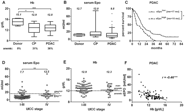
The median Hb level in patients with PDAC was 15% lower than in donors (p<0.001, Figure 1A and Table 1). Likewise, in patients with CP, median Hb level was significantly decreased (p<0.05). A third of patients with PDAC or CP were mildly anemic (≤12 g/dl) prior to surgical resection. 95% of the sEpo values in PDAC patients were within the known normal range (5–34 mU/ml) and did not differ significantly from those in donor and CP groups (median sEPO: 9.8 mU/ml vs. 12.7 and 12.0 mU/ml, respectively; p = 0.15; Figure 1B). Nevertheless, multivariate analysis revealed that the level of endogenous sEpo –but not Hb–was an independent negative prognostic factor for PDAC patients (hazard ratio, HR 2.20, p = 0.01). Patients with sEpo>16 mU/ml (sEpohigh, upper quartile) had a significantly shorter median survival (10.0 vs. 16.7 months; p = 0.03) and increased 3-year mortality rates (100% vs. 75%, Figure 1C). The proportions of sEpohigh individuals in the subgroups with localized (M0, UICC stages I–III) and metastatic (M1, UICC stage IV) disease stages were comparable (M0 vs. M1: 20.3% vs. 30.4%; p = 0.39), even though tumor stage (but not grade, G3 vs. G1/2, p = 0.74) was another independent prognostic factor in PDAC (UICC I+IIa vs. IIb: HR 0.43, p = 0.030; III+IV vs. IIb: HR 2.09, p = 0.020).
Figure 1. Level of sEpo but not Hb was associated with a worse prognosis in PDAC.
The differences in preoperative levels of Hb (A) and sEpo (B) in blood of donors (n = 9), CP (n = 13) and PDAC (n = 87) patients (see table 1A and 1B for full information regarding studied sub-cohorts). (C) Kaplan-Meier analysis of survival data showing that a higher level of sEpo (upper quartile in panel B, ≥16 mU/ml) was associated with shorter survival of PDAC patients; ms = median survival time, in months. (D) Reduced sEpo levels in stage I–III PDAC patients (n = 64) and their restoration in metastatic patients (n = 23). (E) Prevalence of anemia and Hb values were equally distributed during PDAC progression. (F) sEpo inversely correlated with Hb in PDAC. The numbers in panels A, B, D and E depict median levels. Statistically significant differences are labeled by asterisks: p≤0.05 (*), p<0.01(**) or p<0.001(***).
Median sEpo level in patients with stage IV was approximately double that of stage I–III patients (12.5 and 7.7 mU/ml; p<0.01, Figure 1D) but comparable to donor and CP groups (Figure 1B). Lower Epo levels in stage I–III patients could be attributed either to the lack of anemia in this group or to an inadequate Epo response to a given degree of anemia. Yet Hb values (Figure 1E) and the prevalence of anemia were comparable between different stages (UICC I–III vs. IV: 33% vs. 43%, p = 0.45; Table 1). Despite an inverse correlation between sEPO and Hb in PDAC patients (Spearman r = −0.46, p<0.001; Figure 1F), the Epo response in PDAC patients was lower than expected (mean O/P ratio: 0.85±0.24; 95% confidence interval [CI]: 0.80–0.91). However, this impaired Epo response was caused primarily by lower sEpo level consequently leading to reduced O/P ratios in stage I–III patients (mean observed to predicted ratio (O/P) = 0.82±0.26, 95%CI 0.76–0.89) as contrasted by higher sEpo values yielding an adequate Epo responses in CP (O/P = 0.99±0.23, 95%CI 0.85–1.13; p<0.05) and PDAC/stage IV cohorts (OP = 0.96±0.22; 95%CI 0.89–1.08; p<0.01).
Aberrant ectopic expression of Epo in pancreatic diseases
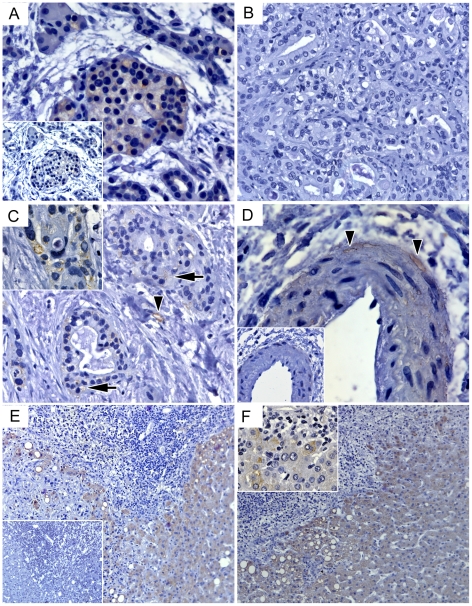
The restoration of EPO response in PDAC/stage IV patients might reflect a ‘passive’ elevation of sEPO levels through ectopic (Hb- and kidney-independent) Epo release. Quantitative mRNA analysis confirmed the ability of the pancreas to produce Epo, but disclosed a progressive loss of Epo mRNA expression in CP by >70% and in PDAC by >95% (Figure 2A). This pattern suggested that Epo-producing islets [37] were disappearing together with the vanishing parenchyma (Figure 3A), and that accumulating PDAC cells were unable to compensate for islet-related losses. Indeed, immunohistochemical staining of PDAC samples showed that Epo-specific antibodies did not bind to the tumor cells (Figure 3B) - except for the rare foci which displayed Epo-positive intracellular vacuoles (Figure 3C). Furthermore, constitutive or hypoxia-stimulated Epo transcription was barely detectable in established PDAC cell lines, except in T3M4 cells showing robust Epo mRNA transcription and its 3-fold up-regulation under hypoxia (Figure 2B). This indicated that the PDAC cells were an unlikely source of ectopic Epo. In contrast, Epo-positive endothelium [21] was frequently found in peripheral capillaries and vasa vasorum of the bigger vessels in PDAC and also in CP (Figure 3D) but not in donor pancreata (not shown).
Figure 2. Aberrant expression of Epo mRNA in pancreatic tissues and tumor cells.
(A) qRT-PCR analysis of pancreatic tissues revealed gradual loss of Epo mRNA expression in diseased pancreata. (B) Except for T3M4, low levels of Epo mRNA expression were found in pancreatic tumor cell lines exposed to or not exposed to 0.75%-hypoxia. Nx = normoxia; Hx = Hypoxia. (C) Barely detectable yet elevated Epo mRNA expression was found in pancreatic specimens obtained from stage IV PDAC patients (n = 12) compared to stage I–III (n = 92) patients. The numbers in panels A and C depict median levels. Statistically significant differences are labeled by asterisks: p≤0.05 (*), p<0.01(**) or p<0.001(***).
Figure 3. Ectopic sources of Epo in tissues of patients with pancreatic diseases.
(A) Remnants of Epo-producing islets in degrading CP-parenchyma. (B) Epo-negativity of tumor cells in primary pancreatic lesion, except for sporadic focal staining observed in intracellular vacuoles of tumor cells (C, arrows and inset). Peripheral capillaries (C, arrowheads) and vasa vasorum (D, arrowheads) of bigger vessels frequently demonstrated Epo positivity in PDAC and CP. (E, F) In liver, cytoplasmic staining of hepatocytes was strong in areas directly bordering Epo-negative metastatic tumor cells and inflammatory infiltrates, but faded away distally, thus pointing to the spatially regulated de novo synthesis and creation of multiple Epo-rich niches. Insets in A, D and E depict negative (isotype IgG) controls; insets in C and F show high-magnification (×630) images of staining patterns in intracellular vacuoles of tumor cells and cytoplasm in hepatocytes.
Although pancreatic tumor samples of stage IV patients showed statistically significant elevation of Epo mRNA levels compared to the non-metastatic group, the measured values were extremely low (Figure 2C). Furthermore, no significant correlation could be observed between pancreatic Epo mRNA and sEpo in PDAC patients (n = 41; Spearman r = −0.18, p = 0.26). Thus, we investigated whether a distant organ affected by metastasis could serve as source for ectopic Epo. Metastasized to the liver tumor cells rarely expressed Epo. However, Epo-negative metastases were frequently outlined by Epo-positive hepatic parenchyma featuring a remarkable gradient of the coloration that faded away in distal hepatocytes (Figure 3E–F). In non-metastatic parenchyma, the cytoplasmic Epo signal was detected in hepatocytes located near portal areas or inflammatory infiltrates (not shown). qRT-PCR analysis confirmed the ability of normal (25±15 copies/10 kCPB, n = 6 organ donor specimens) and metastatic (12±4 copies/10 kCPB, n = 12 biopsies) livers to express Epo. Thus, non-malignant hepatocytes emerged as a potential ectopic source of Epo, possibly being spatially controlled by the tumor cells.
Variability of the EpoR expression in cancerous pancreata
Apparently, increased production, release, and delivery of Epo by non-malignant structures could create multiple Epo-rich niches. Their potential direct impact on tumor cells was assessed by studying i) the expression of EpoR in PDAC cells, ii) Epo-induced down-stream signaling, and iii) functional consequences of exposure to Epo.
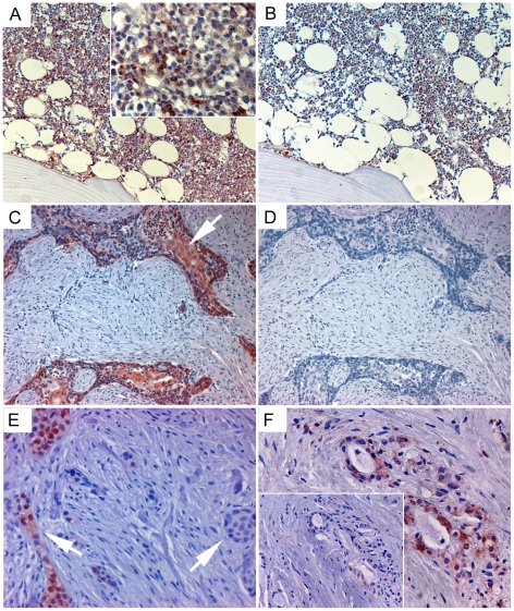
In agreement with previous observations reporting that most of the EpoR was retained in the endoplasmic reticulum [38], we detected strong cytoplasmic staining of erythroid cells in bone marrow, with decrease of positivity after pre-incubation of antibody with sol-EpoR (Figure 4A–B). The staining of PDAC tissues revealed highly heterogeneous patterns of immunopositivity (Figure 4C–E). Whereas 37% (7/19) of specimens showed no staining, cytoplasmic signal was detected in the remaining cases, ranging from a weak sporadic to a strong abundant EpoR-positivity in 26% and 37% of cases, respectively. We could not identify characteristic distinctions between EpoR-positive and EpoR-negative cells (e.g., specific localization within the tumor or near vessels), nor was there a correlation with lymph node-positive N1 status of tumor or grade of tumor differentiation. In addition, few non-malignant structures within PDAC tissue samples also demonstrated EpoR immunopositivity (Figure 4F). The tissue expression of EpoR in normal pancreas and PDAC tissues was further confirmed through mRNA analysis (Table 1).
Figure 4. Detection of EpoR-positive tumor cells in PDAC pancreata.
(A) Staining of erythroid precursor cells in human bone marrow with anti-EpoR 3D10 antibody (inset, ×400) and (B) loss of staining if the anti-EpoR antibody has been pre-incubated with soluble EpoR (sol-EpoR). (C, E) Varying intensity and focal character of EpoR-immunopositivity of tumor cells in PDAC tissues (arrows) and (D) blocking of EpoR signal with sol-EpoR. (F) Sporadic EpoR-immunopositivity of non-malignant structures within a PDAC sample and blocking of EpoR signal with sol-EpoR (inset).
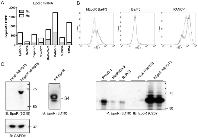
The variability of EpoR expression was also observed in established pancreatic tumor cell lines. All eight studied cell lines expressed EpoR mRNA and showed a 2- to 10-fold up-regulation in hypoxic conditions (Figure 5A). The level of EpoR expression under normoxia ranged from 10 copies/10 kCPB in BxPC3 cells to over 300 copies/10 kCPB in PANC-1 cells. PANC-1 was the only cell line in which EpoR protein was detectable at the cell surface by flow cytometry using anti-EpoR antibodies (Figure 5B), whereas immunoprecipitation with the 3D10 antibody followed by immunoblotting with the C-20 antibody also allowed detection of EpoR protein in the cells with lower mRNA expression, such as MiaPaCa-2 and BxPC3 (Figure 5C).
Figure 5. Expression of EpoR in PDAC cells.
(A) qRT-PCR analysis revealed constitutive EpoR mRNA expression in all studied cancer cell lines and its strong up-regulation under hypoxic conditions. Nx = normoxia, Hx = hypoxia. (B) EpoR was detectable on the surface of hEpoR-transduced Ba/F3 cells and PANC-1 cells by flow cytometry analysis using an FITC-labeled antibody. Mouse IgG2b was used as a negative control. (C) Left panel: detection of the full-length EpoR protein in cell lysates of hEpoR-transduced NIH/3T3 cells and the recombinant sol-EpoR protein using the 3D10-antibody by Western blot analysis (with GAPDH as loading control). Right panel: detection of EpoR in pancreatic tumor cells and hEpoR-NIH/3T3 by immunoprecipitation with the 3D10-antibody followed by immunoblotting with the C-20 antibody.
Epo activates PI3K/Akt but not Jak2/STAT5 signaling in PDAC cells
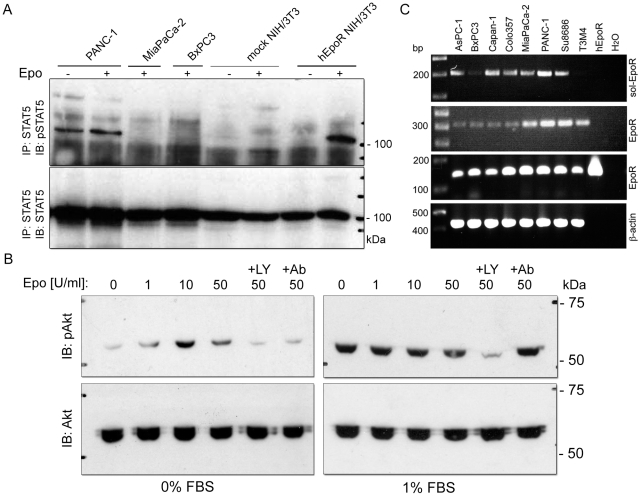
Two principal signaling pathways activated upon binding of Epo to EpoR are the Jak2/STAT5 and PI3K/Akt phosphorylation cascades. Upon stimulation of serum-starved cells with 50 U/ml of Epo, phosphorylation of STAT5 was observed in hEpoR-transduced NIH/3T3 (Figure 6A) and Ba/F3 cells (not shown), but in none of three studied pancreatic tumor cell lines. Epo was unable to either initiate STAT5 activation in MiaPaCa-2 and BxPC3 cells, or to potentiate STAT5 phosphorylation in PANC-1 cells which displayed strong constitutive phosphorylation of STAT5 despite prolonged 24 h serum starvation (Figure 6A). In contrast, dose-dependent phosphorylation of Akt was observed in serum-starved PANC-1 cells showing a very weak level of constitutive Akt activation (Figure 6B, left). This induction could be blocked by the anti-EpoR antibody MAB307 or by the specific PI3K/Akt pathway-inhibitor LY294002. Potentiation of Akt signaling in primed (1% serum) PANC-1 cells was not possible (Figure 6B, right). Thus, the PI3K/Akt pathway but not the Jak2/STAT5 pathway was able to transmit the Epo signal in non-primed PDAC cells.
Figure 6. Activation of PI3K/Akt but not Jak2/STAT5 signaling in PDAC cells exposed to Epo.
(A) Phosphorylation status of Stat5 was assessed in serum-starved pancreatic tumor cells and hEpoR-transduced NIH/3T3 cells stimulated with 50 U/ml erythropoietin (Epo) for 10 min. Clear accumulation of pSTAT5 was observed in hEpoR-NIH/3T3 but not in mock-transduced NIH/3T3 cells or in PDAC cells, independent of the level of constitutive pSTAT5 activation. (B) Phosphorylation status of Akt was assessed in serum-starved (left panel) or non-starved (right panel) PANC-1 cells consequently stimulated with Epo at 0–50 U/ml for 15 min. Epo-enhanced pAkt phosporylation was detected only under conditions of serum starvation and could be specifically inhibited by anti-EpoR antibody (+Ab) or phosphatidylinositol-3-OH kinase (PI3K) inhibitor LY2940020 (+LY). (C) Accumulation of mRNA coding for soluble EpoR isoform (upper panel; primers: EpoR_S5/6, table 2) as compared to mRNAs coding for full-length isoforms (two middle panels) and further related to expression of ß-actin (lower panel). 3′-end-based detection of EpoR mRNA was performed with antisense primers binding after (EpoR_FL1/2) or prior to a stop codon (EpoR_FL3/4) as visualized by a hEpoR plasmid carrying only the coding sequence for a full-length isoform.
Expression of the soluble EpoR- isoform in PDAC cells and lack of the functional responses to rhEPO
The behavior of PDAC cells in an Epo-rich environment (0–50 U/ml) has been studied by assessing the proliferative, invasive and survival potential of three PDAC cell lines (PANC-1, MiaPaCa-2, BxPC3) under conditions of oxygen deprivation, serum starvation and chemotherapeutic treatment (5-fluorouracil and gemcitabine). Neither of these experiments demonstrated any statistically significant pro-malignant or pro-survival effects of exogenously added rhEpo (data not shown). Although we could not exclude the inertness of Epo towards the tumor cells under the studied conditions, we hypothesized that a soluble isoform of EpoR (sol-EpoR) might block Epo signaling by binding of Epo and thereby decreasing receptor-mediated signal transduction. Such a sol-EpoR variant is known to rapidly accumulate up to the nanogram level in the growth medium of tumor cells as soon as 1.5 hours after medium renewal [39]. Probably, sol-EpoR accumulation could occur in the long-term functional studies but not in the short-term signaling experiments. RT-PCR analysis showed varying levels of the alternatively spliced mRNA transcripts coding for the sol-EpoR isoform in all eight studied cell lines (Figure 6C). We concluded that EpoR-positive PDAC cells are equipped to respond to Epo but sol-EpoR might antagonize Epo signaling, with higher Epo having a greater chance of overcoming sol-EpoR and reaching tumor cells in vitro and in sEpohigh patients.
Discussion
Today, the safety of Epo in the oncologic setting is still an issue of concern. Several studies have attempted to elucidate whether and how Epo might stimulate tumor progression, but the data still do not clearly explain the underlying mechanisms [14]. Although the findings of mRNA transcripts coding for multiple EpoR isoforms are abundant and convincing, the observation regarding cell surface expression, down-stream signaling and functional consequences are rather rare and controversial [23], [27]. In the present study, we investigated the effects of endogenous and exogenous Epo on PDAC progression and chemoresistance.
We showed that sEpohigh (≥16 mU/ml)—but not Hb—was an independent, negative prognostic survival marker in PDAC. Although the physiological Epo response seems to be impaired in PDAC, ectopic non-malignant sources of Epo such as hepatocytes and capillaries surfaced as potential candidates for restoring sEpo levels in the circulation and for creating Epo-rich niches at different locations. Since we succeeded in proving EpoR expression at the mRNA and protein level, as well as EpoR initiation of PI3K/Akt signaling in PDAC cells, a direct impact on the tumor cells emerged as a highly probable event to be associated with the negative clinical associations seen in sEpohigh PDAC patients. However, we could not track any effects of Epo on proliferation, invasion or chemoresistance. PI3K/Akt activation by Epo has recently been reported in other malignancies and was associated with either antiapoptotic or proliferative effects or with no impact on cellular proliferation [40]–[42]. Since Epo did not affect PDAC cells even under hostile environmental conditions (hypoxia, serum starvation), one could suggest that neither high sEpo levels creating Epo-rich niches, nor administration of rhEpo will provoke pancreatic malignancy.
The clinical impact and interpretation of these results are complex, however. First, the minimal Epo concentrations needed to demonstrate Akt-phosphorylation (10 U/ml) in vitro were 1000-fold higher than normal blood concentrations in humans, ranging from 5 to 34 mU/ml [18], [31], [32], although we cannot exclude higher levels of Epo accumulating near tumor cells in tissues. PDAC cells also needed to be serum-starved to unmask Epo-mediated Akt-phosphorylation, calling into question whether Epo is even able to activate Akt signaling in vivo. Second, the lack of functional consequences could result from the blockade of Epo-binding to EpoR by sol-EpoR secreted by tumor cells [39]. Since sol-EpoR was found in human plasma, this isoform might emerge as an important modulator of sEpo availability for normal and malignant cells, thus separating sEpohigh patients into a group where sEpo will have higher chances of overcoming neutralizing effects of sol-EpoR while reaching pancreatic or disseminated tumor cells. A recently published mathematical model indicated that Epo signaling is a finely tuned process in which the balance between Epo ligand and available EpoR crucially affects receptor activation and potentially downstream signaling in hematopoietic cells [20]. Thus, an array of interfering factors—variability of EpoR expression, pAkt status, and existence of the neutralizing sol-EpoR isoform—could determine the outcome of signaling in PDAC cells by disturbing the Epo/EpoR balance. As a result, Epo signaling and potential pro-malignant effects would be impeded in tumor cells of patients with a low level of sEpo, and sEPO ≥16 mU/ml could represent a level of saturation sufficient to overcome all existing limitations. This hypothesis would argue to not administer rhEpo if sEpo levels are exceeding a critical threshold. The testing of the available mathematical model in an appropriate experimental system and its validation in clinical material are further steps towards clarifying the role of the Epo/EpoR system in PDAC and towards optimization of rhEpo-based treatments of anemic cancer patients.
The association of high preoperative sEpo levels with decreased survival is in line with recent findings in endometrial, renal and non-small-cell lung cancer patients, in whom high sEpo levels were associated with poorer outcome [15], [43], [44]. However, high sEpo was also a negative prognostic factor in anemic patients with a non-malignant condition such as congestive heart failure [34]. Interestingly, increased mortality in these patients was associated with sEpo levels >22.6 mU/ml—a cut-off close to that observed in the present study (sEpohigh ≥16 mU/ml). Possibly, altered Epo response is a sign of systemic disease accompanying pancreatic disorders. Indeed, both hallmarks of Epo expression in PDAC— ectopic expression and distant delivery—were associated with non-malignant structures and also found in inflammatory pancreatic disease. The inflammatory reaction affecting the pancreas is known to alter a profile of secreted pro-inflammatory Interleukin (IL)-1, IL6, TNFα), pro-angiogenic (IL8, VEGF) and pro-fibrogenic (IL10, TGFβ) cytokines [35], [45]. Systemic action of released cytokines might profoundly change the vascularization, the secretion profile and the functioning of peripheral organs. In cancer, these reactions can be further affected by the tumor cells and yield paraneoplastic syndromes such as cachexia or immune dysfunction [46], [47]. Likewise, a number of factors regulating Epo expression in hepatocytes (IL6, PHD, HIF1 α) could be produced or influenced by primary or metastatic PDAC cells [33], [46], [48]–[50].
Our preliminary observation of an up-regulated Epo mRNA expression in the non-metastatic hepatic specimens (not shown) supports consideration of ectopic Epo production as a systemic syndrome. Still, despite all similarities, the mortality of CP patients is low. A number of recent studies showed preferential expression of EpoR in cancer-initiating (stem) cells [51], [52], enabling a novel explanation for the negative clinical associations of high sEpo (or administered rhEPO) through the direct effects on tumor cells. One could speculate that systemic induction of hepatic or vascular Epo alters the Epo gradient between the circulation and tissues, so provoking an egress of EpoR-positive cancer stem cells from dormant locations (bone marrow or pancreas) into the circulation and/or from the circulation to remote niches (pancreas or liver). Metastasized tumor cells might further upgrade Epo synthesis at distant locations, thus replicating the chain of events and scaling-up metastatic spread. Consequently, the adverse outcome of Epohigh patients could be related to the higher odds of reaching certain Epo thresholds, which are necessary for the mobilization or recruitment of EpoR-positive cancer (stem) cells, notwithstanding sol-EpoR. If ectopic Epo response is inherently capped, patients having a higher Epo response as a result of systemic disease would be pre-disposed to early and faster development of metastasis, thus explaining how the sEPOhigh can be an independent prognostic marker and linked to metastatic status.
In conclusion, the present study demonstrated that PDAC cells are equipped to respond to Epo through functional EpoRs, although conditional activation of PI3K/Akt signaling can possibly be offset by the soluble isoform of EpoR. Despite an impairment of the Epo-response in PDAC patients, sEpo levels could be restored by ectopic production in capillaries and hepatocytes. Elevation of sEPO beyond 16 mU/ml was associated with worse survival independent of metastatic status. Yet higher levels of sEPO could be observed in stage IV patients. Further trials are required to clarify whether high Epo predisposes to or results from metastatic progression and whether the suspected mobilization/recruitment of EpoR-positive cancer-initiating cells is the likely cause of pro-malignant effects for adverse outcome in rhEPO-treated oncologic patients. While high serum Epo levels might be an inherited factor increasing the probability that Epo overcomes the antagonizing effects of sol-EpoR in PDAC patients, elucidation of the threshold determining the outcome of the Epo/EpoR interplay by using mathematical models might help to optimize Epo-based treatments.
Acknowledgments
We would like to thank S. Bauer, M. Meinhardt und B. Bentzinger (Department of General, Visceral and Transplantation Surgery, University of Heidelberg, Germany) and U. Baumann (German Cancer Research Center [DKFZ], Heidelberg, Germany) for their perfect technical assistance. We thank the National Tumor bank/NCT for provision of bone marrow samples and R. Singer, Ch. Tjaden and S. Freirich (Outpatient Unit, European Pancreas Centre) for their help with acquisition of the serum samples.
Footnotes
Competing Interests: The authors have declared that no competing interests exist.
Funding: The work was supported by the grants from the Heidelberger Stiftung Chirurgie to TW, the Helmholtz Alliance on Systems Biology (SBCancer) to VB and UK, the Dietmar-Hopp-Stiftung to NAG and by the German Ministry of Education and Research (NGFNnetwork grants PKB-01GS08178 to BS and PKB-01GS08114 to NAG, who is responsible for the contents of publication). The funders had no role in study design, data collection and analysis, decision to publish, or preparation of the manuscript.
References
- 1.Nowrousian MR. Pathophysiology of anemia in cancer. In: Nowrousian MR, editor. Recombinant Human Erythropoietin (rhEPO) in Clinical Oncology: Scientific and Clinical Aspects of Anemia in Cancer. Wien NewYork: Springer; 1997. pp. 149–188. [Google Scholar]
- 2.Miller CB, Jones RJ, Piantadosi S, Abeloff MD, Spivak JL. Decreased erythropoietin response in patients with the anemia of cancer. N Engl J Med. 1990;322:1689–1692. doi: 10.1056/NEJM199006143222401. [DOI] [PubMed] [Google Scholar]
- 3.Barosi G. Inadequate erythropoietin response to anemia: definition and clinical relevance. Ann Hematol. 1994;68:215–223. doi: 10.1007/BF01737420. [DOI] [PubMed] [Google Scholar]
- 4.Rizzo JD, Brouwers M, Hurley P, Seidenfeld J, Arcasoy MO, et al. American Society of Clinical Oncology/American Society of Hematology clinical practice guideline update on the use of epoetin and darbepoetin in adult patients with cancer. J Clin Oncol. 2010;28:4996–5010. doi: 10.1200/JCO.2010.29.2201. [DOI] [PubMed] [Google Scholar]
- 5.Crawford J, Cella D, Cleeland CS, Cremieux PY, Demetri GD, et al. Relationship between changes in hemoglobin level and quality of life during chemotherapy in anemic cancer patients receiving epoetin alfa therapy. Cancer. 2002;95:888–895. doi: 10.1002/cncr.10763. [DOI] [PubMed] [Google Scholar]
- 6.Leitgeb C, Pecherstorfer M, Fritz E, Ludwig H. Quality of life in chronic anemia of cancer during treatment with recombinant human erythropoietin. Cancer. 1994;73:2535–2542. doi: 10.1002/1097-0142(19940515)73:10<2535::aid-cncr2820731014>3.0.co;2-5. [DOI] [PubMed] [Google Scholar]
- 7.Aapro MS, Link H. September 2007 update on EORTC guidelines and anemia management with erythropoiesis-stimulating agents. Oncologist. 2008;13(Suppl 3):33–36. doi: 10.1634/theoncologist.13-S3-33. [DOI] [PubMed] [Google Scholar]
- 8.Juneja V, Keegan P, Gootenberg JE, Rothmann MD, Shen YL, et al. Continuing reassessment of the risks of erythropoiesis-stimulating agents in patients with cancer. Clin Cancer Res. 2008;14:3242–3247. doi: 10.1158/1078-0432.CCR-07-1872. [DOI] [PubMed] [Google Scholar]
- 9.Aapro M, Scherhag A, Burger HU. Effect of treatment with epoetin-beta on survival, tumour progression and thromboembolic events in patients with cancer: an updated meta-analysis of 12 randomised controlled studies including 2301 patients. Br J Cancer. 2008;99:14–22. doi: 10.1038/sj.bjc.6604408. [DOI] [PMC free article] [PubMed] [Google Scholar]
- 10.Henke M, Laszig R, Rube C, Schafer U, Haase KD, et al. Erythropoietin to treat head and neck cancer patients with anaemia undergoing radiotherapy: randomised, double-blind, placebo-controlled trial. Lancet. 2003;362:1255–1260. doi: 10.1016/S0140-6736(03)14567-9. [DOI] [PubMed] [Google Scholar]
- 11.Szenajch J, Wcislo G, Jeong JY, Szczylik C, Feldman L. The role of erythropoietin and its receptor in growth, survival and therapeutic response of human tumor cells From clinic to bench - a critical review. Biochim Biophys Acta. 2010;1806:82–95. doi: 10.1016/j.bbcan.2010.04.002. [DOI] [PubMed] [Google Scholar]
- 12.Pajonk F, Weil A, Sommer A, Suwinski R, Henke M. The erythropoietin-receptor pathway modulates survival of cancer cells. Oncogene. 2004;23:8987–8991. doi: 10.1038/sj.onc.1208140. [DOI] [PubMed] [Google Scholar]
- 13.Ribatti D, Marzullo A, Gentile A, Longo V, Nico B, et al. Erythropoietin/erythropoietin-receptor system is involved in angiogenesis in human hepatocellular carcinoma. Histopathology. 2007;50:591–596. doi: 10.1111/j.1365-2559.2007.02654.x. [DOI] [PMC free article] [PubMed] [Google Scholar]
- 14.Arcasoy MO. Erythropoiesis-stimulating agent use in cancer: preclinical and clinical perspectives. Clin Cancer Res. 2008;14:4685–4690. doi: 10.1158/1078-0432.CCR-08-0264. [DOI] [PubMed] [Google Scholar]
- 15.Papworth K, Bergh A, Grankvist K, Ljungberg B, Rasmuson T. Expression of erythropoietin and its receptor in human renal cell carcinoma. Tumour Biol. 2009;30:86–92. doi: 10.1159/000216844. [DOI] [PubMed] [Google Scholar]
- 16.Inthal A, Krapf G, Beck D, Joas R, Kauer MO, et al. Role of the erythropoietin receptor in ETV6/RUNX1-positive acute lymphoblastic leukemia. Clin Cancer Res. 2008;14:7196–7204. doi: 10.1158/1078-0432.CCR-07-5051. [DOI] [PMC free article] [PubMed] [Google Scholar]
- 17.Witthuhn BA, Quelle FW, Silvennoinen O, Yi T, Tang B, et al. JAK2 associates with the erythropoietin receptor and is tyrosine phosphorylated and activated following stimulation with erythropoietin. Cell. 1993;74:227–236. doi: 10.1016/0092-8674(93)90414-l. [DOI] [PubMed] [Google Scholar]
- 18.Klingmuller U. The role of tyrosine phosphorylation in proliferation and maturation of erythroid progenitor cells–signals emanating from the erythropoietin receptor. Eur J Biochem. 1997;249:637–647. doi: 10.1111/j.1432-1033.1997.t01-1-00637.x. [DOI] [PubMed] [Google Scholar]
- 19.Richmond TD, Chohan M, Barber DL. Turning cells red: signal transduction mediated by erythropoietin. Trends Cell Biol. 2005;15:146–155. doi: 10.1016/j.tcb.2005.01.007. [DOI] [PubMed] [Google Scholar]
- 20.Becker V, Schilling M, Bachmann J, Baumann U, Raue A, et al. Covering a broad dynamic range: information processing at the erythropoietin receptor. Science. 2010;328:1404–1408. doi: 10.1126/science.1184913. [DOI] [PubMed] [Google Scholar]
- 21.Acs G, Acs P, Beckwith SM, Pitts RL, Clements E, et al. Erythropoietin and erythropoietin receptor expression in human cancer. Cancer Res. 2001;61:3561–3565. [PubMed] [Google Scholar]
- 22.Mirmohammadsadegh A, Marini A, Gustrau A, Delia D, Nambiar S, et al. Role of erythropoietin receptor expression in malignant melanoma. J Invest Dermatol. 2010;130:201–210. doi: 10.1038/jid.2009.162. [DOI] [PubMed] [Google Scholar]
- 23.Sinclair AM, Rogers N, Busse L, Archibeque I, Brown W, et al. Erythropoietin receptor transcription is neither elevated nor predictive of surface expression in human tumour cells. Br J Cancer. 2008;98:1059–1067. doi: 10.1038/sj.bjc.6604220. [DOI] [PMC free article] [PubMed] [Google Scholar]
- 24.Arcasoy MO, Jiang X, Haroon ZA. Expression of erythropoietin receptor splice variants in human cancer. Biochem Biophys Res Commun. 2003;307:999–1007. doi: 10.1016/s0006-291x(03)01303-2. [DOI] [PubMed] [Google Scholar]
- 25.Ribatti D, Marzullo A, Longo V, Poliani L. Schwann cells in neuroblastoma express erythropoietin. J Neurooncol. 2007;82:327–328. doi: 10.1007/s11060-006-9289-8. [DOI] [PubMed] [Google Scholar]
- 26.Elliott S, Busse L, Bass MB, Lu H, Sarosi I, et al. Anti-Epo receptor antibodies do not predict Epo receptor expression. Blood. 2006;107:1892–1895. doi: 10.1182/blood-2005-10-4066. [DOI] [PubMed] [Google Scholar]
- 27.Laugsch M, Metzen E, Svensson T, Depping R, Jelkmann W. Lack of functional erythropoietin receptors of cancer cell lines. Int J Cancer. 2008;122:1005–1011. doi: 10.1002/ijc.23201. [DOI] [PubMed] [Google Scholar]
- 28.Glimelius B, Linne T, Hoffman K, Larsson L, Svensson JH, et al. Epoetin beta in the treatment of anemia in patients with advanced gastrointestinal cancer. J Clin Oncol. 1998;16:434–440. doi: 10.1200/JCO.1998.16.2.434. [DOI] [PubMed] [Google Scholar]
- 29.Dernedde U, Dernedde R, Shepstone L, Barrett A. Three-year single institution audit on transfusion requirements in oncology patients. Clin Oncol (R Coll Radiol) 2007;19:223–227. doi: 10.1016/j.clon.2006.11.011. [DOI] [PubMed] [Google Scholar]
- 30.Welsch T, Degrate L, Zschabitz S, Hofer S, Werner J, et al. The need for extended intensive care after pancreaticoduodenectomy for pancreatic ductal adenocarcinoma. Langenbecks Arch Surg. 2011;396:353–362. doi: 10.1007/s00423-010-0629-y. [DOI] [PubMed] [Google Scholar]
- 31.Beguin Y, Clemons GK, Pootrakul P, Fillet G. Quantitative assessment of erythropoiesis and functional classification of anemia based on measurements of serum transferrin receptor and erythropoietin. Blood. 1993;81:1067–1076. [PubMed] [Google Scholar]
- 32.Roque ME, Sandoval MJ, Aggio MC. Serum erythropoietin and its relation with soluble transferrin receptor in patients with different types of anaemia in a locally defined reference population. Clin Lab Haematol. 2001;23:291–295. doi: 10.1046/j.1365-2257.2001.00413.x. [DOI] [PubMed] [Google Scholar]
- 33.Opasich C, Cazzola M, Scelsi L, De Feo S, Bosimini E, et al. Blunted erythropoietin production and defective iron supply for erythropoiesis as major causes of anaemia in patients with chronic heart failure. Eur Heart J. 2005;26:2232–2237. doi: 10.1093/eurheartj/ehi388. [DOI] [PubMed] [Google Scholar]
- 34.van der Meer P, Voors AA, Lipsic E, Smilde TD, van Gilst WH, et al. Prognostic value of plasma erythropoietin on mortality in patients with chronic heart failure. J Am Coll Cardiol. 2004;44:63–67. doi: 10.1016/j.jacc.2004.03.052. [DOI] [PubMed] [Google Scholar]
- 35.Bartel M, Hansch GM, Giese T, Penzel R, Ceyhan G, et al. Abnormal crosstalk between pancreatic acini and macrophages during the clearance of apoptotic cells in chronic pancreatitis. J Pathol. 2008;215:195–203. doi: 10.1002/path.2348. [DOI] [PubMed] [Google Scholar]
- 36.Welsch T, Keleg S, Bergmann F, Degrate L, Bauer S, et al. Comparative analysis of tumorbiology and CD133 positivity in primary and recurrent pancreatic ductal adenocarcinoma. Clin Exp Metastasis. 2009;26:701–711. doi: 10.1007/s10585-009-9269-4. [DOI] [PubMed] [Google Scholar]
- 37.Samyn I, Fontaine C, Van Tussenbroek F, Pipeleers-Marichal M, De Greve J. Paraneoplastic syndromes in cancer: Case 1. Polycythemia as a result of ectopic erythropoietin production in metastatic pancreatic carcinoid tumor. J Clin Oncol. 2004;22:2240–2242. doi: 10.1200/JCO.2004.10.031. [DOI] [PubMed] [Google Scholar]
- 38.Ketteler R, Heinrich AC, Offe JK, Becker V, Cohen J, et al. A functional green fluorescent protein-erythropoietin receptor despite physical separation of JAK2 binding site and tyrosine residues. J Biol Chem. 2002;277:26547–26552. doi: 10.1074/jbc.M202287200. [DOI] [PubMed] [Google Scholar]
- 39.Westphal G, Braun K, Debus J. Detection and quantification of the soluble form of the human erythropoietin receptor (sEpoR) in the growth medium of tumor cell lines and in the plasma of blood samples. Clin Exp Med. 2002;2:45–52. doi: 10.1007/s102380200006. [DOI] [PubMed] [Google Scholar]
- 40.Hardee ME, Rabbani ZN, Arcasoy MO, Kirkpatrick JP, Vujaskovic Z, et al. Erythropoietin inhibits apoptosis in breast cancer cells via an Akt-dependent pathway without modulating in vivo chemosensitivity. Mol Cancer Ther. 2006;5:356–361. doi: 10.1158/1535-7163.MCT-05-0196. [DOI] [PubMed] [Google Scholar]
- 41.Yin D, Kawabata H, Tcherniamtchouk O, Huynh T, Black KL, et al. Glioblastoma multiforme cells: expression of erythropoietin receptor and response to erythropoietin. Int J Oncol. 2007;31:1193–1198. [PubMed] [Google Scholar]
- 42.Dunlop EA, Maxwell AP, Lappin TR. Impaired downregulation following erythropoietin receptor activation in non-small cell lung carcinoma. Stem Cells. 2007;25:380–384. doi: 10.1634/stemcells.2006-0452. [DOI] [PubMed] [Google Scholar]
- 43.Rades D, Setter C, Dahl O, Schild SE, Noack F. Prognostic Impact of Erythropoietin Expression and Erythropoietin Receptor Expression on Locoregional Control and Survival of Patients Irradiated for Stage II/III Non-Small-Cell Lung Cancer. Int J Radiat Oncol Biol Phys. 2010 doi: 10.1016/j.ijrobp.2010.02.003. [DOI] [PubMed] [Google Scholar]
- 44.Acs G, Xu X, Chu C, Acs P, Verma A. Prognostic significance of erythropoietin expression in human endometrial carcinoma. Cancer. 2004;100:2376–2386. doi: 10.1002/cncr.20244. [DOI] [PubMed] [Google Scholar]
- 45.Zeh HJ, Winikoff S, Landsittel DP, Gorelik E, Marrangoni AM, et al. Multianalyte profiling of serum cytokines for detection of pancreatic cancer. Cancer Biomark. 2005;1:259–269. doi: 10.3233/cbm-2005-1601. [DOI] [PubMed] [Google Scholar]
- 46.Martignoni ME, Kunze P, Hildebrandt W, Kunzli B, Berberat P, et al. Role of mononuclear cells and inflammatory cytokines in pancreatic cancer-related cachexia. Clin Cancer Res. 2005;11:5802–5808. doi: 10.1158/1078-0432.CCR-05-0185. [DOI] [PubMed] [Google Scholar]
- 47.Poch B, Lotspeich E, Ramadani M, Gansauge S, Beger HG, et al. Systemic immune dysfunction in pancreatic cancer patients. Langenbecks Arch Surg. 2007;392:353–358. doi: 10.1007/s00423-006-0140-7. [DOI] [PubMed] [Google Scholar]
- 48.Ramadori P, Ahmad G, Ramadori G. Cellular and molecular mechanisms regulating the hepatic erythropoietin expression during acute-phase response: a role for IL-6. Lab Invest. 2010;90:1306–1324. doi: 10.1038/labinvest.2010.85. [DOI] [PubMed] [Google Scholar]
- 49.Minamishima YA, Kaelin WG., Jr Reactivation of hepatic EPO synthesis in mice after PHD loss. Science. 2010;329:407. doi: 10.1126/science.1192811. [DOI] [PMC free article] [PubMed] [Google Scholar]
- 50.Talar-Wojnarowska R, Gasiorowska A, Smolarz B, Romanowicz-Makowska H, Kulig A, et al. Clinical significance of interleukin-6 (IL-6) gene polymorphism and IL-6 serum level in pancreatic adenocarcinoma and chronic pancreatitis. Dig Dis Sci. 2009;54:683–689. doi: 10.1007/s10620-008-0390-z. [DOI] [PubMed] [Google Scholar]
- 51.Phillips TM, Kim K, Vlashi E, McBride WH, Pajonk F. Effects of recombinant erythropoietin on breast cancer-initiating cells. Neoplasia. 2007;9:1122–1129. doi: 10.1593/neo.07694. [DOI] [PMC free article] [PubMed] [Google Scholar]
- 52.Cao Y, Lathia JD, Eyler CE, Wu Q, Li Z, et al. Erythropoietin Receptor Signaling Through STAT3 Is Required For Glioma Stem Cell Maintenance. Genes Cancer. 2010;1:50–61. doi: 10.1177/1947601909356352. [DOI] [PMC free article] [PubMed] [Google Scholar]


