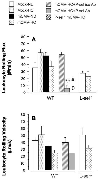
Figure 5.
Leukocyte rolling flux (# of leukocytes rolling past a specific point per minute) (A), and leukocyte rolling velocity (B) in postcapillary venules of WT, P-sel−/− or L-sel−/− mice injected with mock inoculum and maintained on ND or infected with mCMV and placed on HC at 5 wk p.i. Intravital microscopy observation of the venules was performed at 11 wks p.i. Only one out of ten mice had leukocytes that rolled for 100 μm in the P-sel Ab group, and could be considered for measurement of velocity. No leukocyte rolling was detected in the P-sel−/− mice (denoted by “0”). * P<0.05 vs. WT mCMV-HC; # P<0.005 vs. WT mCMV-HC+P-sel iso Ab.

