Abstract
Atrial fibrillation (AF) is an extremely common cardiac rhythm disorder that causes substantial morbidity and contributes to mortality. The mechanisms underlying AF are complex, involving both increased spontaneous ectopic firing of atrial cells and impulse reentry through atrial tissue. Over the past ten years, there has been enormous progress in understanding the underlying molecular pathobiology. This article reviews the basic mechanisms and molecular processes causing AF. We discuss the ways in which cardiac disease states, extracardiac factors, and abnormal genetic control lead to the arrhythmia. We conclude with a discussion of the potential therapeutic implications that might arise from an improved mechanistic understanding.
Introduction
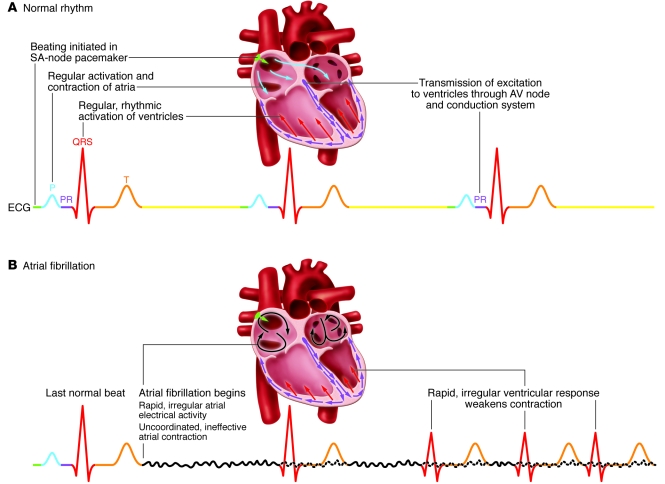
Atrial fibrillation (AF) is the most common sustained cardiac arrhythmia, and its prevalence is increasing with aging of the population (1). Normal cardiac rhythm shows regular rhythm initiation in the sinoatrial (SA) node, followed by atrial and then ventricular activation (Figure 1A). Abnormal spontaneous firing (ectopic activity) from sources other than the SA node is absent. AF is reflected in the ECG recording by the replacement of regular P-waves by an undulating baseline (reflecting continuous, rapid, spatially heterogeneous atrial activation) and irregular ventricular QRS complexes (Figure 1B). Uncoordinated atrial activity prevents effective atrial contraction, leading to clot formation in the blind pouch atrial appendage. Irregular and inappropriately rapid ventricular activity interferes with cardiac contractile function.
Figure 1. ECG recordings of sinus rhythm and AF.
(A) Bottom: A normal ECG recording showing sinus rhythm. Top: Schematics of major events in one cardiac activation cycle: rhythm is initiated by the SA node pacemaker, resulting in atrial activation, followed by atrioventricular conduction via the AV node and His-Purkinje conducting system, leading to ventricular activation. (B) ECG showing onset of AF after one regular sinus beat. Atrial activation is now rapid and irregular, producing an undulating baseline that is visible when not obscured by larger QRS and T waves (continuous atrial activity during this phase is represented by dotted lines). During AF, rapid and uncoordinated atrial activity leads to ineffective atrial contraction. Ventricular activations (QRS complexes) now driven by the fibrillating atria occur rapidly and irregularly, weakening cardiac contraction efficiency and causing clinical symptoms.
AF contributes significantly to population morbidity and mortality, and presently available therapeutic approaches have major limitations, including limited efficacy and potentially serious side effects such as malignant ventricular arrhythmia induction (2). An improved understanding of the mechanisms underlying AF is needed for the development of novel therapeutic approaches (3). A detailed review nine years ago highlighted progress in understanding AF pathophysiology and outlined important unresolved issues (4); since then, knowledge has greatly increased. The purpose of the present article is to summarize these recent findings, particularly in the area of molecular pathophysiology.
Pathophysiological mechanisms
Pathophysiological mechanisms and relation to AF forms
To understand the molecular mechanisms underlying AF, it is necessary to place them in a pathophysiological context. Because of their importance, these mechanisms will be discussed briefly here (for more detailed treatments, see refs. 4, 5).
Focal ectopic activity.
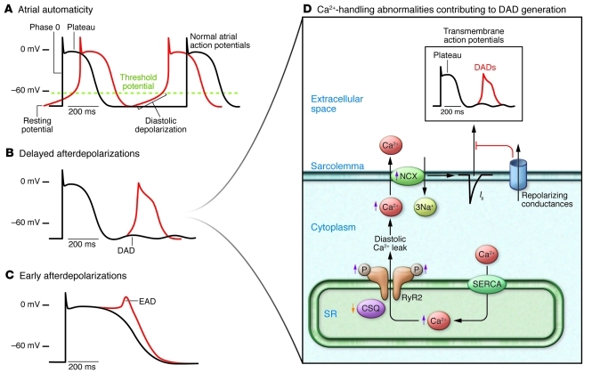
The mechanisms believed to produce ectopic activity from atrial foci are illustrated in Figure 2. Normal atrial cells (“Normal atrial action potentials” in Figure 2A) display typical voltage changes over time. They start at a negative intracellular membrane potential (the resting potential), become very positive when fired (depolarized) during a period called phase 0, then go through a series of repolarizing steps to get back to the resting potential, at which they remain until the next action potential. Automatic activity occurs when an increase in time-dependent depolarizing inward currents carried by Na+ or Ca2+ (making the cell interior more positive) or a decrease in repolarizing outward currents carried by K+ (which keep the cell interior negative) causes progressive time-dependent cell depolarization. When threshold potential is reached, the cell fires, producing automatic activity. If automatic firing occurs before the next normal (sinus) beat, an ectopic atrial activation results.
Figure 2. Cellular mechanisms underlying focal ectopic activity.
(A) The normal atrial action potential (transmembrane potential as a function of time, in black) has a stable resting value close to –80 mV. Cell firing causes rapid depolarization (phase 0) to a positive value. Following initial repolarization, there is a flat (plateau) phase and then repolarization back to the resting potential. Normal atrial cells remain at the resting potential until they are fired through the SA node pacemaking system. Abnormal atrial automaticity results from spontaneous diastolic depolarization to a threshold value for activation. (B and C) Afterdepolarizations: abnormal membrane depolarizations after completion of the AP. DADs occur after full repolarization (B); EADs precede full repolarization (C). (D) Fundamental mechanisms leading to DADs, the most important source of ectopic activity in AF. DADs result from spontaneous diastolic SR Ca2+ releases through channels called RyR2s. RyR2s are sensitive to intra-SR free Ca2+ concentration. Abnormal diastolic RyR2 Ca2+ releases can result from excess SR intraluminal Ca2+ (pumped into the SR by SERCA) or reduced SR Ca2+ binding by the principal SR Ca2+ buffer, calsequestrin (CSQ). RyR2 hyperphosphorylation increases sensitivity to SR Ca2+, causing abnormal RyR2 Ca2+ release events. Diastolic RyR2 Ca2+ release increases cytosolic Ca2+, which has to be removed by the NCX. NCX moves three Na+ ions into the cell in exchange for each Ca2+ ion moved out, creating an inward movement of positive charges that produces a depolarizing Iti. Repolarizing conductances oppose Iti, protecting against excessive diastolic membrane voltage oscillations.
Delayed afterdepolarizations (DADs; Figure 2B) constitute the most important mechanism of focal atrial arrhythmias. They result from abnormal diastolic leak of Ca2+ from the main cardiomyocyte Ca2+ storage organelle, the sarcoplasmic reticulum (SR). The principal Ca2+-handling mechanisms governing DAD-related firing (triggered activity) are shown in Figure 2D. Ca2+ enters cardiomyocytes through voltage-dependent Ca2+ channels during the action potential plateau, triggering Ca2+ release from the SR via Ca2+ release channels known as ryanodine receptors (RyRs; RyR2 is the cardiac form). This systolic Ca2+ release is responsible for cardiac contraction. Following action potential repolarization, diastolic cardiac relaxation occurs when Ca2+ is removed from the cytosol back into the SR by a Ca2+ uptake pump, the SR Ca2+ ATPase (SERCA). DADs result from abnormal diastolic Ca2+ leak through RyR2 from the SR to the cytoplasm (6). Excess diastolic Ca2+ is handled by the cell membrane Na+,Ca2+-exchanger (NCX), which transports three Na+ ions into the cell per single Ca2+ ion extruded, creating a net depolarizing current (called transient inward current, or Iti) that produces DADs. DADs that are large enough to reach threshold cause ectopic firing. Repetitive DADs cause focal atrial tachycardias (tachycardia is a heart rhythm >100 bpm). RyR2s are Ca2+ sensitive, and RyR2 leak results from SR Ca2+ overload or intrinsic RyR2 dysfunction. RyR2 function is modulated by channel phosphorylation: hyperphosphorylation makes RyRs leaky and arrhythmogenic (7). Loss or dysfunction of calsequestrin (CSQ), the main SR Ca2+-binding protein, exposes RyRs to excess free SR Ca2+ (7).
When action potential duration (APD) is excessively prolonged, cell membrane Ca2+ currents recover from inactivation and allow Ca2+ to move inward, causing early afterdepolarizations (EADs; Figure 2C). APD prolongation is spatially variable (4). Cells that generate EADs adjacent to more normally repolarizing cells raise the latter to threshold, causing them to fire and to initiate focal activity (4).
It must be emphasized that the mechanisms depicted in Figure 2 are based on concepts developed previously in ventricular tissue, and that the evidence for their involvement in atrial arrhythmias, particularly clinical AF, remains fragmentary. Particularly limiting is the paucity of reliable clinically relevant animal models of spontaneous AF occurrence.
Reentry.
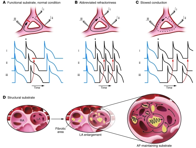
Reentry requires appropriate tissue properties, a vulnerable “substrate.” Reentry substrates can be caused by altered electrical properties or by fixed structural changes. Cardiac tissue exhibits a discrete refractory period (inexcitable interval following the last firing, governed by APD). Reentry initiation usually requires a premature ectopic beat that acts as a trigger. Figure 3, A–C, shows a premature beat arising at a branch point (labeled “i”). The resulting impulse conducts through the pathway leading to recording point ii, which is no longer refractory, but blocks in the pathway leading to recording point iii because of its longer refractory period. The premature impulse arrives at the distal end of previously refractory site iii and attempts to reenter. Under normal conditions without a reentry substrate (Figure 3A), the conduction time from point i around the circuit through point ii and back through point iii is shorter than the refractory period, and the impulse cannot reenter. When APD is decreased, reducing the refractory period sufficiently (Figure 3B), excitability is recaptured earlier and the reentering impulse can now sustain itself throughout the circuit. Slowed conduction can similarly allow the impulse to reenter (without APD abbreviation), because the more slowly conducting impulse leaves additional time for refractoriness to dissipate (Figure 3C). Figure 3D illustrates the development of a fixed structural reentry substrate. A combination of atrial dilation and fibrosis creates longer potential conduction pathways for reentry, slows conduction, and imposes conduction barriers that favor the initiation and maintenance of multiple irregular reentry circuits that sustain AF (5).
Figure 3. Factors promoting reentry.
Reentry occurs via interactions between interconnected zones of tissue, initiated by a premature beat (*). i, ii, and iii indicate microelectrode recordings in three zones of a potential reentry circuit. (A) Normal atrial tissue is unlikely to maintain reentry. Reentry maintenance can result from either a shortened refractory period (B) or slowed conduction (C). (D) A variety of cardiac conditions cause structural reentry substrates characterized by atrial enlargement and fibrosis. LA, left atrium; RA, right atrium.
Relation of basic mechanisms to clinical forms
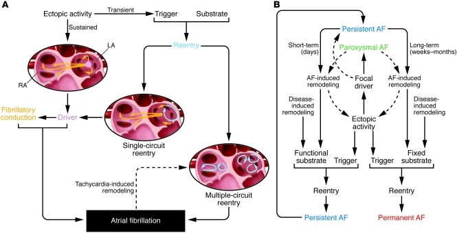
Figure 4A shows how basic mechanisms relate to the pathophysiology of AF. Ectopic activity can be transient, manifesting as isolated ectopic beats, or sustained, causing tachycardia. Any source of sustained rapid atrial tachycardia, whether an ectopic focus or regularly discharging reentry circuit, is called a driver. Drivers that discharge rapidly and regularly can cause irregular activity characteristic of AF if the emanating propagation waves break up in functionally heterogeneous atrial tissue, leading to fibrillatory conduction (5, 8). AF-related reentry occurs in two general forms: (a) single-circuit reentry, involving one primary reentry circuit driver; and (b) multiple-circuit reentry, involving multiple simultaneous dyssynchronous reentry circuits. The very rapid activation caused by AF produces atrial tachycardia remodeling (ATR) of atrial electrical properties, promoting functional reentry (4). ATR effects vary in different atrial regions, causing spatial heterogeneity that promotes multiple-circuit reentry (4).
Figure 4. Tissue mechanisms leading to AF and clinical forms.
(A) Ectopic activity can act as a driver maintaining AF or as a trigger on a vulnerable substrate resulting in reentry (single- or multiple-circuit). Local driver mechanisms (ectopic or single-circuit reentrant) produce irregular fibrillatory activity via fibrillatory conduction. Rapid atrial activity (tachycardia) causes atrial remodeling, promoting multiple-circuit reentry. (B) Clinical AF can manifest as paroxysmal AF (self-terminating), persistent AF (requires drug therapy or electrical cardioversion to terminate), and permanent AF (non-terminating). Focal ectopic drivers are principally associated with paroxysmal forms, functional reentrant substrates with persistent AF, and increasingly fixed substrates with permanent forms.
Clinical AF can be paroxysmal (self-terminating), persistent (requiring medical intervention to terminate), or permanent (Figure 4B). Focal drivers, particularly ectopic sites in cardiomyocyte sleeves around the pulmonary veins (9), produce paroxysmal AF forms. Functional reentry substrates cause persistent AF that can be terminated, restoring normal rhythm. AF becomes permanent as the substrate becomes fixed and irreversible because of structural remodeling (10). AF itself induces remodeling (4, 11): ATR, which promotes functional reentry, as well as structural remodeling.
Etiological contributors
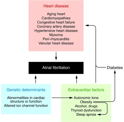
A variety of etiological factors contribute to AF occurrence (Figure 5). In most patients, AF results from interactions among multiple factors operating simultaneously.
Figure 5. Schematic overview of factors governing AF occurrence.
Over 70% of AF cases have associated heart disease (12). Aging is a major risk factor, largely via structural remodeling (3, 11). Congestive heart failure (CHF), hypertension, valvular heart disease, and coronary artery disease (CAD) are common contributors (11). Less common predisposing conditions include peri- or myocarditis, atrial myxomas, and hypertrophic cardiomyopathy. Extracardiac conditions also promote AF occurrence. Sufficiently powered studies suggest that heavy alcohol consumption promotes AF (13). Hyperthyroidism is a well-recognized contributor (14), and the roles of sleep apnea and obesity are increasingly recognized (15). Autonomic tone is a well-established factor (16). Ten years ago, genetic determinants of AF were largely unknown (4), but there has since been an explosion of information (17, 18). Many disease-causing mutations have been identified and their pathophysiology analyzed (17). While new insights have been provided, these findings raise as many questions as they answer (17). The recent rapid development of large-scale genome-wide association studies (GWASs) has provided exciting leads about genetic predisposition to AF, while raising challenging pathophysiological issues (18).
We will now consider how each of the three principal categories of etiological contributors in Figure 5 (heart disease, extracardiac factors, and genetic determinants) act through the pathophysiological mechanisms in Figures 2–4 (focal ectopic activity, functional reentry substrates, and fixed reentry substrates).
Mechanisms underlying AF-promoting ectopic activity
Heart disease–related ectopic activity
Atrial tissue samples from AF patients provide insights into pathophysiology caused by AF and/or underlying heart disease (19). AF atria show abnormal SR Ca2+ handling (20–23), which causes spontaneous diastolic SR Ca2+ release events (20, 22, 23). SR Ca2+ load is not increased (20, 23), suggesting that spontaneous Ca2+ releases occur because of altered RyR2 function.
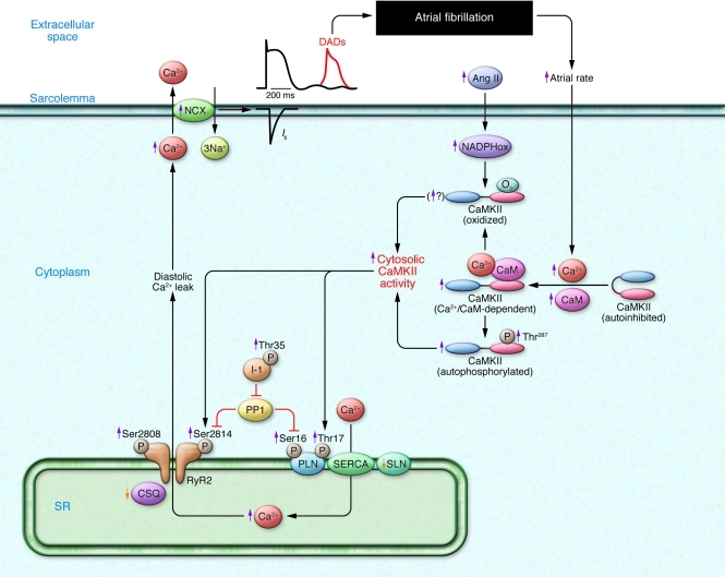
The detailed molecular pathobiology of AF-related DADs is summarized in Figure 6. RyR2 phosphorylation by PKA at Ser2808 (21) and by Ca2+/calmodulin-dependent kinase II (CaMKII) at Ser2814 is increased in AF (23–25). CaMKII activity is normally autoinhibited. Ca2+-calmodulin binding removes autoinhibition, activating CaMKII and causing autophosphorylation that makes CaMKII Ca2+-independent (23). Similar activation may result from CaMKII oxidation. Changes in PKA and CaMKII RyR2 phosphorylation state may result not only from changed kinase activity, but also from alterations in phosphatases (6). RyR2 phosphorylation increases its Ca2+ sensitivity, enhancing channel open probability (24). Mice lacking RyR2-inhibitory FK506-binding protein 12.6 or with gain-of-function RyR2 mutations exhibit increased susceptibility to AF, along with increased atrial cell SR Ca2+ leak and triggered activity (24, 26). Angiotensin promotes AF, perhaps via CaMKII oxidation and enhanced CaMKII phosphorylation of RyR2 (27), in addition to its well-recognized effect of promoting structural remodeling (10). RyR2 dysfunction can also be induced by Ca2+ overload resulting from phospholamban hyperphosphorylation, which removes phospholamban inhibition of SERCA and enhances SR Ca2+ uptake. Phospholamban hyperphosphorylation can result from enhanced PKA and/or CaMKII activity or from decreased phosphatase function due to increased activity of a phosphatase-inhibitory protein, I-1, caused by I-1 hyperphosphorylation (6, 28).
Figure 6. Factors promoting AF by inducing SR diastolic Ca2+ leak.
RyR dysfunction may result from hyperphosphorylation or exposure to excess intraluminal Ca2+. SR Ca2+ overload can result from phospholamban hyperphosphorylation or reduced sarcolipin (SLN) expression, which disinhibit SERCA and enhance SR Ca2+ uptake. Increased cellular Ca2+ entry due to high atrial rate facilitates Ca2+/calmodulin (CaM) binding to the regulatory domain of CaMKII, resulting in disinhibition of the catalytic subunit. After initial activation of the holoenzyme by Ca2+/CaM, oxidation at Met281/282 or phosphorylation at Thr287 blocks reassociation of the catalytic domain, yielding persistent CaMKII activity. PP1 suppression by enhanced SR compartment inhibitor–1 (I-1 activity) contributes to increased phospholamban (PLN) and RyR phosphorylation. NADPHox, oxidized NADPH.
Increased NCX expression and/or function are commonly observed in AF (23, 28, 29), suggesting that Iti resulting from given amounts of diastolic SR Ca2+ leak may be amplified, enhancing the risk of DADs/triggered activity (6). Cardiac inositol 1,4,5-trisphosphate (IP3) receptors (IP3R2) act as Ca2+-transporting pathways that facilitate arrhythmogenic SR Ca2+ leak (30). IP3R2 expression is increased by ATR (31). IP3R2-coupled amplification of atrial SR Ca2+ release events and related arrhythmogenesis (32) may thus contribute to AF-related ectopic activity.
AF is very common in CHF patients, with a prevalence varying from approximately 10% in patients in New York Heart Association (NYHA) heart failure classes II–III to approximately 50% in NYHA class IV (33). Focal drivers and triggered activity contribute to CHF-related AF (34, 35). CHF increases atrial SR Ca2+ load and reduces CSQ expression, thereby promoting spontaneous SR Ca2+ release (36). Mice overexpressing tumor necrosis factor develop CHF along with abnormal atrial cell diastolic Ca2+ release upon Ca2+ loading (37).
CAD increases AF risk approximately 3.5-fold (38). Atrial ischemia promotes AF (39, 40), in part by causing spontaneous SR Ca2+ release events and increasing NCX function, leading to triggered activity and atrial ectopy (39).
Extracardiac factors contributing to ectopic activity
Adrenergic-dependent RyR2 phosphorylation promotes spontaneous SR Ca2+ leak (41). Clinically relevant conditions that cause abnormal DAD-promoting Ca2+ handling may require adrenergic stimulation to elicit Ca2+ sparks and triggered arrhythmias (39). Autonomic hyperinnervation causes spontaneous paroxysmal AF in dogs (42), with sympathovagal nerve discharge immediately preceding AF paroxysms (43). Vagal activation may promote spontaneous arrhythmogenesis by reducing APD, allowing adrenergically induced afterdepolarizations to induce ectopic activity in susceptible regions such as pulmonary veins (44).
Genetic contributors to ectopic activity
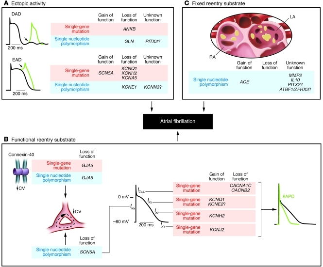
Genetic factors fall into two broad groups: rare genetic variants with strong effects and a clear phenotype (single-gene mutations) and common genetic variants with weaker effects and a less overt phenotype (single nucleotide polymorphisms [SNPs]). Figure 7 provides a broad overview of the mutations/SNPs predisposing to AF and their potential pathophysiological mechanisms. Gene variants believed to cause ectopic activity are indicated in Figure 7A. A mutation in the gene encoding the adapter protein ankyrin-B (long-QT syndrome-4 [LQT4]), which impairs targeting of multiple proteins to the cell membrane, alters Ca2+ handling, and leads to DADs/triggered activity (45). A predicted loss-of-function SNP in the gene encoding the SERCA-inhibitory protein sarcolipin is also associated with AF (46).
Figure 7. Genetic variants predisposing to AF.
Genetic variants (single-gene mutations: red; single nucleotide polymorphisms: blue) are displayed in relation to presumed AF-promoting mechanisms: (A) ectopic activity; (B) reentry with functional substrates; (C) reentry with fixed structural substrates.
The first potential genetic cause of AF linked to an EAD mechanism was a loss-of-function SNP in KCNE1, which encodes the slow delayed-rectifier K+ channel (IKs) β-subunit minK involved in LQT5 (47). The classical EAD-promoting congenital LQTS mutations also predispose to AF (48, 49). A single-gene mutation of the KCNA5 gene encoding the Kv1.5 α-subunit of the ultrarapid delayed-rectifier IKur channel identified in an individual with idiopathic AF promotes EADs under adrenergic stimulation (50). Other gene variants in KNCA5 suggest a role for tyrosine kinases in AF (51). Recent work in a genetic LQTS mouse model (52) directly implicated EAD mechanisms in AF (53).
Recently, two gain-of-function Na+ channel (SCN5A) mutations associated with AF, but with no evidence for EAD-promoting APD prolongation, have been reported (54, 55). These mutations may favor AF initiation by increasing Na+ channel availability and thereby promoting ectopic activity (54).
Genetic loci on chromosomes 4q25, 16q22, and 1q21 have been associated with AF by GWAS. The first locus on chromosome 4q25 (56) has since been replicated in two additional European cohorts (18). The closest gene is the pair-like homeodomain-2 gene (PITX2), which encodes a transcription factor that is crucial for cardiac development and pulmonary vein formation (57, 58). This gene variant may therefore be implicated in pulmonary vein ectopic sources of AF. An AF-associated SNP of the KCNN3 gene on chromosome 1q21 affects a Ca2+-activated K+ channel (59). Based on prior work, this mutation may act by abbreviating APD in pulmonary veins, promoting microreentry (60), or cause ectopic activity via EAD mechanisms (61).
Molecular mechanisms underlying functional substrates for AF-maintaining reentry
Heart disease–related functional reentry
AF-induced APD shortening.
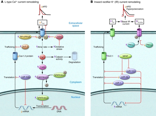
AF (62), and indeed all very rapid atrial tachyarrhythmias (63), promote AF initiation and maintenance via ATR. A major AF-promoting component of ATR is refractory period reduction due to APD abbreviation (Figure 3B). Decreased depolarizing L-type Ca2+ current (ICa,L), along with increased repolarizing inward-rectifier K+ currents, background (IK1), and constitutively active acetylcholine-dependent current (IK,AChc), underlie ATR-induced APD shortening (64–71). The molecular basis of ATR-induced ICa,L reduction is complex (Figure 8A). Rapid atrial activation causes Ca2+ loading, which activates the Ca2+/calmodulin/calcineurin/NFAT system, causing transcriptional downregulation of the Cav1.2 α-subunit (72, 73). Other potential contributors include downregulation of accessory β1-, β2a-, β2b-, β3-, and α2δ2-subunits (19, 66, 74, 75), Ca2+ channel dephosphorylation due to type 1 (PP1) and type 2A (PP2A) serine/threonine protein phosphatase activation (28, 66), and enhanced Cav1.2 α-subunit S-nitrosylation (76). MicroRNAs (miRNAs) appear to play a major role in AF (77). Recent work implicates increased miR-328 in ATR-induced ICa,L downregulation (78). Finally, impaired Cav1.2 protein trafficking induced by a zinc-binding protein (ZnT-1) may contribute to ICa,L reduction (79).
Figure 8. Remodeling of ICa,L and inward-rectifier K+ currents by AF/tachycardia.
(A) The high atrial rate in AF increases intracellular Ca2+ load, activating calcineurin via the Ca2+/calmodulin system. Calcineurin stimulates nuclear translocation of nuclear factor of activated T lymphocytes (NFAT), reducing transcription of the principal ICa,L subunit, Cav1.2. Increased mRNA degradation/impaired protein translation of Cav1.2 and breakdown of Cav1.2 protein by calpains may also contribute. Increased expression of zinc transporter–1 (ZnT-1) impairs membrane trafficking of Cav1.2. Reduced Cav1.2 phosphorylation due to increased protein phosphatase (PP) activity and increased channel nitrosylation may also decrease ICa,L. GSH, glutathione. (B) Increased IK1 density results from upregulation of the principle Kir2.1 subunit, likely due to reduced levels of inhibitory miRNAs (miR-101, miR-26, miR-1). Increased IK,AChc is caused by altered PKC regulation: increased membrane abundance of stimulatory PKCε and reduced expression of inhibitory PKCα.
Inward-rectifier K+ currents play an important role in AF maintenance, by both reducing APD and accelerating arrhythmia-maintaining rotors by hyperpolarizing atrial cells, thereby removing voltage-dependent INa inactivation (5, 80). Figure 8B shows mechanisms underlying AF-related changes in IK1 and IK,AChc. IK1 increases because of increased expression of the underlying Kir2.1 subunit (19, 67–71, 81, 82), likely due to reduced levels of Kir2.1-inhibitory miRNAs, particularly miR-1, miR-26, and miR-101 (82, 83). Stronger IK1 dephosphorylation (and thus activation) due to increased PP1 and PP2A activity (28, 66, 84) might also contribute.
Muscarinic cholinergic receptor–mediated IK,ACh activation is reduced in AF (69, 70, 85). Loss of receptor-mediated IK,ACh channel control is associated with increased agonist-independent (“constitutive”) IK,AChc (69, 71, 79, 85). Increased IK,AChc is due to enhanced single IK,ACh channel opening frequency, without changes in single-channel conductance, density, or voltage dependence (85). Enhanced open probability is related to altered PKC isoform balance, with increased IK,ACh-stimulatory novel isoform function and reduced inhibitory classical isoform influence (86). Blockade of IK,AChc suppresses ATR-induced APD abbreviation and AF promotion (70).
AF-associated atrial conduction abnormalities.
Conduction slowing promotes reentry (Figure 3C). Gap junctions are crucial for cell-to-cell coupling and conduction; however, information in the literature about gap-junctional remodeling in AF is inconsistent (19, 87). Connexin alterations likely vary with AF duration, underlying pathology, and species (88). Spatially heterogeneous connexin-40 remodeling occurs in the well-controlled goat AF remodeling system (89), consistent with clinical evidence that connexin-40 gene variants predispose to AF (90, 91). The importance of atrial connexin-43 dephosphorylation and lateralization in CHF is unclear, since CHF-induced conduction slowing and AF promotion remain following CHF recovery, despite full resolution of connexin abnormalities (92).
Sodium current (INa) is a primary determinant of conduction velocity. INa density is reduced in canine ATR, with corresponding decreases in Nav1.5 subunit mRNA and protein expression (64). Gaborit et al. did not observe atrial INa changes at the genomic level in AF patients (19). Atrial myocytes from AF patients show either unchanged (93) or only slightly reduced (94) INa.
Atrial ischemia/infarction produces a substrate for AF via functional reentry circuits stabilized by a line of conduction block in the ischemic zone (39, 40). With acute ischemia, the conduction block is likely related to gap junction uncoupling (40).
Extracardiac factors contributing to functional reentry
Clinical AF is more likely under vagotonic conditions, with AF in some patients clearly vagally dependent (95). Vagal nerve endings release acetylcholine, stimulating cardiac muscarinic cholinergic receptors that activate IK,ACh. Vagal innervation is patchy, so the effects of vagal activation are spatially heterogeneous, promoting the initiation and stabilization of multiple AF-maintaining reentrant rotors (95). Knockout of the IK,ACh pore-forming subunit Kir3.1 prevents cholinergic AF in mice (96).
Genetic factors contributing to functional reentry
APD shortening.
Perhaps the most common AF-promoting genetic paradigm is APD shortening caused by gain-of-function K+ channel mutations (Figure 7B). The first gene mutation linked to lone AF caused gain of function in the α-subunit (KCNQ1) of the slow delayed rectifier IKs (97). Other genes with AF-inducing gain-of-function K+ channel mutations include KCNH2 (98), KCNJ2 (99), and KCNE2 (100), corresponding to ion channel subunits of IKr, IK1, and possibly IKs, respectively. Loss-of-function mutations in ICa,L subunits would also be expected to decrease APD and promote AF. In 82 patients with Brugada syndrome/short-QT ECG phenotypes, loss-of-function mutations of the CACNA1C and CACNB2 genes, encoding ICa,L α- and β-subunits, were associated with AF (101). Patients with short-QT syndromes have reduced APDs and are predisposed to AF (17). Despite the correlation between these pathophysiological mechanisms and functional alterations, many questions remain unanswered; for example, why mutations that appear to be associated with gain of function at the atrial level leave ventricular repolarization unaltered or even delayed (17).
Conduction slowing.
Several gene variants promote AF by targeting ion channels that govern conduction velocity. The GJA5 gene encodes connexin-40, a gap junction ion channel that is particularly important in the atria. Mice lacking connexin-40 demonstrate conduction abnormalities and atrial arrhythmias (102). Missense somatic mutations in GJA5 were identified in 4 of 15 patients with idiopathic AF (91). GJA5 promoter sequence variants that decrease gene transcription are associated with increased AF vulnerability (90).
Gene variants impairing Na+ channel function promote AF, presumably via conduction slowing that favors reentry. Loss-of-function Na+ channel α-subunit (SCN5A) mutations were first associated with AF in a family with dilated cardiomyopathy, AF, impaired automaticity, and conduction slowing (103). Subsequently, additional loss-of-function SCN5A mutations and SNPs were identified in idiopathic AF subjects (104, 105). Loss-of-function SCN5A mutations are the most common genetically defined cause of Brugada syndrome, which classically produces sudden death due to ventricular fibrillation, but which also frequently causes AF (106). More recently, loss-of-function mutations in the cardiac Na+ channel β-subunits SCN1B, SCN2B, and SCN3B have been associated with AF (107, 108).
AF has been associated with a SNP of the NOS3 gene encoding eNOS (109). Experimental data suggest that eNOS can regulate cardiac vagal activity and ICa,L, providing plausible links to reentry substrates (110, 111).
Molecular mechanisms leading to a fixed substrate for reentry
Atrial fibrosis plays an important role in many AF-promoting pathologies (10). Once formed, atrial fibrosis reverses slowly if at all (10); prevention therefore becomes very important. Considerable effort is being invested in targeting mechanisms leading to atrial fibrosis to develop effective prophylactic therapy (112).
Heart disease–related atrial fibrosis
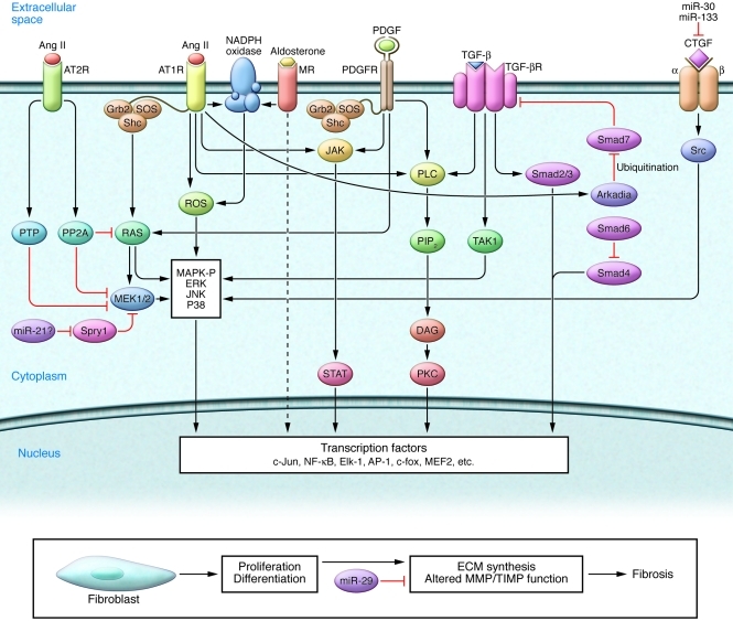
Atrial fibrosis occurs in many pathophysiological settings, including senescence, cardiac dysfunction, valve disease, and ischemic heart disease (10, 39, 113, 114). Profibrotic signaling pathways (Figure 9) demonstrate extensive crosstalk; fibrosis likely results from multiple factors working simultaneously rather than any single individual component. There are extensive interactions between cardiomyocytes and fibroblasts (10). Fibroblasts produce fibrotic ECM proteins and cardioactive mediators that affect cardiomyocyte phenotype, whereas cardiomyocyte-derived products such as ROS, TGF-β, PDGF, and connective tissue growth factor (CTGF) modulate fibroblast properties and ECM synthesis.
Figure 9. Molecular mechanisms leading to atrial fibrosis.
Major profibrotic signaling pathways involved in fibrosis generation are shown. MR, mineralocorticoid receptor.
Ang II, a well-characterized profibrotic molecule, plays an important role in AF (115). Ang II type 1 receptors (AT1Rs) have profibrotic actions mediated via enhanced expression of TGF-β, Smad2/3, Smad4, Arkadia, and activated ERK MAPK (116). Arkadia reduces Smad7 expression by marking it for ubiquitination, increasing TGF-β signaling by removing Smad7 antagonism at the TGF-β receptor (117). In addition, AT1R signaling through the Shc/Grb2/SOS adapter-protein complex activates the GTPase protein Ras, which enhances profibrotic MAPK phosphorylation (118). AT1R stimulation activates phospholipase C (PLC), further acting through phosphatidylinositol 4,5-bisphosphate (PIP2), diacylglycerol (DAG), and IP3. DAG activates PKC, while IP3 causes intracellular Ca2+ release, both contributing to remodeling. The AT1R-stimulated JAK/STAT pathway activates transcription factors such as activating protein–1 (AP-1) and NF-κB, which promote cardiomyocyte remodeling. On the other hand, AT2R activation counteracts MAPK activation by increasing dephosphorylation through PP2A and phosphotyrosine phosphatase (PTP) (118).
TGF-β1, secreted by both fibroblasts and cardiomyocytes, is an important profibrotic mediator (119). Cardiac overexpression of constitutively active TGF-β1 causes selective atrial fibrosis, conduction heterogeneity, and AF susceptibility (120). TGF-β1 functions as a downstream mediator of Ang II in both paracrine and autocrine ways (119). It acts primarily through the SMAD pathway to stimulate fibroblast activation and collagen production (10, 117). Rapid atrial firing causes atrial cardiomyocytes to produce substances, recently identified as Ang II and ROS acting via enhanced TGF-β production, that cause fibroblast differentiation into ECM-secretory myofibroblasts (121, 122). This effect likely contributes to progressive structural remodeling during long-standing AF.
PDGF stimulates proliferation and differentiation of fibroblasts (10, 119). PDGF receptors contain two transmembrane domains, which dimerize on stimulation and activate a tyrosine kinase. This tyrosine kinase autophosphorylates other intracellular domains that activate the PDGF receptor and initiate signaling via Ras/MEK1/2 and MAPK, JAK/STAT, and PLC. PDGF overexpression causes cardiac fibrosis and cardiomyopathy (123). PDGF is also secreted by mast cells, which may contribute to profibrotic fibroblast responses associated with low-grade atrial inflammation (124). The genes encoding PDGF and its receptor are more strongly expressed in atrial than ventricular fibroblasts, and PDGF receptor tyrosine kinase inhibition suppresses atrial fibroblast hyperresponsiveness, suggesting that PDGF may contribute to the more intense fibrotic responses of atria versus ventricles (119).
CTGF, a member of the CCN (cyr61/ctgf/nov) protein family, is a downstream mediator of TGF-β1 and Ang II profibrotic signaling. CTGF activates fibroblasts via Src kinase and MAPKs (125). Genomic analysis points to CTGF as a central player in atrial structural remodeling (126). Recent work implicates CTGF as a key signaling molecule downstream of Rac1 (127).
miRNAs are emerging as potentially important players in atrial fibrosis. miR-29 targets and inhibits collagen genes (128). miR-29 downregulation in atrial tissue precedes and likely contributes to profibrillatory atrial fibrosis (129). Similarly, miR-30 and miR-133, which suppress CTGF production (130), are downregulated in the AF-promoting, atrial profibrotic CHF substrate (131). Atrial miR-21 is upregulated in CHF (131). Elegant work suggests profibrotic effects of miR-21 via suppression of sprouty 1 (Spry1), which enhances fibroblast MAPK phosphorylation and survival (132); however, these findings have recently been questioned (133).
Extracardiac factors contributing to atrial fibrosis
Patients with sleep apnea, obesity, and diabetes are at increased risk of AF (15), but exploration of the underlying mechanisms has just begun. Sleep apnea increases atrial pressure, causing atrial stretch that could promote remodeling (134). Diabetes causes atrial fibrosis, along with increased expression of the receptor for advanced glycosylation end-products (AGEs) (RAGE) and CTGF (135). Suppression of AGE production prevented diabetes-related atrial remodeling and CTGF upregulation in a rat model (135). Adrenergic stimulation activates MAPKs and profibrotic remodeling paradigms; however, no clear role has yet been demonstrated in atrial fibrotic remodeling.
Genetic factors contributing to atrial fibrosis
SNPs in genes contributing to atrial structural integrity, and affecting inflammation and neurohumoral pathways, have been associated with AF by candidate gene approaches. Noteworthy are polymorphisms in genes encoding angiotensin-converting enzyme (ACE) (136), MMP2, and interleukin-10 (137). GWASs have identified SNPs on chromosome 4q25, with PITX2c suspected to be the target gene, and a SNP on chromosome 16q22 near the AT-binding factor (ATBF) gene, also known as zinc finger homeobox 3 (ZFHX3) (138). In addition to a possible mechanism involving pulmonary vein ectopy, PITX2c’s role in cardiac development (57, 58) raises the possibility that its dysfunction could cause atrial structural remodeling. ATBF/ZFHX3 is a tumor-suppressor gene (139) and promotes survival of neurons by inducing PDGF receptor β expression and protecting against oxidative stress (140). These properties are consistent with a role in atrial structural remodeling processes.
Potential therapeutic implications
AF is a problem of growing proportions that has been termed “epidemic” (141). Present therapeutic options have limited efficacy and disconcerting adverse-effect risks (2, 3). Consequently, a variety of novel, mechanism-based approaches are in development (2). One approach has been to target atrial remodeling, with the use of “upstream therapy” interventions that interfere with putative signaling pathways. Interventions such as statin drugs, omega-3 fatty acids, and renin-angiotensin system inhibitors prevent atrial remodeling in experimental models (142). While retrospective analyses of clinical data have been promising, the few prospective randomized clinical trials completed to date have been negative or inconclusive (142). Even though the notion of remodeling prevention is very attractive, its feasibility remains to be demonstrated.
Knowledge of AF mechanisms may allow for more direct targeting of specific pathophysiological contributors. The importance of APD as a determinant of reentry led to widespread use of APD-prolonging “class III” agents in AF; however, most of these agents target IKr, and their use is limited by potentially life-threatening ventricular arrhythmia induction due to EAD mechanisms (2). Several approaches are being studied to treat AF by prolonging atrial APD without affecting the ventricles. One is to target IK,ACh, which is prominently involved in AF (70, 95) but absent in the ventricles. A highly selective IK,ACh inhibitor has recently been shown effective in several experimental AF models (143). Another mechanism-based target under study is the Ca2+-dependent K+ current (IKCa), which appears to be important in AF (59–61). A selective small-conductance IKCa channel inhibitor suppresses AF in a range of animal models (144). Still another approach is to use gene transfer to deliver dominant-negative K+ channel subunits to selectively prolong atrial APD, which suppresses AF in a porcine model (145).
Ectopic activity due to diastolic RyR2 Ca2+ release is also a potential target. Stabilizers of RyR2 show value in AF prevention (146, 147). CaMKII hyperphosphorylation plays a role in AF-related RyR2 dysfunction, and interventions targeting CaMKII hold promise (6).
The emerging role of miRNAs in AF-inducing atrial remodeling presents potentially exciting therapeutic opportunities. miRNAs are stable in blood, and interventions have been developed to enhance or suppress the expression of miRNAs involved in disease progression (148). The apparent participation of miRNAs in a wide range of AF-inducing mechanisms, including abnormal SR Ca2+ release, APD reduction, connexin modulation, and tissue fibrosis, points to potential target mechanisms (77).
Recent insights into the molecular pathophysiology of AF open new opportunities in risk assessment and monitoring of therapeutic responses. Novel biomarkers under investigation include noninvasive indices of atrial fibrosis (149) and plasma biomarkers reflecting underlying biochemical mechanisms or responses (150).
A great deal of information about the molecular pathophysiology of AF has been obtained over the past 10 years. This information has provided insights, raised many new questions, and provided novel therapeutic opportunities. It is hoped that concrete improvements in AF management will result, but only time and a great deal of additional research will tell.
Acknowledgments
The authors thank France Thériault for excellent secretarial help. This work was supported by the Canadian Institutes of Health Research (MGP6957 and MOP44365); the Quebec Heart and Stroke Foundation, the Foundation Leducq (European/North American Atrial Fibrillation Research Alliance [ENAFRA]); the MITACS Network; the German Federal Ministry of Education and Research Atrial Fibrillation Competence Network (grant 01Gi0204); the Deutsche Forschungsgemeinschaft (Do 769/1-3); the European Union (European Network for Translational Research in Atrial Fibrillation [EUTRAF]); Friedrich-Baur-Foundation; “Förderprogramm für Forschung und Lehre der LMU” (FöFoLe), University of Munich; National Genome Research Network (NGFN) Plus; and Ludwig-Maximilians-Universität (LMU) Excellence Initiative.
Footnotes
Authorship note: Reza Wakili and Niels Voigt contributed equally to this work.
Conflict of interest: The authors have declared that no conflict of interest exists.
Citation for this article: J Clin Invest. 2011;121(8):2955–2968. doi:10.1172/JCI46315
References
- 1.Miyasaka Y, et al. Secular trends in incidence of atrial fibrillation in Olmsted County, Minnesota, 1980 to 2000, and implications on the projections for future prevalence. Circulation. 2006;114(2):119–125. doi: 10.1161/CIRCULATIONAHA.105.595140. [DOI] [PubMed] [Google Scholar]
- 2.Dobrev D, Nattel S. New antiarrhythmic drugs for treatment of atrial fibrillation. Lancet. 2010;375(9721):1212–1223. doi: 10.1016/S0140-6736(10)60096-7. [DOI] [PubMed] [Google Scholar]
- 3.Nattel S. From guidelines to bench: implications of unresolved clinical issues for basic investigations of atrial fibrillation mechanisms. Can J Cardiol. 2011;27(1):19–26. doi: 10.1016/j.cjca.2010.11.004. [DOI] [PubMed] [Google Scholar]
- 4.Nattel S. New ideas about atrial fibrillation 50 years on. Nature. 2002;415(6868):219–226. doi: 10.1038/415219a. [DOI] [PubMed] [Google Scholar]
- 5.Nattel S, Burstein B, Dobrev D. Atrial remodeling and atrial fibrillation: mechanisms and implications. Circ Arrhythm Electrophysiol. 2008;1(1):62–73. doi: 10.1161/CIRCEP.107.754564. [DOI] [PubMed] [Google Scholar]
- 6.Dobrev D, Voigt N, Wehrens XH. The ryanodine receptor channel as a molecular motif in atrial fibrillation: pathophysiological and therapeutic implications. Cardiovasc Res. 2011;89(4):734–743. doi: 10.1093/cvr/cvq324. [DOI] [PMC free article] [PubMed] [Google Scholar]
- 7.MacLennan DH, Chen SR. Store overload-induced Ca2+ release as a triggering mechanism for CPVT and MH episodes caused by mutations in RYR and CASQ genes. J Physiol. 2009;587(pt 13):3113–3115. doi: 10.1113/jphysiol.2009.172155. [DOI] [PMC free article] [PubMed] [Google Scholar]
- 8.Berenfeld O, Zaitsev AV, Mironov SF, Pertsov AM, Jalife J. Frequency-dependent breakdown of wave propagation into fibrillatory conduction across the pectinate muscle network in the isolated sheep right atrium. Circ Res. 2002;90(11):1173–1180. doi: 10.1161/01.RES.0000022854.95998.5C. [DOI] [PubMed] [Google Scholar]
- 9.Haissaguerre M, et al. Spontaneous initiation of atrial fibrillation by ectopic beats originating in the pulmonary veins. N Engl J Med. 1998;339(10):659–666. doi: 10.1056/NEJM199809033391003. [DOI] [PubMed] [Google Scholar]
- 10.Burstein B, Nattel S. Atrial fibrosis: mechanisms and clinical relevance in atrial fibrillation. J Am Coll Cardiol. 2008;51(8):802–809. doi: 10.1016/j.jacc.2007.09.064. [DOI] [PubMed] [Google Scholar]
- 11.Allessie MA, et al. Pathophysiology and prevention of atrial fibrillation. Circulation. 2001;103(5):769–777. doi: 10.1161/01.cir.103.5.769. [DOI] [PubMed] [Google Scholar]
- 12.Kozlowski D, et al. Lone atrial fibrillation: what do we know? Heart. 2010;96(7):498–503. doi: 10.1136/hrt.2009.176321. [DOI] [PubMed] [Google Scholar]
- 13.Mukamal KJ, Tolstrup JS, Friberg J, Jensen G, Gronbaek M. Alcohol consumption and risk of atrial fibrillation in men and women: the Copenhagen City Heart Study. Circulation. 2005;112(12):1736–1742. doi: 10.1161/CIRCULATIONAHA.105.547844. [DOI] [PubMed] [Google Scholar]
- 14.Auer J, Scheibner P, Mische T, Langsteger W, Eber O, Eber B. Subclinical hyperthyroidism as a risk factor for atrial fibrillation. Am Heart J. 2001;142(5):838–842. doi: 10.1067/mhj.2001.119370. [DOI] [PubMed] [Google Scholar]
- 15.Schoonderwoerd BA, Smit MD, Pen L, Van Gelder IC. New risk factors for atrial fibrillation: causes of “not–so–lone atrial fibrillation.”. Europace. 2008;10(1):668–673. doi: 10.1093/europace/eun124. [DOI] [PubMed] [Google Scholar]
- 16.Chou CC, Chen PS. New concepts in atrial fibrillation: neural mechanisms and calcium dynamics. Cardiol Clin. 2009;27(1):35–43, viii. doi: 10.1016/j.ccl.2008.09.003. [DOI] [PMC free article] [PubMed] [Google Scholar]
- 17.Mahida S, Lubitz SA, Rienstra M, Milan DJ, Ellinor PT. Monogenic atrial fibrillation as pathophysiological paradigms. Cardiovasc Res. 2011;89(4):692–700. doi: 10.1093/cvr/cvq381. [DOI] [PMC free article] [PubMed] [Google Scholar]
- 18.Sinner M, Ellinor PT, Meitinger T, Benjamin EJ, Kääb S. Genome-wide association studies: past, present, and future. Cardiovasc Res. 2011;89(4):701–709. doi: 10.1093/cvr/cvr001. [DOI] [PMC free article] [PubMed] [Google Scholar]
- 19.Gaborit N, et al. Human atrial ion channel and transporter subunit gene-expression remodeling associated with valvular heart disease and atrial fibrillation. Circulation. 2005;112(4):471–481. doi: 10.1161/CIRCULATIONAHA.104.506857. [DOI] [PubMed] [Google Scholar]
- 20.Hove-Madsen L, et al. Atrial fibrillation is associated with increased spontaneous calcium release from the sarcoplasmic reticulum in human atrial myocytes. Circulation. 2004;110(11):1358–1363. doi: 10.1161/01.CIR.0000141296.59876.87. [DOI] [PubMed] [Google Scholar]
- 21.Vest JA, et al. Defective cardiac ryanodine receptor regulation during atrial fibrillation. Circulation. 2005;111(16):2025–2032. doi: 10.1161/01.CIR.0000162461.67140.4C. [DOI] [PubMed] [Google Scholar]
- 22.Liang X, et al. Ryanodine receptor-mediated Ca2+ events in atrial myocytes of patients with atrial fibrillation. Cardiology. 2008;111(2):102–110. doi: 10.1159/000119697. [DOI] [PubMed] [Google Scholar]
- 23.Neef S, et al. CaMKII-dependent diastolic SR Ca2+ leak and elevated diastolic Ca2+ levels in right atrial myocardium of patients with atrial fibrillation. . Circ Res. 2010;106(6):1134–1144. doi: 10.1161/CIRCRESAHA.109.203836. [DOI] [PubMed] [Google Scholar]
- 24.Chelu MG, et al. Calmodulin kinase II-mediated sarcoplasmic reticulum Ca2+ leak promotes atrial fibrillation in mice. . J Clin Invest. 2009;119(7):1940–1951. doi: 10.1172/JCI37059. [DOI] [PMC free article] [PubMed] [Google Scholar]
- 25.Greiser M, et al. Distinct contractile and molecular differences between two goat models of atrial dysfunction: AV block-induced atrial dilatation and atrial fibrillation. J Mol Cell Cardiol. 2009;46(3):385–394. doi: 10.1016/j.yjmcc.2008.11.012. [DOI] [PubMed] [Google Scholar]
- 26.Sood S, et al. Intracellular calcium leak due to FKBP12.6 deficiency in mice facilitates the inducibility of atrial fibrillation. Heart Rhythm. 2008;5(7):1047–1054. doi: 10.1016/j.hrthm.2008.03.030. [DOI] [PMC free article] [PubMed] [Google Scholar]
- 27.Gassanov N, Brandt MC, Michels G, Lindner M, Er F, Hoppe UC. Angiotensin II–induced changes of calcium sparks and ionic currents in human atrial myocytes: potential role for early remodeling in atrial fibrillation. Cell Calcium. 2006;39(2):175–186. doi: 10.1016/j.ceca.2005.10.008. [DOI] [PubMed] [Google Scholar]
- 28.El-Armouche A, et al. Molecular determinants of altered Ca2+ handling in human chronic atrial fibrillation. . Circulation. 2006;114(7):670–680. doi: 10.1161/CIRCULATIONAHA.106.636845. [DOI] [PubMed] [Google Scholar]
- 29.Lenaerts I, et al. Ultrastructural and functional remodeling of the coupling between Ca2+ influx and sarcoplasmic reticulum Ca2+ release in right atrial myocytes from experimental persistent atrial fibrillation. . Circ Res. 2009;105(9):876–885. doi: 10.1161/CIRCRESAHA.109.206276. [DOI] [PubMed] [Google Scholar]
- 30.Li X, Zima AV, Sheikh F, Blatter LA, Chen J. Endothelin-1-induced arrhythmogenic Ca2+ signaling is abolished in atrial myocytes of inositol-1,4,5-trisphosphate(IP3)-receptor type 2-deficient mice. . Circ Res. 2005;96(12):1274–1281. doi: 10.1161/01.RES.0000172556.05576.4c. [DOI] [PubMed] [Google Scholar]
- 31.Zhao ZH, et al. Inositol-1,4,5-trisphosphate and ryanodine-dependent Ca2+ signaling in a chronic dog model of atrial fibrillation. Cardiology. 2007;107(4):269–276. doi: 10.1159/000095517. [DOI] [PubMed] [Google Scholar]
- 32.Zima AV, Blatter LA. Inositol-1,4,5-trisphosphate-dependent Ca2+ signalling in cat atrial excitation-contraction coupling and arrhythmias. . J Physiol. 2004;555(pt 3):607–615. doi: 10.1113/jphysiol.2003.058529. [DOI] [PMC free article] [PubMed] [Google Scholar]
- 33.Ehrlich JR, Nattel S, Hohnloser SH. Atrial fibrillation and congestive heart failure: specific considerations at the intersection of two common and important cardiac disease sets. J Cardiovasc Electrophysiol. 2002;13(4):399–405. doi: 10.1046/j.1540-8167.2002.00399.x. [DOI] [PubMed] [Google Scholar]
- 34.Ryu K, Shroff SC, Sahadevan J, Martovitz NL, Khrestian CM, Stambler BS. Mapping of atrial activation during sustained atrial fibrillation in dogs with rapid ventricular pacing induced heart failure: evidence for a role of driver regions. J Cardiovasc Electrophysiol. 2005;16(12):1348–1358. doi: 10.1111/j.1540-8167.2005.00266.x. [DOI] [PubMed] [Google Scholar]
- 35.Stambler BS, Fenelon G, Shepard RK, Clemo HF, Guiraudon CM. Characterization of sustained atrial tachycardia in dogs with rapid ventricular pacing-induced heart failure. J Cardiovasc Electrophysiol. 2003;14(5):499–507. doi: 10.1046/j.1540-8167.2003.02519.x. [DOI] [PubMed] [Google Scholar]
- 36.Yeh YH, et al. Calcium-handling abnormalities underlying atrial arrhythmogenesis and contractile dysfunction in dogs with congestive heart failure. Circ Arrhythm Electrophysiol. 2008;1(2):93–102. doi: 10.1161/CIRCEP.107.754788. [DOI] [PubMed] [Google Scholar]
- 37.Saba S, et al. Atrial contractile dysfunction, fibrosis, and arrhythmias in a mouse model of cardiomyopathy secondary to cardiac-specific overexpression of tumor necrosis factor-{alpha}. Am J Physiol Heart Circ Physiol. 2005;289(4):H1456–H1467. doi: 10.1152/ajpheart.00733.2004. [DOI] [PubMed] [Google Scholar]
- 38.Krahn AD, Manfreda J, Tate RB, Mathewson FA, Cuddy TE. The natural history of atrial fibrillation: incidence, risk factors, and prognosis in the Manitoba Follow-Up Study. Am J Med. 1995;98(5):476–484. doi: 10.1016/S0002-9343(99)80348-9. [DOI] [PubMed] [Google Scholar]
- 39.Nishida K, et al. Mechanisms of atrial tachyarrhythmias associated with coronary artery occlusion in a chronic canine model. Circulation. 2011;123(2):137–146. doi: 10.1161/CIRCULATIONAHA.110.972778. [DOI] [PubMed] [Google Scholar]
- 40.Sinno H, Derakhchan K, Libersan D, Merhi Y, Leung TK, Nattel S. Atrial ischemia promotes atrial fibrillation in dogs. Circulation. 2003;107(14):1930–1936. doi: 10.1161/01.CIR.0000058743.15215.03. [DOI] [PubMed] [Google Scholar]
- 41.Ogrodnik J, Niggli E. Increased Ca2+ leak and spatiotemporal coherence of Ca2+ release in cardiomyocytes during beta-adrenergic stimulation. . J Physiol. 2010;588(pt 1):225–242. doi: 10.1113/jphysiol.2009.181800. [DOI] [PMC free article] [PubMed] [Google Scholar]
- 42.Tan AY, et al. Neural mechanisms of paroxysmal atrial fibrillation and paroxysmal atrial tachycardia in ambulatory canines. Circulation. 2008;118(9):916–925. doi: 10.1161/CIRCULATIONAHA.108.776203. [DOI] [PMC free article] [PubMed] [Google Scholar]
- 43.Choi EK, et al. Intrinsic cardiac nerve activity and paroxysmal atrial tachyarrhythmia in ambulatory dogs. Circulation. 2010;121(24):2615–2623. doi: 10.1161/CIRCULATIONAHA.109.919829. [DOI] [PMC free article] [PubMed] [Google Scholar]
- 44.Chou CC, et al. Intracellular calcium dynamics and acetylcholine-induced triggered activity in the pulmonary veins of dogs with pacing-induced heart failure. Heart Rhythm. 2008;5(8):1170–1177. doi: 10.1016/j.hrthm.2008.04.009. [DOI] [PMC free article] [PubMed] [Google Scholar]
- 45.Mohler PJ, et al. Ankyrin-B mutation causes type 4 long-QT cardiac arrhythmia and sudden cardiac death. Nature. 2003;421(6923):634–639. doi: 10.1038/nature01335. [DOI] [PubMed] [Google Scholar]
- 46.Nyberg MT, Stoevring B, Behr ER, Ravn LS, McKenna WJ, Christiansen M. The variation of the sarcolipin gene (SLN) in atrial fibrillation, long QT syndrome and sudden arrhythmic death syndrome. Clin Chim Acta. 2007;375(1–2):87–91. doi: 10.1016/j.cca.2006.06.020. [DOI] [PubMed] [Google Scholar]
- 47.Ehrlich JR, Zicha S, Coutu P, Hebert TE, Nattel S. Atrial fibrillation-associated minK38G/S polymorphism modulates delayed rectifier current and membrane localization. Cardiovasc Res. 2005;67(3):520–528. doi: 10.1016/j.cardiores.2005.03.007. [DOI] [PubMed] [Google Scholar]
- 48.Johnson JN, Tester DJ, Perry J, Salisbury BA, Reed CR, Ackerman MJ. Prevalence of early-onset atrial fibrillation in congenital long QT syndrome. Heart Rhythm. 2008;5(5):704–709. doi: 10.1016/j.hrthm.2008.02.007. [DOI] [PMC free article] [PubMed] [Google Scholar]
- 49.Zellerhoff S, et al. Atrial Arrhythmias in long-QT syndrome under daily life conditions: a nested case control study. J Cardiovasc Electrophysiol. 2009;20(4):401–407. doi: 10.1111/j.1540-8167.2008.01339.x. [DOI] [PubMed] [Google Scholar]
- 50.Olson TM, et al. Kv1.5 channelopathy due to KCNA5 loss-of-function mutation causes human atrial fibrillation. Hum Mol Genet. 2006;15(14):2185–2191. doi: 10.1093/hmg/ddl143. [DOI] [PubMed] [Google Scholar]
- 51.Yang T, Yang P, Roden DM, Darbar D. Novel KCNA5 mutation implicates tyrosine kinase signaling in human atrial fibrillation. Heart Rhythm. 2010;7(9):1246–1252. doi: 10.1016/j.hrthm.2010.05.032. [DOI] [PMC free article] [PubMed] [Google Scholar]
- 52.Blana A, et al. Knock-in gain-of-function sodium channel mutation prolongs atrial action potentials and alters atrial vulnerability. Heart Rhythm. 2010;7(12):1862–1869. doi: 10.1016/j.hrthm.2010.08.016. [DOI] [PubMed] [Google Scholar]
- 53.Lemoine MD, Qi XY, Chartier D, Fabritz L, Kirchhof P, Nattel S. Electrophysiological basis for atrial arrhythmia in a Long QT Syndrome mouse model. Circulation. 2010;122(1):A11401. [Google Scholar]
- 54.Li Q, et al. Gain-of-function mutation of Nav1.5 in atrial fibrillation enhances cellular excitability and lowers the threshold for action potential firing. Biochem Biophys Res Commun. 2009;380(1):132–137. doi: 10.1016/j.bbrc.2009.01.052. [DOI] [PubMed] [Google Scholar]
- 55.Makiyama T, et al. A novel SCN5A gain-of-function mutation M1875T associated with familial atrial fibrillation. J Am Coll Cardiol. 2008;52(16):1326–1334. doi: 10.1016/j.jacc.2008.07.013. [DOI] [PubMed] [Google Scholar]
- 56.Gudbjartsson DF, et al. Variants conferring risk of atrial fibrillation on chromosome 4q25. Nature. 2007;448(7151):353–357. doi: 10.1038/nature06007. [DOI] [PubMed] [Google Scholar]
- 57.Mommersteeg MT, et al. Pitx2c and Nkx2-5 are required for the formation and identity of the pulmonary myocardium. Circ Res. 2007;101(9):902–909. doi: 10.1161/CIRCRESAHA.107.161182. [DOI] [PubMed] [Google Scholar]
- 58.Mommersteeg MT, et al. Molecular pathway for the localized formation of the sinoatrial node. Circ Res. 2007;100(3):354–362. doi: 10.1161/01.RES.0000258019.74591.b3. [DOI] [PubMed] [Google Scholar]
- 59.Ellinor PT, et al. Common variants in KCNN3 are associated with lone atrial fibrillation. Nat Genet. 2010;42(3):240–244. doi: 10.1038/ng.537. [DOI] [PMC free article] [PubMed] [Google Scholar]
- 60.Ozgen N, et al. Early electrical remodeling in rabbit pulmonary vein results from trafficking of intracellular SK2 channels to membrane sites. Cardiovasc Res. 2007;75(4):758–769. doi: 10.1016/j.cardiores.2007.05.008. [DOI] [PMC free article] [PubMed] [Google Scholar]
- 61.Li N, et al. Ablation of a Ca2+-activated K+ channel (SK2 channel) results in action potential prolongation in atrial myocytes and atrial fibrillation. . J Physiol. 2009;587(pt 5):1087–1100. doi: 10.1113/jphysiol.2008.167718. [DOI] [PMC free article] [PubMed] [Google Scholar]
- 62.Wijffels MC, Kirchhof CJ, Dorland R, Allessie MA. Atrial fibrillation begets atrial fibrillation. A study in awake chronically instrumented goats. Circulation. 1995;92(7):1954–1968. doi: 10.1161/01.cir.92.7.1954. [DOI] [PubMed] [Google Scholar]
- 63.Shiroshita-Takeshita A, Mitamura H, Ogawa S, Nattel S. Rate-dependence of atrial tachycardia effects on atrial refractoriness and atrial fibrillation maintenance. Cardiovasc Res. 2009;81(1):90–97. doi: 10.1093/cvr/cvn249. [DOI] [PubMed] [Google Scholar]
- 64.Yue L, Feng J, Gaspo R, Li GR, Wang Z, Nattel S. Ionic remodeling underlying action potential changes in a canine model of atrial fibrillation. Circ Res. 1997;81(4):512–525. doi: 10.1161/01.res.81.4.512. [DOI] [PubMed] [Google Scholar]
- 65.Van Wagoner DR, Pond AL, Lamorgese M, Rossie SS, McCarthy PM, Nerbonne JM. Atrial L-type Ca2+ currents and human atrial fibrillation. . Circ Res. 1999;85(5):428–436. doi: 10.1161/01.res.85.5.428. [DOI] [PubMed] [Google Scholar]
- 66.Christ T, et al. L-type Ca2+ current downregulation in chronic human atrial fibrillation is associated with increased activity of protein phosphatases. . Circulation. 2004;110(17):2651–2657. doi: 10.1161/01.CIR.0000145659.80212.6A. [DOI] [PubMed] [Google Scholar]
- 67.Ehrlich JR, et al. Characterization of a hyperpolarization-activated time-dependent potassium current in canine cardiomyocytes from pulmonary vein myocardial sleeves and left atrium. J Physiol. 2004;557(pt 2):583–597. doi: 10.1113/jphysiol.2004.061119. [DOI] [PMC free article] [PubMed] [Google Scholar]
- 68.Dobrev D, et al. Molecular basis of downregulation of G-protein-coupled inward rectifying K+ current IK,ACh in chronic human atrial fibrillation: decrease in GIRK4 mRNA correlates with reduced IK,ACh and muscarinic receptor-mediated shortening of action potentials. . Circulation. 2001;104(21):2551–2557. doi: 10.1161/hc4601.099466. [DOI] [PubMed] [Google Scholar]
- 69.Dobrev D, et al. The G protein-gated potassium current IK,ACh is constitutively active in patients with chronic atrial fibrillation. . Circulation. 2005;112(24):3697–3706. doi: 10.1161/CIRCULATIONAHA.105.575332. [DOI] [PubMed] [Google Scholar]
- 70.Cha TJ, Ehrlich JR, Chartier D, Qi XY, Xiao L, Nattel S. Kir3-based inward rectifier potassium current: potential role in atrial tachycardia remodeling effects on atrial repolarization and arrhythmias. Circulation. 2006;113(14):1730–1737. doi: 10.1161/CIRCULATIONAHA.105.561738. [DOI] [PubMed] [Google Scholar]
- 71.Voigt N, et al. Differential phosphorylation-dependent regulation of constitutively active and muscarinic receptor-activated IK,ACh channels in patients with chronic atrial fibrillation. Cardiovasc Res. 2007;74(3):426–437. doi: 10.1016/j.cardiores.2007.02.009. [DOI] [PubMed] [Google Scholar]
- 72.Yue L, Melnyk P, Gaspo R, Wang Z, Nattel S. Molecular mechanisms underlying ionic remodeling in a dog model of atrial fibrillation. Circ Res. 1999;84(7):776–784. doi: 10.1161/01.res.84.7.776. [DOI] [PubMed] [Google Scholar]
- 73.Qi XY, et al. Cellular signaling underlying atrial tachycardia remodeling of L-type calcium current. Circ Res. 2008;103(8):845–854. doi: 10.1161/CIRCRESAHA.108.175463. [DOI] [PubMed] [Google Scholar]
- 74.Bosch RF, et al. Molecular mechanisms of early electrical remodeling: transcriptional downregulation of ion channel subunits reduces ICa,L and Ito in rapid atrial pacing in rabbits. . J Am Coll Cardiol. 2003;41(5):858–869. doi: 10.1016/S0735-1097(02)02922-4. [DOI] [PubMed] [Google Scholar]
- 75.Grammer JB, Zeng X, Bosch RF, Kuhlkamp V. Atrial L-type Ca2+-channel, beta-adrenorecptor, and 5-hydroxytryptamine type 4 receptor mRNAs in human atrial fibrillation. . Basic Res Cardiol. 2001;96(1):82–90. doi: 10.1007/s003950170081. [DOI] [PubMed] [Google Scholar]
- 76.Carnes CA, et al. Atrial glutathione content, calcium current, and contractility. J Biol Chem. 2007;282(38):28063–28073. doi: 10.1074/jbc.M704893200. [DOI] [PubMed] [Google Scholar]
- 77.Wang Z, Lu Y, Yang B. MicroRNAs and atrial fibrillation: new fundamentals. Cardiovasc Res. 2011;89(4):710–721. doi: 10.1093/cvr/cvq350. [DOI] [PubMed] [Google Scholar]
- 78.Lu Y, et al. MicroRNA-328 contributes to adverse electrical remodeling in atrial fibrillation. Circulation. 2010;122(23):2378–2387. doi: 10.1161/CIRCULATIONAHA.110.958967. [DOI] [PubMed] [Google Scholar]
- 79.Levy S, et al. Molecular basis for zinc transporter 1 action as an endogenous inhibitor of L-type calcium channels. J Biol Chem. 2009;284(47):32434–32443. doi: 10.1074/jbc.M109.058842. [DOI] [PMC free article] [PubMed] [Google Scholar]
- 80.Pandit SV, et al. Ionic determinants of functional reentry in a 2-D model of human atrial cells during simulated chronic atrial fibrillation. Biophys J. 2005;88(6):3806–3821. doi: 10.1529/biophysj.105.060459. [DOI] [PMC free article] [PubMed] [Google Scholar]
- 81.Cha TJ, Ehrlich JR, Zhang L, Chartier D, Leung TK, Nattel S. Atrial tachycardia remodeling of pulmonary vein cardiomyocytes: comparison with left atrium and potential relation to arrhythmogenesis. Circulation. 2005;111(6):728–735. doi: 10.1161/01.CIR.0000155240.05251.D0. [DOI] [PubMed] [Google Scholar]
- 82.Voigt N, et al. Changes in I K, ACh single-channel activity with atrial tachycardia remodelling in canine atrial cardiomyocytes. Cardiovasc Res. 2008;77(1):35–43. doi: 10.1093/cvr/cvm051. [DOI] [PubMed] [Google Scholar]
- 83.Girmatsion Z, et al. Changes in microRNA-1 expression and IK1 up-regulation in human atrial fibrillation. Heart Rhythm. 2009;6(12):1802–1809. doi: 10.1016/j.hrthm.2009.08.035. [DOI] [PubMed] [Google Scholar]
- 84.Luo X, et al. Critical role of microRNAs miR-26 and miR-101 in atrial electrical remodeling in experimental atrial fibrillation. Circulation. 2010;122:A19435. [Google Scholar]
- 85.Karle CA, et al. Human cardiac inwardly-rectifying K+ channel Kir(2.1b) is inhibited by direct protein kinase C-dependent regulation in human isolated cardiomyocytes and in an expression system. Circulation. 2002;106(12):1493–1499. doi: 10.1161/01.CIR.0000029747.53262.5C. [DOI] [PubMed] [Google Scholar]
- 86.Makary S, et al. Mechanisms underlying activation of constitutive acetylcholine-regulated potassium channels by atrial tachycardia remodeling. Circulation. 2009;120:S664. [Google Scholar]
- 87.Dhein S, et al. Improving cardiac gap junction communication as a new antiarrhythmic mechanism: the action of antiarrhythmic peptides. Naunyn Schmiedebergs Arch Pharmacol. 2010;381(3):221–234. doi: 10.1007/s00210-009-0473-1. [DOI] [PubMed] [Google Scholar]
- 88.Nishida K, Michael G, Dobrev D, Nattel S. Animal models for atrial fibrillation: clinical insights and scientific opportunities. Europace. 2010;12(2):160–172. doi: 10.1093/europace/eup328. [DOI] [PubMed] [Google Scholar]
- 89.Ausma J, et al. Reverse structural and gap-junctional remodeling after prolonged atrial fibrillation in the goat. Circulation. 2003;107(15):2051–2058. doi: 10.1161/01.CIR.0000062689.04037.3F. [DOI] [PubMed] [Google Scholar]
- 90.Firouzi M, et al. Association of human connexin40 gene polymorphisms with atrial vulnerability as a risk factor for idiopathic atrial fibrillation. Circ Res. 2004;95(4):e29–e33. doi: 10.1161/01.RES.0000141134.64811.0a. [DOI] [PubMed] [Google Scholar]
- 91.Gollob MH, et al. Somatic mutations in the connexin 40 gene (GJA5) in atrial fibrillation. N Engl J Med. 2006;354(25):2677–2688. doi: 10.1056/NEJMoa052800. [DOI] [PubMed] [Google Scholar]
- 92.Burstein B, et al. Changes in connexin expression and the atrial fibrillation substrate in congestive heart failure. Circ Res. 2009;105(12):1213–1222. doi: 10.1161/CIRCRESAHA.108.183400. [DOI] [PubMed] [Google Scholar]
- 93.Bosch RF, Zeng X, Grammer JB, Popovic K, Mewis C, Kuhlkamp V. Ionic mechanisms of electrical remodeling in human atrial fibrillation. Cardiovasc Res. 1999;44(1):121–131. doi: 10.1016/S0008-6363(99)00178-9. [DOI] [PubMed] [Google Scholar]
- 94.Sossalla S, et al. Altered Na+ currents in atrial fibrillation effects of ranolazine on arrhythmias and contractility in human atrial myocardium. . J Am Coll Cardiol. 2010;55(21):2330–2342. doi: 10.1016/j.jacc.2009.12.055. [DOI] [PubMed] [Google Scholar]
- 95.Kneller J, Zou R, Vigmond EJ, Wang Z, Leon LJ, Nattel S. Cholinergic atrial fibrillation in a computer model of a two-dimensional sheet of canine atrial cells with realistic ionic properties. Circ Res. 2002;90(9):E73–E87. doi: 10.1161/01.RES.0000019783.88094.BA. [DOI] [PubMed] [Google Scholar]
- 96.Kovoor P, et al. Evaluation of the role of IKACh in atrial fibrillation using a mouse knockout model. . J Am Coll Cardiol. 2001;37(8):2136–2143. doi: 10.1016/S0735-1097(01)01304-3. [DOI] [PubMed] [Google Scholar]
- 97.Chen YH, et al. KCNQ1 gain-of-function mutation in familial atrial fibrillation. Science. 2003;299(5604):251–254. doi: 10.1126/science.1077771. [DOI] [PubMed] [Google Scholar]
- 98.Hong K, Bjerregaard P, Gussak I, Brugada R. Short QT syndrome and atrial fibrillation caused by mutation in KCNH2. J Cardiovasc Electrophysiol. 2005;16(4):394–396. doi: 10.1046/j.1540-8167.2005.40621.x. [DOI] [PubMed] [Google Scholar]
- 99.Xia M, et al. A Kir2.1 gain-of-function mutation underlies familial atrial fibrillation. Biochem Biophys Res Commun. 2005;332(4):1012–1019. doi: 10.1016/j.bbrc.2005.05.054. [DOI] [PubMed] [Google Scholar]
- 100.Yang Y, et al. Identification of a KCNE2 gain-of-function mutation in patients with familial atrial fibrillation. Am J Hum Genet. 2004;75(5):899–905. doi: 10.1086/425342. [DOI] [PMC free article] [PubMed] [Google Scholar]
- 101.Antzelevitch C, et al. Loss-of-function mutations in the cardiac calcium channel underlie a new clinical entity characterized by ST-segment elevation, short QT intervals, and sudden cardiac death. Circulation. 2007;115(4):442–449. doi: 10.1161/CIRCULATIONAHA.106.668392. [DOI] [PMC free article] [PubMed] [Google Scholar]
- 102.Hagendorff A, Schumacher B, Kirchhoff S, Luderitz B, Willecke K. Conduction disturbances and increased atrial vulnerability in Connexin40-deficient mice analyzed by transesophageal stimulation. Circulation. 1999;99(11):1508–1515. doi: 10.1161/01.cir.99.11.1508. [DOI] [PubMed] [Google Scholar]
- 103.Olson TM, et al. Sodium channel mutations and susceptibility to heart failure and atrial fibrillation. JAMA. 2005;293(4):447–454. doi: 10.1001/jama.293.4.447. [DOI] [PMC free article] [PubMed] [Google Scholar]
- 104.Chen LY, Ballew JD, Herron KJ, Rodeheffer RJ, Olson TM. A common polymorphism in SCN5A is associated with lone atrial fibrillation. Clin Pharmacol Ther. 2007;81(1):35–41. doi: 10.1038/sj.clpt.6100016. [DOI] [PMC free article] [PubMed] [Google Scholar]
- 105.Ellinor PT, Nam EG, Shea MA, Milan DJ, Ruskin JN, MacRae CA. Cardiac sodium channel mutation in atrial fibrillation. Heart Rhythm. 2008;5(1):99–105. doi: 10.1016/j.hrthm.2007.09.015. [DOI] [PubMed] [Google Scholar]
- 106.Francis J, Antzelevitch C. Atrial fibrillation and Brugada syndrome. J Am Coll Cardiol. 2008;51(12):1149–1153. doi: 10.1016/j.jacc.2007.10.062. [DOI] [PMC free article] [PubMed] [Google Scholar]
- 107.Watanabe H, et al. Mutations in sodium channel beta1- and beta2-subunits associated with atrial fibrillation. Circ Arrhythm Electrophysiol. 2010;2(3):268–275. doi: 10.1161/CIRCEP.108.779181. [DOI] [PMC free article] [PubMed] [Google Scholar]
- 108.Olesen MS, et al. Mutations in sodium channel {beta}-subunit SCN3B are associated with early-onset lone atrial fibrillation. Cardiovasc Res. 2011;89(4):786–793. doi: 10.1093/cvr/cvq348. [DOI] [PubMed] [Google Scholar]
- 109.Fatini C, et al. Analysis of minK and eNOS genes as candidate loci for predisposition to non-valvular atrial fibrillation. Eur Heart J. 2006;27(14):1712–1718. doi: 10.1093/eurheartj/ehl087. [DOI] [PubMed] [Google Scholar]
- 110.Elvan A, Rubart M, Zipes DP. NO modulates autonomic effects on sinus discharge rate and AV nodal conduction in open-chest dogs. Am J Physiol. 1997;272(1 pt 2):H263–H271. doi: 10.1152/ajpheart.1997.272.1.H263. [DOI] [PubMed] [Google Scholar]
- 111.Mery PF, Pavoine C, Belhassen L, Pecker F, Fischmeister R. Nitric oxide regulates cardiac Ca2+ current. Involvement of cGMP-inhibited and cGMP-stimulated phosphodiesterases through guanylyl cyclase activation. . J Biol Chem. 1993;268(35):26286–26295. [PubMed] [Google Scholar]
- 112.Benjamin EJ, et al. Prevention of atrial fibrillation: report from a national heart, lung, and blood institute workshop. Circulation. 2009;119(4):606–618. doi: 10.1161/CIRCULATIONAHA.108.825380. [DOI] [PMC free article] [PubMed] [Google Scholar]
- 113.Li D, Fareh S, Leung TK, Nattel S. Promotion of atrial fibrillation by heart failure in dogs: atrial remodeling of a different sort. Circulation. 1999;100(1):87–95. doi: 10.1161/01.cir.100.1.87. [DOI] [PubMed] [Google Scholar]
- 114.Lamirault G, et al. Gene expression profile associated with chronic atrial fibrillation and underlying valvular heart disease in man. J Mol Cell Cardiol. 2006;40(1):173–184. doi: 10.1016/j.yjmcc.2005.09.004. [DOI] [PubMed] [Google Scholar]
- 115.Ehrlich JR, Hohnloser SH, Nattel S. Role of angiotensin system and effects of its inhibition in atrial fibrillation: clinical and experimental evidence. Eur Heart J. 2006;27(5):512–518. doi: 10.1093/eurheartj/ehi668. [DOI] [PubMed] [Google Scholar]
- 116.He X, et al. Atrial fibrillation induces myocardial fibrosis through angiotensin II type 1 receptor-specific arkadia-mediated downregulation of Smad7. Circ Res. 2011;108(2):164–175. doi: 10.1161/CIRCRESAHA.110.234369. [DOI] [PMC free article] [PubMed] [Google Scholar]
- 117.Koinuma D, et al. Arkadia amplifies TGF-beta superfamily signalling through degradation of Smad7. EMBO J. 2003;22(24):6458–6470. doi: 10.1093/emboj/cdg632. [DOI] [PMC free article] [PubMed] [Google Scholar]
- 118.Hunyady L, Catt KJ. Pleiotropic AT1 receptor signaling pathways mediating physiological and pathogenic actions of angiotensin II. Mol Endocrinol. 2006;20(5):953–970. doi: 10.1210/me.2004-0536. [DOI] [PubMed] [Google Scholar]
- 119.Burstein B, Libby E, Calderone A, Nattel S. Differential behaviors of atrial versus ventricular fibroblasts: a potential role for platelet-derived growth factor in atrial-ventricular remodeling differences. Circulation. 2008;117(13):1630–1641. doi: 10.1161/CIRCULATIONAHA.107.748053. [DOI] [PubMed] [Google Scholar]
- 120.Verheule S, et al. Increased vulnerability to atrial fibrillation in transgenic mice with selective atrial fibrosis caused by overexpression of TGF-beta1. Circ Res. 2004;94(11):1458–1465. doi: 10.1161/01.RES.0000129579.59664.9d. [DOI] [PMC free article] [PubMed] [Google Scholar]
- 121.Burstein B, Qi XY, Yeh YH, Calderone A, Nattel S. Atrial cardiomyocyte tachycardia alters cardiac fibroblast function: a novel consideration in atrial remodeling. Cardiovasc Res. 2007;76(3):442–452. doi: 10.1016/j.cardiores.2007.07.013. [DOI] [PubMed] [Google Scholar]
- 122.Tsai CT, et al. Tachycardia of atrial myocytes induces collagen expression in atrial fibroblasts through transforming growth factor {beta}1. Cardiovasc Res. 2011;89(4):805–815. doi: 10.1093/cvr/cvq322. [DOI] [PubMed] [Google Scholar]
- 123.Ponten A, Folestad EB, Pietras K, Eriksson U. Platelet-derived growth factor D induces cardiac fibrosis and proliferation of vascular smooth muscle cells in heart-specific transgenic mice. Circ Res. 2005;97(10):1036–1045. doi: 10.1161/01.RES.0000190590.31545.d4. [DOI] [PubMed] [Google Scholar]
- 124.Liao CH, et al. Cardiac mast cells cause atrial fibrillation through PDGF-A-mediated fibrosis in pressure-overloaded mouse hearts. J Clin Invest. 2004;120(1):242–253. doi: 10.1172/JCI39942. [DOI] [PMC free article] [PubMed] [Google Scholar]
- 125.Ahmed MS, et al. Connective tissue growth factor — a novel mediator of angiotensin II-stimulated cardiac fibroblast activation in heart failure in rats. J Mol Cell Cardiol. 2004;36(3):393–404. doi: 10.1016/j.yjmcc.2003.12.004. [DOI] [PubMed] [Google Scholar]
- 126.Cardin S, et al. Contrasting gene expression profiles in two canine models of atrial fibrillation. Circ Res. 2007;100(3):425–433. doi: 10.1161/01.RES.0000258428.09589.1a. [DOI] [PubMed] [Google Scholar]
- 127.Adam O, et al. Rac1-induced connective tissue growth factor regulates connexin 43 and N-cadherin expression in atrial fibrillation. J Am Coll Cardiol. 2010;55(5):469–480. doi: 10.1016/j.jacc.2009.08.064. [DOI] [PubMed] [Google Scholar]
- 128.van Rooij E, et al. Dysregulation of microRNAs after myocardial infarction reveals a role of miR-29 in cardiac fibrosis. Proc Natl Acad Sci U S A. 2008;105(35):13027–13032. doi: 10.1073/pnas.0805038105. [DOI] [PMC free article] [PubMed] [Google Scholar]
- 129.Dawson K, et al. Potential role of MicroRNA-29b in atrial fibrillation-promoting fibrotic remodeling. Circulation. 2010;122:A12545. [Google Scholar]
- 130.Duisters RF, et al. miR-133 and miR-30 regulate connective tissue growth factor: implications for a role of microRNAs in myocardial matrix remodeling. Circ Res. 2009;104(2):170–178. doi: 10.1161/CIRCRESAHA.108.182535. [DOI] [PubMed] [Google Scholar]
- 131.Chen Y, et al. MicroRNA changes and atrial arrhythmogenic remodeling in tachycardiomyopathic heart failure. Circulation. 2010;122:A12988. [Google Scholar]
- 132.Thum T, et al. MicroRNA-21 contributes to myocardial disease by stimulating MAP kinase signalling in fibroblasts. Nature. 2008;456(7224):980–984. doi: 10.1038/nature07511. [DOI] [PubMed] [Google Scholar]
- 133.Patrick DM, et al. Stress-dependent cardiac remodeling occurs in the absence of microRNA-21 in mice. J Clin Invest. 2010;120(11):3912–3916. doi: 10.1172/JCI43604. [DOI] [PMC free article] [PubMed] [Google Scholar]
- 134.Kannel WB, Benjamin EJ. Current perceptions of the epidemiology of atrial fibrillation. Cardiol Clin. 2009;27(1):13–24, vii. doi: 10.1016/j.ccl.2008.09.015. [DOI] [PMC free article] [PubMed] [Google Scholar]
- 135.Kato T, et al. AGEs-RAGE system mediates atrial structural remodeling in the diabetic rat. J Cardiovasc Electrophysiol. 2008;19(4):415–420. doi: 10.1111/j.1540-8167.2007.01037.x. [DOI] [PubMed] [Google Scholar]
- 136.Fatini C, et al. Lone and secondary nonvalvular atrial fibrillation: role of a genetic susceptibility. Int J Cardiol. 2007;120(1):59–65. doi: 10.1016/j.ijcard.2006.08.079. [DOI] [PubMed] [Google Scholar]
- 137.Kato K, et al. Genetic factors for lone atrial fibrillation. Int J Mol Med. 2007;19(6):933–939. [PubMed] [Google Scholar]
- 138.Benjamin EJ, et al. Variants in ZFHX3 are associated with atrial fibrillation in individuals of European ancestry. Nat Genet. 2009;41(8):879–881. doi: 10.1038/ng.416. [DOI] [PMC free article] [PubMed] [Google Scholar]
- 139.Kim CJ, et al. Down-regulation of ATBF1 is a major inactivating mechanism in hepatocellular carcinoma. Histopathology. 2008;52(5):552–559. doi: 10.1111/j.1365-2559.2008.02980.x. [DOI] [PubMed] [Google Scholar]
- 140.Kim TS, et al. The ZFHX3 (ATBF1) transcription factor induces PDGFRB, which activates ATM in the cytoplasm to protect cerebellar neurons from oxidative stress. Dis Model Mech. 2010;3(11–12):752–762. doi: 10.1242/dmm.004689. [DOI] [PubMed] [Google Scholar]
- 141.Chen LY, Shen WK. Epidemiology of atrial fibrillation: a current perspective. Heart Rhythm. 2007;4(3 suppl):S1–S6. doi: 10.1016/j.hrthm.2006.12.018. [DOI] [PubMed] [Google Scholar]
- 142.Savelieva I, Kakouros N, Kourliouros A, Camm AJ. Upstream therapies for management of atrial fibrillation: review of clinical evidence and implications for European Society of Cardiology guidelines. Part I: primary prevention. Europace. 2011;13(3):308–328. doi: 10.1093/europace/eur002. [DOI] [PubMed] [Google Scholar]
- 143.Machida T, et al. Effects of a highly selective acetylcholine-activated K+ channel blocker on experimental atrial fibrillation. . Circ Arrhythm Electrophysiol. 2011;4(1):94–102. doi: 10.1161/CIRCEP.110.951608. [DOI] [PubMed] [Google Scholar]
- 144.Diness JG, et al. Inhibition of small-conductance Ca2+-activated K+ channels terminates and protects against atrial fibrillation. . Circ Arrhythm Electrophysiol. 2010;3(4):380–390. doi: 10.1161/CIRCEP.110.957407. [DOI] [PubMed] [Google Scholar]
- 145.Amit G, Kikuchi K, Greener ID, Yang L, Novack V, Donahue JK. Selective molecular potassium channel blockade prevents atrial fibrillation. Circulation. 2010;121(21):2263–2270. doi: 10.1161/CIRCULATIONAHA.109.911156. [DOI] [PMC free article] [PubMed] [Google Scholar]
- 146.Cheng Y, Zhan Q, Zhao J, Xiao J. Stabilizing ryanodine receptor type 2: a novel strategy for the treatment of atrial fibrillation. Med Sci Monit. 2010;16(7):HY23–JY26. [PubMed] [Google Scholar]
- 147.Kushnir A, Marks AR. The ryanodine receptor in cardiac physiology and disease. Adv Pharmacol. 2010;59:1–30. doi: 10.1016/S1054-3589(10)59001-X. [DOI] [PMC free article] [PubMed] [Google Scholar]
- 148.Seok HY, Wang DZ. The emerging role of microRNAs as a therapeutic target for cardiovascular disease. BioDrugs. 2010;24(3):147–155. doi: 10.2165/11535860-000000000-00000. [DOI] [PubMed] [Google Scholar]
- 149.Daccarett M, McGann CJ, Akoum NW, MacLeod RS, Marrouche NF. MRI of the left atrium: predicting clinical outcomes in patients with atrial fibrillation. Expert Rev Cardiovasc Ther. 2011;9(1):105–111. doi: 10.1586/erc.10.177. [DOI] [PMC free article] [PubMed] [Google Scholar]
- 150.Latini R, et al. , on the behalf of the GISSI-AF Investigators. Circulating cardiovascular biomarkers in recurrent atrial fibrillation: data from the GISSI-atrial fibrillation trial. J Intern Med. 2011;269(2):160–171. doi: 10.1111/j.1365-2796.2010.02287.x. [DOI] [PubMed] [Google Scholar]


