Abstract
Background
The understanding of non-genetic regulation of platelet indices - platelet count, plateletcrit, mean platelet volume, and platelet distribution width - is limited. The association of these platelet indices with a number of biochemical, environmental and clinical variables was studied in a large cohort of the general population.
Design and Methods
Men and women (n=18,097, 52% women, 56±12 years) were randomly recruited from various villages in Molise (Italy) in the framework of the population-based cohort study “Moli-sani”. Hemochromocytometric analyses were performed using an automatic analyzer (Beckman Coulter, IL, Milan, Italy). Associations of platelet indices with dependent variables were investigated by multivariable linear regression analysis.
Results
Full models including age, sex, body mass index, blood pressure, smoking, menopause, white and red blood cell counts, mean corpuscular volume, D-dimers, C-reactive protein, high-density lipoproteins, low-density lipoproteins, triglycerides, glucose, and drug use explained 16%, 21%, 1.9% and 4.7% of platelet count, plateletcrit, mean platelet volume and platelet distribution width variability, respectively; variables that appeared to be most strongly associated were white blood cell count, age, and sex. Platelet count, mean platelet volume and plateletcrit were positively associated with white blood cell count, while platelet distribution width was negatively associated with white blood cell count. Platelet count and plateletcrit were also positively associated with C-reactive protein and D-dimers (P<0.0001).
Each of the other variables, although associated with platelet indices in a statistically significant manner, only explained less than 0.5% of their variability.
Platelet indices varied across Molise villages, independently of any other platelet count determinant or characteristics of the villages.
Conclusions
The association of platelet indices with white blood cell count, C-reactive protein and D-dimers in a general population underline the relation between platelets and inflammation.
Keywords: platelet, inflammation, C-reactive protein, white blood cells, age, gender
Introduction
Platelets are essential for primary hemostasis and endothelial repair, but also play a key role in atherogenesis and thrombus formation.1 Epidemiological studies suggest that platelet indices are related to the risk of cardiovascular disease. Platelet count has been associated with vascular and non-vascular death2,3 and a recent meta-analysis showed that mean platelet volume is a predictor of cardiovascular risk.4 However, our understanding of the regulation of platelet indices at a population level is limited.
Genetics may contribute to a certain variation in platelet count and mean platelet volume. Various studies found that inherited components explain a large part of the variability of such indices in normal people.5–8 A recent meta-analysis identified 15 loci associated with variation in platelet count and mean platelet volume, but these explained only a minor part of trait variance.9
The understanding of the non-genetic regulation of platelet indices is even more limited. A few studies have identified only age, sex and ethnicity as variables influencing platelet count.10–13 Bain first showed that platelet count varied according to age, gender and ethnicity,11 findings which were subsequently confirmed by Segal.12 More recently, Biino et al.13 showed, in a Sardinia geographic isolate population including subjects with a large age range, that platelet count progressively decreased during aging, with a consequent increase of cases with thrombocytopenia and a decrease of cases with thrombocytosis in the elderly.
We took advantage of a large adult population-based survey we performed in the Molise region, Italy14,15 to study the association of each of the four major platelet indices –namely, platelet count, plateletcrit, mean platelet volume, and platelet distribution width – with the other three indices and with a number of biochemical, environmental and clinical variables. Moreover, since Biino et al.13 found large differences in platelet count levels across Sardinia villages, we also studied the levels of platelet indices across Molise villages.
Design and Methods
Population
The cohort of the Moli-Sani Project was recruited in the Molise region from city hall registries by a multistage sampling. First, townships were sampled in major areas by cluster sampling; then, within each township, participants aged 35 years or over were selected by simple random sampling. Exclusion criteria were pregnancy at the time of recruitment, disturbances in understanding or willingness, current multiple trauma or coma, or refusal to sign the informed consent. Thirty percent of subjects refused to participate; based on a short questionnaire on risk factors and major diseases administered by telephone, those who refused were older and had a higher prevalence of cardiovascular disease and cancer.
Subjects (n=24,318) from 30 Molise cities and villages of different sizes attended either of the two recruiting centers: the Catholic University in Campobasso (n=19,211; 79%) and San Timoteo Hospital in Termoli (n=5,107; 21%). The recruitment strategies were carefully defined and standardized across the two centers. The Moli-sani study was approved by the Catholic University ethical committee. All participants enrolled provided written informed consent.
Structured questionnaires were administered by research staff, carefully trained to collect personal and clinical information, including socio-economic status, physical activity, medical history, drug use and dietary habits, risk factors for cardiovascular disease and other diseases, including cancer, liver disorders, hematologic and thrombotic events and family/personal history of cardiovascular disease and/or cancer.
The Italian European Prospective Investigation into Cancer and Nutrition (EPIC) Food Frequency questionnaire was used to determine daily nutritional intakes consumed in the past year.16
After the exclusion of subjects with incomplete questionnaires (n=188, 1%), or missing platelet count, plateletcrit, mean platelet volume and platelet distribution width values (n=926, 5%), and all subjects recruited in the Termoli center, because a different cell counter was used (n=5,107), 18,097 subjects were finally analyzed.
Anthropometric and blood pressure measurements
The anthropometric and blood pressure measurements are described in the Online Supplementary Design and Methods.
Definition of risk factors
Hypertension, diabetes and dyslipidemia were defined as present when an individual self-reported a health professional’s diagnosis of one of these conditions and was using anti-hypertensive, anti-diabetics or lipid-lowering medication. Subjects were classified as non-smokers if they had smoked less than 100 cigarettes in their lifetime or they had never smoked cigarettes, ex-smokers if they had smoked cigarettes in the past and had stopped smoking for at least 1 year, and current smokers if they reported having smoked at least 100 cigarettes in their lifetime and still smoked or had quit smoking within the preceding year.17 Socio-economic status was defined by a score based on eight variables [(income, education, employment, housing, ratio between the number of live-in partners and the number of rooms (both current and in childhood) and availability of hot water at home during childhood)]; the higher the score, the higher the socio-economic level.15 Physical activity was assessed by a structured questionnaire and expressed as daily energy expenditure in metabolic equivalent task-hours (MET-h).18 Metabolic syndrome was defined using the ATP III criteria.19
Biochemical measurements
Blood samples were obtained between 07:00 and 09:00 from participants who had fasted overnight and had refrained from smoking for at least 6 h. Biochemical analyses were performed in the centralized Moli-sani laboratory. All hemochromocyto-metric analyses were performed using the same cell counter (Coulter HMX, Beckman Coulter, IL, Milan, Italy) within 1 hour of venipuncture. Coefficients of variation (CV) were 4.0 %, for platelet count, 5.0% for plateletcrit (calculated by multiplying platelet count by mean platelet volume), 1.4% for mean platelet volume and 2.0% for platelet distribution width. Thrombocytopenia was defined as a platelet count less than 150×109/L; thrombocytosis as a platelet count higher than 400×109/L; anemia as hemoglobin levels lower than 14.0 g/dL in men and 12.3 g/dL in women; and leukopenia as a white blood cell count lower than 4×10⁹/L.
Serum lipids and glucose were assayed by enzymatic reaction methods using an automatic analyzer (ILab 350, Instrumentation Laboratory, Milan, Italy). The concentration of low-density lipoprotein (LDL) cholesterol was calculated using Friedewald’s formula. High sensitivity C-reactive protein was measured in fresh serum, by a latex particle-enhanced immunoturbidimetric assay (IL Coagulation Systems on ACL9000). Inter- and intra-day CV were 5.5% and 4.2%, respectively. D-dimer concentrations were measured in fresh citrate plasma, utilizing HemosIL, an automated latex immunoassay on an IL coagulation System ACL9000. Inter and intra-day CV were 5.4% and 7.6%, respectively.
Statistical analysis
All continuous variables were tested for normality using Shapiro’s test and are reported as means± standard deviation (SD) or standard error of the mean (SEM). Categorical variables are reported as frequencies and percentages. Correlations among platelet parameters were calculated using Pearson’s approach. Differences in platelet parameter distribution among potential determinants were investigated by a series of linear regression analyses. Factors considered for association with platelet indices were age, sex, smoking, physical activity, total cholesterol, high-density lipoprotein (HDL) cholesterol, LDL cholesterol, triglycerides, glucose, systolic and diastolic blood pressure, red blood cells, white blood cells, mean corpuscular volume, C-reactive protein, D-dimers, cancer, hepatitis B/C, hematologic diseases, coronary heart disease (angina, myocardial infarction, revascularisation procedures), cardiovascular disease (coronary heart disease, stroke, transient ischemic attacks, peripheral arterial disease), diabetes, dyslipidemia, hypertension, metabolic syndrome, use of oral contraception, hormone replacement therapy, non-steroidal anti-inflammatory drugs, anti-platelets drugs, ticlopidine, clopidogrel or aspirin, predicted risk of cardiovascular disease, body mass index, menopause, alcohol consumption, total calorie consumption and dietary pattern. Continuous determinants were categorized in quintiles, separately for men and women. For each platelet parameter, a specific multivariable model was built, including in the full model the parameters associated with the dependent variable with a P value less than 0.10 in a first step analysis adjusted for age. Principal component analysis, conducted on the correlation matrix of 45 food groups derived from the EPIC questionnaire, was used to identify dietary patterns and to further reduce food groups. Two-sided 95% confidence intervals (95% CI) and P values were calculated. P values less than 0.05 were considered statistically significant. The data were analyzed using SAS/STAT software, version 9.1.3 of the SAS System for Windows©2009 (SAS Institute Inc., Cary, NC, USA).
Results
The major characteristics of the study population are shown in Online Supplementary Table S1. Among 18,097 subjects (aged more than 35 years), 52% were women. The crude prevalences of anemia (P=0.12), cancer (P<0.0001), leukopenia (P<0.0001) and hematologic diseases (P=0.002) were higher in women than in men, while hepatitis (B or C) (P=0.001), coronary heart disease (P<0.0001) and cardiovascular disease (P<0.0001) were more frequent in men. The prevalence of thrombocytopenia (P<0.0001) was 4.7% in men and 2.2% in women, whereas the prevalence of thrombocytosis (P<0.0001) was 1.5% in men and 2.8% in women (Online Supplementary Table S1).
Distribution of platelet indices and mutual correlations
Online Supplementary Figure S1 shows the distribution of platelet parameters in men and women. All four parameters considered were normally distributed and followed a Gaussian trend. The distribution in men and women was comparable.
Platelet count and plateletcrit were strongly positively correlated (r=0.90, P<0.0001), platelet count instead was inversely correlated with both mean platelet volume (r=−0.36, P<0.0001) and platelet distribution width (r=−0.24, P<0.0001), while plateletcrit was inversely correlated with platelet distribution width (r=−0.22, P<0.0001). No correlation was apparent between mean platelet volume and either platelet distribution width (r=0.095) or plateletcrit (r=0.048).
Platelet indices and environmental, biochemical or clinical variables
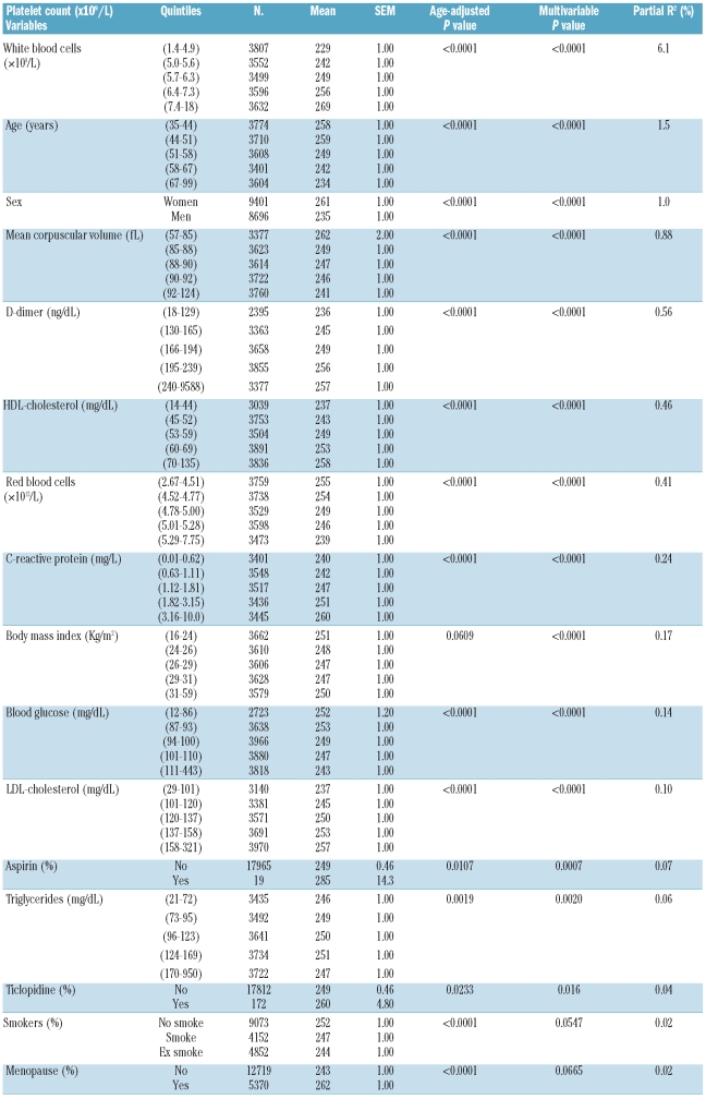
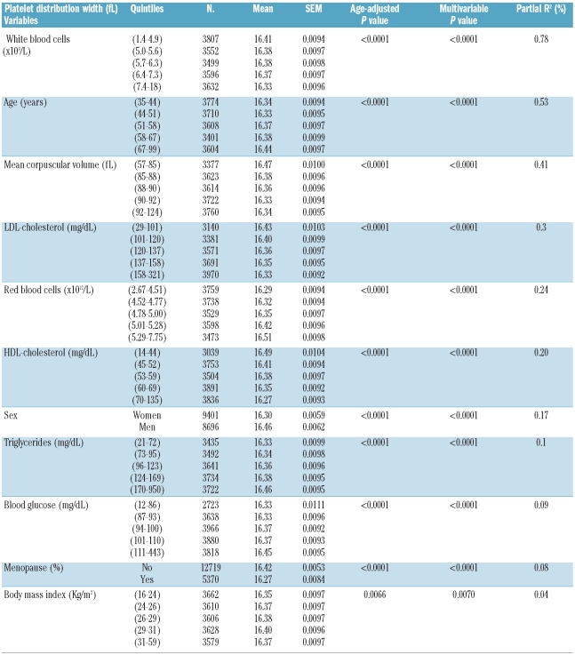
Numerous variables were associated with platelet indices in the simple linear regression analysis (data not shown). We only report here the variables that remained associated with platelet indices in the analyses adjusted for age (Tables 1–3, Online Supplementary Table S2). The full model explained 16%, 21%, 1.9% and 4.7% of the variability of platelet count, plateletcrit, mean platelet volume and platelet distribution width, in the whole sample. The determinants that explained most of the variability of all four platelet indices were white blood cell count, age and sex (except for platelet distribution width).
Table 1.
Variables associated with platelet count in the Moli-sani population.
Table 3.
Variables associated with platelet distribution width in the MOLI-SANI population.
Platelet indices and white blood cell count
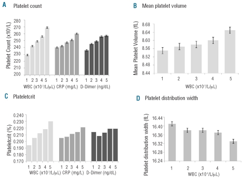
Platelet count, plateletcrit and platelet distribution width were associated with white blood cell count, which explained 6.1%, 9.1% and 0.78% of their variability, respectively. Platelet count, mean platelet volume and plateletcrit were positively, and platelet distribution width was negatively associated with white blood cell count (Figure 1A–D, Tables 1–3, Online Supplementary Table S2). Mean platelet volume was also directly associated with white blood cell count, which explained 0.26% of the 1.9% of its variability (Table 2). Platelet count and platelet-crit were significantly associated with C-reactive protein and D-dimer levels in a way similar to that with the white blood cell count (Figure 1A–C, Table 1, Online Supplementary Table S2).
Figure 1.
A-D. Platelet parameters by white blood cell count, C-reactive protein and D-dimer levels. (A) Platelet count. (B) Mean platelet volume. (C) Plateletcrit. (D) Platelet distribution width.
Table 2.
Variables associated with mean platelet volume in the Moli-sani population.
Platelet indices: association with gender and age
Tables 1–3 and Online Supplementary Table S2 report the four platelet indices by gender: women had significantly higher platelet counts, plateletcrit and mean platelet volume than men, 261±64 versus 235±59×109/L (P≤0.0001), 0.22±0.05 versus 0.20±0.04 % (P≤0.0001) and 8.67±0.94 versus 8.50 ±0.92 fL (P≤0.0001), respectively, while platelet distribution width was slightly higher in men (16.3±0.57 versus 16.4±0.58 fL (P≤0.0001), respectively.
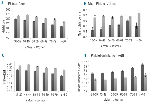
The sex difference was present in all age ranges (Figure 2A–D). Figure 2A shows a progressive decline of platelet number during aging in both men and women and Online Supplementary Figure S2 illustrates the relationship of thrombocytopenia and thrombocytosis frequency with age: the former increased while the latter decreased with age. On average, a 10-year increase in age corresponds to a sex-adjusted decrease of 10×109/L in the platelet count. Like the platelet count, plateletcrit decreased with age in both men and women (Figure 2C). While it was not possible to identify a clear relation between mean platelet volume and age in women, in men it increased with age until 79 years and then decreased (Figure 2B). Finally platelet distribution width increased with age in both men and women (Figure 2D).
Figure 2.
A-D. Platelet parameters by age and sex. (A) Platelet count. (B) Mean platelet volume. (C) Plateletcrit. (D) Platelet distribution width. The difference between men and women holds in any age range. The P values for the differences according to age were P<0.0001 for all platelet parameters in men, whereas in women they were P<0.0001 for platelet count and plateletcrit, P=0.77 for mean platelet volume and P=0.0024 for platelet distribution width.
Platelet indices and other variables
Variables such as body mass index, HDL cholesterol, LDL cholesterol, glucose, triglycerides, smoking habit, systolic or diastolic blood pressure and antiplatelet drug use, although associated in a statistically significant manner, each explained less than 0.5% of the variability in platelet indices (Tables 1–3, Online Supplementary Table S2).
Platelet indices across Molise villages
Average platelet indices were significantly different across Molise villages (Online Supplementary Figure S3). Platelet count ranged from 228×109/L to 269×109/L, plateletcrit from 0.19 fL to 0.23 fL, mean platelet volume from 8.0 fL to 8.9 fL and platelet distribution width from 16.3% to 16.5%. In particular, average platelet count differed by more than 50×109/L between Gildone and Sant’Angelo Limosano. Correspondingly, the prevalence of thrombocytopenia differed from 0.7% to 6.8% between these two villages. This variation could not be explained by differences in either other platelet count determinants, or liver disorders or cancer, the altitude of the villages above sea level or their distance from the recruitment center (data not shown).
Within villages, increasing average platelet volume was associated with a decrease in platelet count, whereas an increase in platelet count was associated with an increase in plateletcrit. Villages whose inhabitants had the highest platelet counts also showed the highest levels of plateletcrit but the lowest levels of mean platelet volume.
Adjustment for age, sex, other platelet count determinants, liver disorders, cancer, altitude of the villages above sea level or their distance from the recruitment center did not modify what whas observed across Molise villages (data not shown).
Discussion
Platelet indices are receiving increasing attention as potential markers of platelet activation, since they are easily measurable in the context of epidemiological studies given the widespread availability of reliable automated blood cell counters. These counters provide platelet count as part of the full blood count, in addition to derived indices related to platelet size, such as plateletcrit (total volume of platelets in a given volume of blood), mean platelet volume (plateletcrit divided by total platelet number) and platelet distribution width, which directly measures the variability in platelet size.
In a large population sample recruited at random from a general adult population we studied the relation of platelet indices with each other and with a number of non-genetic variables. We found a significant inverse correlation between platelet count and mean platelet volume, in agreement with other studies,20–22 or platelet distribution width. This finding could be explained by the possibility that, in order to maintain constant platelet functional mass,23 platelet count would be decreased in the presence of bigger platelets. This could be the case of increased production by the bone marrow of young (reticulated) platelets that are larger than older (exhausted) platelets.24–25
Platelets, hematologic cells and inflammation
By analyzing a large number of variables, we observed, apparently for the first time, that both platelet count and plateletcrit were strongly associated with white blood cell count, explaining 6.2 out of 16% and 9.0 out of 21% of their total variability, respectively. Although our model explained a smaller part of mean platelet volume and platelet distribution width heterogeneity, white blood cell count was again a major associated variable.
These findings support the hypothesis of a common regulation of blood cells, as recently suggested by the presence of a common genetic component.9 The link between platelets and inflammation, suggested by previous studies26–28 is also supported by our present findings. Indeed platelet indices are not only associated with white blood cell count, but are also significantly and independently related to a soluble inflammatory marker such as C-reactive protein.
Platelets are considered as inflammatory cells since their count increases in many inflammatory states and platelet count has been associated with the level of other acute-phase proteins.27 During inflammation higher thrombopoietin levels have also been observed,28 which could, at least in part, represent the link to the increase in platelet production. In turn, platelets are a source of inflammatory mediators and can be activated by several inflammatory triggers during the process of atherothrombosis.26,29 The positive significant association between platelet count and plateletcrit with D-dimer levels further underlies the link between inflammation, platelets and activation of blood coagulation.30
Sex and age
Women had significantly higher platelet indices than men (except for platelet distribution width, which was slightly higher in men), suggesting a hormonal influence in their regulation. The process by which megakaryocytes proceed to proplatelet formation and platelet production is reportedly under the influence of autocrine estrogen.31 Additionally, estrogen-receptor antagonists inhibit platelet production in vivo, supporting a role of estrogens in platelet production.12 In agreement with a previous study on platelet count,11 we found that the differences between men and women persisted for all platelet indices at any age range, as well as after the menopause. Contraceptives and hormone therapy, used by only a relatively small proportion of women in our sample, did not significantly influence any platelet parameter.
Although the higher platelet count in women might be the result of greater inflammation, even in the setting of a lower white cell count, differences between men and women were not attenuated after adjustment for inflammatory variables.
All platelet indices varied with age. In particular platelet count and plateletcrit decreased with age in both men and women. A similar trend was observed for the prevalence of thrombocytosis, which progressively decreased with age, and the prevalence of thrombocytopenia, which, in contrast, increased, up to the level of 9% in subjects over 80 years old. In a recent publication, Biino et al.13 provided for the first time an estimate of the prevalences of thrombocytosis and thrombocytopenia in the general population of the Ogliastra region, in Sardinia, clearly showing that the prevalence of mild thrombocytopenia was higher in older people. Although differences in platelet count with age and sex have been previously reported,10–13 normal laboratory ranges of platelet count are not usually distinguished for sex and age. This might possibly lead to some overestimation of platelet count defects in the elderly. The decline in platelet counts we observed in older age is difficult to interpret because of the cross-sectional nature of our data: it may reflect a reduction in hematopoietic stem-cell reserve during aging or a survival advantage in those subjects with lower platelet counts.
Platelet distribution width increased with age in both men and women, while the relation between mean platelet volume and age differed by sex: it increased with age until 79 years and then decreased in women over 80 years, while it remained constant over age in men. Previous studies yielded contrasting results on the association between mean platelet volume and age, but a sex-specific analysis was not performed in any of them.32–34
Other variables
Many other biological and clinical variables were associated with platelet indices, such as body mass index, HDL cholesterol, LDL cholesterol, triglycerides, glucose, diastolic blood pressure and antiplatelet drug use; however, although statistically significant, each explained less than 0.5% of the variability of platelet indices. As the association of these variables was mainly directed towards a risk profile for cardiovascular disease, such an association might become stronger in pathological conditions, such as diabetes, obesity and hypertension.35–37
Environmental variables, including dietary habits, did not show any relevant association with platelet indices.
Molise villages
Since a recent Italian study13 found significant variations in both platelet number and prevalence of thrombocytopenia among some Sardinian villages, we investigated the distribution of platelet indices across our villages, finding that they were significantly different across the villages. This variation could not be explained by differences in other platelet count determinants, in liver disorders or cancer distribution or in the altitude of the villages above the sea or in their distance from the recruitment center.
That platelet count might differ by ethnicity has already been reported;10–12,38 our findings, together with Biino’s data,13 from a large Caucasian population, indicate that there is a micro-heterogeneity in platelet parameters even among apparently ethnically homogeneous subjects living in the same country and even in the same region.
Conclusions
As our data show that a large number of non-genetic variables explain a small proportion of the heterogeneity in platelet indices, it appears that genetic factors might play the major role. That both platelet count and platelet volume are highly inherited traits is already known.5–8 We have also recently found in large pedigrees derived from the same population studied here that the genetic component explained 69.0%, 52.1% and 34.0% of mean platelet volume, platelet count and platelet distribution width, respectively, with a small contribution being made by shared household (<10%), while the environmental component was minimal (data not shown).8
A recent meta-analysis9 identified 12 loci linked to mean platelet volume or platelet count. These single nucleotide polymorphisms did, however, only explain 8.6% of mean platelet volume variance upon 75% of heritability, suggesting that other genetic or epigenetic factors could be involved. The availability of DNA samples stored in the large Moli-sani biological research bank will help better understanding of the genetic regulation of platelets in the near future.
Acknowledgments
The Authors thank Professor Jozef Vermylen, Catholic University, Leuven, Belgium for his critical review of the manuscript and Associazione Cuore-Sano (Campobasso, Italy), Instrumentation Laboratory (IL, Milano, Italy), DerbyBlue (San Lazzaro di Savena, Bologna, Italy), Caffè Monforte (Campobasso, Italy) for their generous support to the Moli-sani Project. Part of the data included in this paper were first presented at the 10th Meeting of the Gruppo di Studio delle Piastrine, Termoli, Italy, October 2009.
Footnotes
Funding: the Moli-sani Project was supported by research grants from Pfizer Foundation (Rome, Italy) and the Italian Ministry of University and Research (MIUR, Rome, Italy)–Programma Triennale di Ricerca, Decreto no.1588.
The online version of this article has a Supplementary Appendix.
Authorship and Disclosures
The information provided by the authors about contributions from persons listed as authors and in acknowledgments is available with the full text of this paper at www.haematologica.org.
Financial and other disclosures provided by the authors using the ICMJE (www.icmje.org) Uniform Format for Disclosure of Competing Interests are also available at www.haematologica.org.
References
- 1.de Gaetano G. Historical overview of the role of platelets in hemostasis and thrombosis. Haematologica. 2001;86(4):349–56. [PubMed] [Google Scholar]
- 2.van der Bom JG, Heckbert SR, Lumley T, Holmes CE, Cushman M, Folsom AR, et al. Platelet count and the risk for thrombosis and death in the elderly. J Thromb Hemost. 2009;7(3):399–405. doi: 10.1111/j.1538-7836.2008.03267.x. [DOI] [PMC free article] [PubMed] [Google Scholar]
- 3.Thaulow E, Erikssen J, Sandvik L, Stormorken H, Cohn PF. Blood platelet count and function are related to total and cardiovascular death in apparently healthy men. Circulation. 1991;84(2):613–7. doi: 10.1161/01.cir.84.2.613. [DOI] [PubMed] [Google Scholar]
- 4.Chu SG, Becker RC, Berger PB, Bhatt DL, Eikelboom JW, Konkle B, et al. Mean platelet volume as a predictor of cardiovascular risk: a systematic review and meta-analysis. J Thromb Haemost. 2009;8(1):148–56. doi: 10.1111/j.1538-7836.2009.03584.x. [DOI] [PMC free article] [PubMed] [Google Scholar]
- 5.Kunicki TJ, Nugent DJ. The genetics of normal platelet reactivity. Blood. 2010;116(15):2627–34. doi: 10.1182/blood-2010-04-262048. [DOI] [PMC free article] [PubMed] [Google Scholar]
- 6.Garner C, Tatu T, Reittie JE, Littlewood T, Darley J, Cervino S, et al. Genetic influences on F cells and other hematologic variables: a twin heritability study. Blood. 2000;95(1):342–6. [PubMed] [Google Scholar]
- 7.Traglia M, Sala C, Masciullo C, Cverhova V, Lori F, Pistis G, et al. Heritability and demographic analyses in the large isolated population of Val Borbera suggest advantages in mapping complex traits genes. PLoS One. 2009;4(10):e7554. doi: 10.1371/journal.pone.0007554. [DOI] [PMC free article] [PubMed] [Google Scholar]
- 8.Vohnout B, Gianfagna F, Lorenzet R, Cerletti C, de Gaetano G, Donati MB, et al. Genetic regulation of inflammation-mediated activation of haemostasis: family-based approaches in population studies. Nutr Metab Cardiovasc Dis. 2010 Aug 5; doi: 10.1016/j.numecd.2010.03.002. [Epub ahead of print] [DOI] [PubMed] [Google Scholar]
- 9.Soranzo N, Spector TD, Mangino M, Kühnel B, Rendon A, Teumer A, et al. A genome-wide meta-analysis identifies 22 loci associated with eight hematological parameters in the HaemGen consortium. Nat Genet. 2009;41(11):1182–90. doi: 10.1038/ng.467. [DOI] [PMC free article] [PubMed] [Google Scholar]
- 10.Saxena S, Cramer AD, Weiner JM, Carmel R. Platelet in three racial groups. Am J Clin Pathol. 1987;88(1):106–9. doi: 10.1093/ajcp/88.1.106. [DOI] [PubMed] [Google Scholar]
- 11.Bain BJ. Ethnic and sex differences in the total and differential white cell count and platelet count. J Clin Pathol. 1996;49(8):664–6. doi: 10.1136/jcp.49.8.664. [DOI] [PMC free article] [PubMed] [Google Scholar]
- 12.Segal JB, Moliterno AR. Platelet counts differ by sex, ethnicity, and age in the United States. Ann Epidemiol. 2006;16(2):123–30. doi: 10.1016/j.annepidem.2005.06.052. [DOI] [PubMed] [Google Scholar]
- 13.Biino G, Balduini CL, Casula L, Cavallo P, Vaccargiu S, Parracciani D, et al. Analysis of 12, 517 inhabitants of Sardinian geographic isolate reveals that propensity to develop mild thrombocytopenia during ageing and to present mild, transient thrombocytosis in youth are new genetic traits. Haematologica. 2011;96(1):96–101. doi: 10.3324/haematol.2010.029934. [DOI] [PMC free article] [PubMed] [Google Scholar]
- 14.Iacoviello L, Bonanni A, Costanzo S, De Curtis A, Di Castelnuovo A, Olivieri M, et al. The Moli-Sani Project, a randomized, prospective cohort study in the Molise region in Italy; design, rationale and objectives. Ital J Public Health. 2007;4(2):110–8. [Google Scholar]
- 15.Centritto F, Iacoviello L, di Giuseppe R, De Curtis A, Costanzo S, Zito F, et al. Dietary patterns, cardiovascular risk factors and C-reactive protein in a healthy Italian population. Nutr Metab Cardiovasc Dis. 2009;19(10):697–706. doi: 10.1016/j.numecd.2008.11.009. [DOI] [PubMed] [Google Scholar]
- 16.Pala V, Sieri S, Palli D, Salvini S, Berrino F, Bellegotti M, et al. Diet in the Italian EPIC cohorts: presentation of data and methodological issues. Tumori. 2003;89(6):594–607. doi: 10.1177/030089160308900603. [DOI] [PubMed] [Google Scholar]
- 17.Nuorti JP, Butler JC, Farley MM, Harrison LH, McGeer A, Kolczak MS, et al. Cigarette smoking and invasive pneumococcal disease. Active Bacterial Core Surveillance Team. N Engl J Med. 2000;342(10):681–9. doi: 10.1056/NEJM200003093421002. [DOI] [PubMed] [Google Scholar]
- 18.Ainsworth BE, Haskell WL, Whitt MC, Irwin ML, Swartz AM, Strath SJ, et al. Compendium of physical activities: an update of activity codes and MET intensities. Med Sci Sports Exerc. 2000;32(9 Suppl):S498–504. doi: 10.1097/00005768-200009001-00009. [DOI] [PubMed] [Google Scholar]
- 19.Third Report of the National Cholesterol Education Program (NCEP) expert panel on detection, evaluation, treatment of high blood cholesterol in adults (Adult Treatment Panel III). Final report. Circulation. 2002;106(25):3143–421. [PubMed] [Google Scholar]
- 20.Huczek Z, Kochman J, Filipiak KJ, Horszczaruk GJ, Grabowski M, Piatkowski R, et al. Mean platelet volume on admission predicts impaired reperfusion and long-term mortality in acute myocardial infarction treated with primary percutaneous coronary intervention. J Am Coll Cardiol. 2005;46(2):284–90. doi: 10.1016/j.jacc.2005.03.065. [DOI] [PubMed] [Google Scholar]
- 21.Hendra TJ, Oswald GA, Yudkin JS. Increased mean platelet volume after acute myocardial infarction relates to diabetes and to cardiac failure. Diabetes Res Clin Pract. 1988;5(1):63–9. doi: 10.1016/s0168-8227(88)80080-9. [DOI] [PubMed] [Google Scholar]
- 22.Yang A, Pizzulli L, Luderitz B. Mean platelet volume as marker of restenosis after percutaneous transluminar coronary angioplasty in patients with stable and unstable angina pectoris. Thromb Res. 2006;117(4):371–7. doi: 10.1016/j.thromres.2005.04.004. [DOI] [PubMed] [Google Scholar]
- 23.Thompson CB, Jakubowski JA. The patho-physiology and clinical relevance of platelet heterogeneity. Blood. 1988;72(1):1–8. [PubMed] [Google Scholar]
- 24.Smith NM, Pathansali R, Bath PM. Altered megakaryocyte-platelet-haemostatic axis in patients with acute stroke. Platelets. 2002;13(2):113–20. doi: 10.1080/09537100120111559. [DOI] [PubMed] [Google Scholar]
- 25.De Luca G, Venegoni L, Iorio S, Secco GG, Cassetti E, Verdoia M, et al. Platelet distribution width and the extent of coronary artery disease: Results from a large prospective study. Platelets. 2010;21(7):508–14. doi: 10.3109/09537104.2010.494743. [DOI] [PubMed] [Google Scholar]
- 26.de Gaetano G, Cerletti C, Nanni-Costa MP, Poggi A. The blood platelet as an inflammatory cell. Eur Respir J Suppl. 1989;6(6):441s–445s. [PubMed] [Google Scholar]
- 27.Evangelista V, Celardo A, Dell’Elba G, Manarini S, Mironov A, de Gaetano G, et al. Platelet contribution to leukotriene production in inflammation: in vivo evidence in the rabbit. Thromb Haemost. 1999;81(3):442–8. [PubMed] [Google Scholar]
- 28.Ceresa IF, Noris P, Ambaglio C, Pecci A, Balduini CL. Thrombopoietin is not uniquely responsible for thrombocytosis in inflammatory disorders. Platelets. 2007;18(8):579–82. doi: 10.1080/09537100701593601. [DOI] [PubMed] [Google Scholar]
- 29.de Gaetano G, Donati MB, Cerletti C. Prevention of thrombosis and vascular inflammation: benefits and limitations of selective or combined COX-1, COX-2 and 5-LOX inhibitors. Trends Pharmacol Sci. 2003;24(5):245–52. doi: 10.1016/S0165-6147(03)00077-4. [DOI] [PubMed] [Google Scholar]
- 30.Napoleone E, Di Santo A, Bastone A, Peri G, Mantovani A, de Gaetano G, et al. Long pentraxin PTX3 upregulates tissue factor expression in human endothelial cells: a novel link between vascular inflammation and clotting activation. Arterioscler Thromb Vasc Biol. 2002;22(5):782–7. doi: 10.1161/01.atv.0000012282.39306.64. [DOI] [PubMed] [Google Scholar]
- 31.Nagata Y, Yoshikawa J, Hashimoto A, Yamamoto M, Payne AH, Todokoro K. Proplatelet formation of megakaryocytes is triggered by autocrine estradiol. Genes Dev. 2003;17(23):2864–9. doi: 10.1101/gad.1128003. [DOI] [PMC free article] [PubMed] [Google Scholar]
- 32.Huczek Z, Kochman J, Filipiak KJ, Horszczaruk GJ, Grabowski M, Piatkowski R, et al. Mean platelet volume on admission predicts impaired reperfusion and long-term mortality in acute myocardial infarction treated with primary percutaneous coronary intervention. J Am Coll Cardiol. 2005;46(2):284–90. doi: 10.1016/j.jacc.2005.03.065. [DOI] [PubMed] [Google Scholar]
- 33.Braekkan SK, Mathiesen EB, Njølstad I, Wilsgaard T, Størmer J, Hansen JB. Mean platelet volume is a risk factor for venous thromboembolism: the Tromsø study. 2010;8(1):157–62. doi: 10.1111/j.1538-7836.2009.03498.x. [DOI] [PubMed] [Google Scholar]
- 34.Berger JS, Eraso LH, Xie D, Sha D, Mohler ER., III Mean platelet volume and prevalence of peripheral artery disease, the National Health and Nutrition Examination Survey 1999–2004. Atherosclerosis. 2010;213(2):586–91. doi: 10.1016/j.atherosclerosis.2010.09.010. [DOI] [PMC free article] [PubMed] [Google Scholar]
- 35.Samocha Bonet D, Justo D, Rogowski O, Saar N, Abu-Abeid S, Shenkerman G, et al. Platelet counts and platelet activation markers in obese subjects. Mediators Inflamm. 2008;2008:834153. doi: 10.1155/2008/834153. [DOI] [PMC free article] [PubMed] [Google Scholar]
- 36.Tavil Y, Sen N, Yazici H, Turfan M, Hizal F, Cengel A, et al. Coronary heart disease is associated with mean platelet volume in type 2 diabetic patients. Platelets. 2010;21(5):368–72. doi: 10.3109/09537101003628421. [DOI] [PubMed] [Google Scholar]
- 37.Yazici M, Kaya A, Kaya Y, Albayrak S, Cinemre H, Ozhan H. Lifestyle modification decreases the mean platelet volume in prehypertensive patients. Platelets. 2009;20(1):58–63. doi: 10.1080/09537100802613449. [DOI] [PubMed] [Google Scholar]
- 38.Lugada ES, Mermin J, Kaharuza F, Ulvestad E, Were W, Langeland N, et al. Population-based hematologic and immunologic reference values for a healthy Uganda population. Clin Diagn Lab Immunol. 2004;11(1):29–34. doi: 10.1128/CDLI.11.1.29-34.2004. [DOI] [PMC free article] [PubMed] [Google Scholar]


