Abstract
Tumours of neurogenic origin are rare in parotid gland. The authors are presenting here a case of neurofibroma in a 40-year-male who presented with slow growing tumour in preauricular region of 1 year duration.
Background
Neurofibromas of salivary gland are very rare and constitute only 0.4% of all salivary neoplasms.1
Case presentation
A 40-year-male presented with a slow growing painless swelling in right side of face. On examination, a right sided preauricular swelling measuring 6×4 cm, freely mobile, non-tender and overlying skin was normal. No other swelling, skin pigmentation or axillary `swellings were present on the body.
Investigations
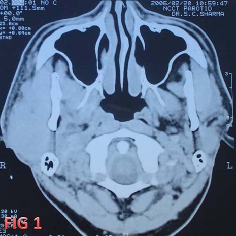
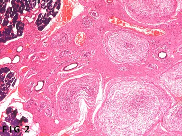
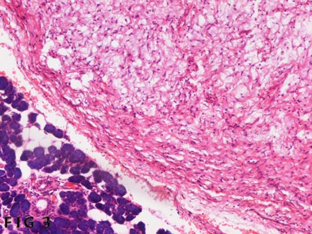
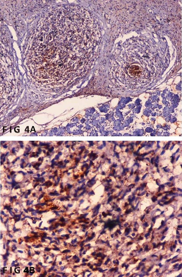
Routine blood investigations were unremarkable. CT scan showed a mass with well defined margins arising from right parotid gland without involvement of bone (figure 1). Fine needle aspiration cytology was inconclusive. Surgery was done and tumour was removed. Gross examination showed a greyish white mass measuring 5×4 cm. Cut section was solid and homogenous. Microscopic examination revealed the tumour consists of cells with ill-defined margins and oval to spindle shape nuclei of uniform size and shape in a loose collagenous stroma along with serous salivary gland and duct (figures 2 and 3). S-100 immunostain showed cytoplasmic positivity (figure 4). On the basis of these findings a diagnosis of neurofibroma of parotid gland was rendered.
Figure 1.

CT scan showing mass arising from right parotid.
Figure 2.

Section showing tumour consisting of spindle shape in loose collagenous stroma and serous salivary gland.
Figure 3.

Section showing tumour consisting of spindle shape in loose collagenous stroma and serous salivary gland along with duct.
Figure 4.

(A,B)Section showing S-100 positivity.
Outcome and follow-up
After surgery the patient recovered well and discharged in good conditions.
Discussion
Majority of tumours of parotid gland are benign and the most common tumour of parotid gland is pleomorphic adenoma.2–4 Tumours of nerve tissue origin are extremely rare. Neurofibroma and schwannoma are common tumour which arises from nerve tissue.5
Neurofibromas are tumours that originate from nerve tissue. They may be solitary or multiple, sporadic or associated with neurofibromatosis I or II syndromes. Neurofibroma can arise from extratemporal part of facial nerve which traverses in between superficial and deep lobe of parotid. It is a slow growing tumour which is usually asymptomatic. Unlike schwannoma neurofibroma is intimately attached to nerve of origin. Histopatholopgical examination shows proliferation of all elements of nerve which include axons, Schwann cells and fibroblasts. Treatment is surgical removal of tumour.
Learning points.
-
▶
Rarely neurofibroma can be seen in parotid gland.
-
▶
Preoperative diagnosis is rare.
Footnotes
Competing interests None.
Patient consent Obtained.
References
- 1.Seifert G, Miehlke A, Haubrich J, et al. Pathology, Diagnosis, Treatment, Facial Nerve Surgery. Stuttgart, Germany: Georg Thieme Verlag; 1986:171–301 [Google Scholar]
- 2.Ellis GL, Auclair PL. Tumors of the salivary glands. In: Ellis GL, Auclair PL, eds. Atlas of Tumor Pathology. Third series, fascicle 17 Washington, DC: Armed Forces Institute of Pathology; 1996:318–24 [Google Scholar]
- 3.Speight PM, Barrett AW. Salivary gland tumours. Oral Dis 2002;8:229–40 [DOI] [PubMed] [Google Scholar]
- 4.Ledesma-Montes C, Garces-Ortiz M. Salivary gland tumours in a Mexican sample. A retrospective study. Med Oral 2002;7:324–30 [PubMed] [Google Scholar]
- 5.McGuirt WF, Sr, Johnson PE, McGuirt WT. Intraparotid facial nerve neurofibromas. Laryngoscope 2003;113:82–4 [DOI] [PubMed] [Google Scholar]


