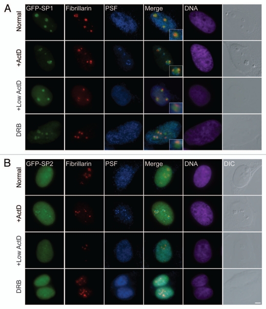
Figure 3.
NOL7-SP1 is found in nucleolar caps during nucleolar segregation. (A) GFP-NOL7-SP1 (green), endogenous fibrillarin (red) and PSF (blue) show different nuclear localization. During nucleolar segregation by actinomycin D treatment (high 5 µg/ml or low 0.05 µg/ml), NOL7-SP1 is found in nucleolar caps containing fibrillarin, while some of the protein remains in the central body. PSF is localized to a second and larger subset of nucleolar caps (dark nucleolar caps). Inhibition of RNA Pol II only by DRB shows segregation of NOL7 and fibrillarin signals. (B) GFP-NOL7-SP2 is mostly nucleoplasmic with some nucleolar fraction. Upon nucleolar segregation (low and high ActD or DRB treatment) some of the protein is found in fibrillarin containing nucleolar caps. DNA is pseudo-colored in purple, DIC is grey. Insets show enlarged nucleolar areas. Bar, 5 µm.

