Abstract
Functional MRI is used to study the effects of acupuncture on the BOLD response and the functional connectivity of the human brain. Results demonstrate that acupuncture mobilizes a limbic-paralimbic-neocortical network and its anti-correlated sensorimotor/paralimbic network at multiple levels of the brain and that the hemodynamic response is influenced by the psychophysical response. Physiological monitoring may be performed to explore the peripheral response of the autonomic nerve function. This video describes the studies performed at LI4 (hegu), ST36 (zusanli) and LV3 (taichong), classical acupoints that are commonly used for modulatory and pain-reducing actions. Some issues that require attention in the applications of fMRI to acupuncture investigation are noted.
Protocol
Part 1: Informed consent and subject preparation
Healthy adult volunteers are recruited for the study according to Institutional Review Board approved procedures. Informed consent by the subject is signed before beginning the study.
Part 2: Setting up physiological monitoring equipment
The AD Instruments PowerLab system allows for physiological data to be synchronized with the fMRI scan by using the fMRI trigger signal recorded on an input channel. This is a laptop based system with inputs for ECG, respiration, skin conductance response, pulse oximetry, and any other physiological signal that would need to be recorded.
If a respiratory belt system is being used, the transducer must be plugged into a power outlet in the room adjacent to the scanner, with the tube leading to the belt fed via inlet into the scan room and a BNC connector leading to the PowerLab in the scan control room.
If a trigger is being used to synchronize the physiological data to the fMRI data, then a BNC cable must also connect the MRI scanner trigger signal to the PowerLab in the scan control room.
Gather monitoring devices. Four ECG electrodes will be needed, as well as an ECG cable connected to an MR-compatible In Vivo system rollcart.
If skin conductance is being measured, plug the skin conductance connectors into the breakout box in the scan room, and have skin conductance gel ready in the scan room.
Part 3: Preparing subject for MR scanning
It is important to rule out undue anxiety and fatigue on the day of the scan because basic state can affect the response of the brain to acupuncture stimulation. Suggest the subject eat a light meal prior to the procedure so that weakness and untoward reactions can be avoided.
Instruct the subject to lie supine in the machine wearing ear plugs to suppress scanner noise and with the head immobilized by cushioned supports. Since the signal/noise ratio of acupuncture stimulation scans is low compared with common motor or visual tasks, the data tends to be more easily corrupted by head motion. A support is placed under the knees to keep the heels form the surface of the stretcher to reduce body motion. The head coil is fastened and cushions are adjusted for comfort. For an average scanning session of 2 hours, comfort is vital for uninterrupted scanning.
If physiological monitoring is desired, subject should be prepared according to the previous section before being put into the scanner. It is desirable to monitor the wakefulness of the subject with eye-tracking during the scans if available.
Attach 4 ECG electrodes to the subject's left upper chest (for female subjects, take privacy precautions such as lowering the blinds on the window to the bay in the scan control room and shutting the scanner door). Attach cable to the electrodes and to the In Vivo device. For safety this cable and all other cables leading to the subject should run directly along the scan table, without forming any loops. Turn on the In Vivo device and check for ECG signal quality. Adjust electrode positioning if necessary.
Fill the wells of the SCR electrodes with conductive gel. This special SCR gel should be isotonic to the skin. Perform open circuit zero in Chart software. Attach SCR electrodes to 2 adjacent distal finger pads of the subject's hand by wrapping the velcro around as tightly as is comfortable.
Part 4: Functional MR imaging sequences tailored for imaging acupuncture stimulation.
We use a 1.5 or 3.0 Tesla Siemens Trio MRI system (Siemens, Erlangen, Germany) equipped for echo planar imaging with a standard quadratic head coil.
Our studies over the years consistently demonstrated that acupuncture had extensive action over functional systems at multiple levels of the brain. The structures located at the base of the brain such as the amygdala, hippocampus, ventral medial prefrontal cortex and subgenual areas are particularly susceptible to susceptibility artifacts. It is important to check each EPI functional scan to rule out signal loss in these regions as we found differences in loss of signals between two models of a Siemens scanner with similar field strength and with a similar MRI sequence for data collection.
Thinner brain slices are preferable for reducing the susceptibility artifacts. Slices 3 mm thick with 0.6 mm or 0.75 mm gap in the sagittal or axial orientation are used to cover the entire brain including the brainstem and cerebellum depending on the memory capacity of the imaging system. Other orientations and slice thickness can be used for partial brain imaging. The special attention we pay to these issues contribute to the more consistent findings of acupuncture effects on the structures at the base of the brain.
The functional scans are acquired with a T2*-weighted gradient echo sequence (TR/TE 4000/30 ms, FOV 200 mm, matrix 64x64, flip angle 90°, slice thickness 3.0 mm, gap 0.6 mm). We acquired a total of 150 time points in 10 min. Image collection is preceded by 4 dummy scans to allow for equilibration of the fMRI signal. The shorter TE reduces susceptibility artifacts. The relatively long TR permits whole brain coverage with high spatial resolution.
Part 5: The initial localizer and anatomic scans
Before functional scans with acupuncture are run, a localizer scan, a high resolution anatomical scan, and the T1 weighted 3D MP-RAGE are acquired to assure correct positioning and anatomical soundness. General observation at this point can assure that unnecessary scans are not run. The localizer, a very short scan, assures that the whole brain is included in the scan and allows you to line it up with the AC-PC line. If there are any suspect anomalies, researchers will send images to a trained radiologist but should not make any claims about what is on the scan to subjects.
The initial scans take about 20 minutes total. Brief viewing can tell the investigator if it will be useful to continue scanning. This is a good time to speak with the subject and make sure that they are still comfortable and aren't experiencing any adverse effects from being in the Magnet.
Part 6: Acupuncture with fMRI monitoring
Of the two forms of treatment (manual needle manipulation and electric stimulation), this video will be limited to manual acupuncture, which has a much longer history of clinical use. Acupuncture stimulation is administered in duplicate runs to classical acupoints LI4 on the hand, LV3 on the foot or ST36 on the leg, employing sterile, one-time use only acupuncture needles.
Instruct subjects to relax, keep their eyes closed and refrain from motions during the scanning. To avoid noxious stimulation, we specifically instruct the subject to raise one finger if any of the acupuncture sensations approaches a score of 8, and raise two fingers in case of sharp pain of any degree.
After inserting the needle, pretest the subject's sensitivity and tolerance to needle manipulation to help choose the depth of insertion and the force of manipulation for data collection.
During the 10 min functional imaging, the needle is rotated for 2 min during M1 and M2, and left in place during the rest periods R1 (2min) ), R2 (3min), R3 (1 min). The acupuncturist makes sure not to approach or leave the subject directly before or after the stimulation period, as even that motion could affect the results. (Paradigm, Fig 1)
 Figure 1. Acupuncture fMRI Paradigm.
Upon the completion of acupuncture stimulation, the acupuncturist records the force with which the needle resists manipulation. At the end of each scan, another staff investigator asks the subject to grade the sensations experienced on a scale of 1-10. The list, aching, soreness, pressure, fullness, heaviness, numbness, tingling, dull pain, sharp pain, is based on the experience of the acupuncturist community and previous experimental data rather than pain questionnaires in the literature. The psychophysical response is categorized as a typical deqi group and another group of deqi mixed with sharp pain based on these records.
Speak to the subjects between scans to make sure they feel comfortable and they stay awake. If any part of their body begins to cramp or feel uncomfortable, the research staff on site should take appropriate measures to relieve the discomfort. Movements generated by the subject would cause data corruption. Occasional subjects may fall asleep during scanning, which would affect the hemodynamic response.
Part 7: Data Processing
Multiple levels of analysis can be run. A general linear model uses rest periods as a baseline to determine the % BOLD MR signal change at every voxel of the brain during the task. It also determines the statistical significance of these changes and removes BOLD data that do not reach 3 contiguous voxels at a specific p-value.
Perform seed-based cross correlation analysis to show the functional connectivity between one neurological region and the rest of the brain. This is done by selecting a reference regions and comparing its time-course with that of every other voxel in the brain. Other analyses such as independent component analysis, fuzzy cluster analysis, structural equation modeling, Granger Causality can be applied.
Physiological data can be analyzed by averaging response within windows related to acupuncture needle stimulation. For instance, ECG data can be annotated for QRS complex timing followed by analysis of heart rate and heart rate variability.
Part 8: Results
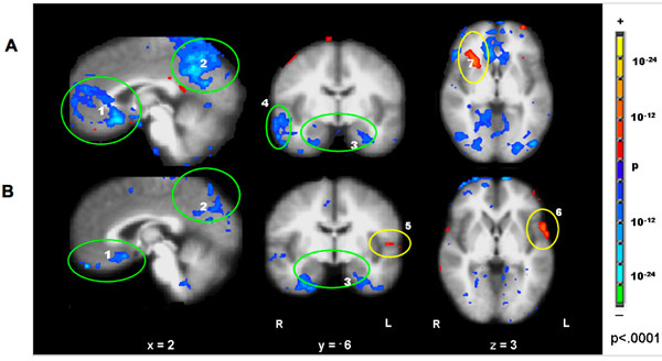
 Figure 2. Hemodynamic response in acupuncture deqi vs. tactile stimulation at right LI4, ST36 and LV3 (52 runs/37 subjects). The clusters of deactivated structures at the MPFC (medial prefrontal cortex) (1), MPC (medial parietal cortex) (2), MTL (medial temporal lobe) (3), were more prominent in acupuncture (Fig A) than in tactile stimulation (Fig B). Extensive deactivation of the right BA22/21 (4) in acupuncture was not observed in tactile stimulation. The sensorimotor BA43 (5) and association cortex (6) showed more activation in tactile stimulation than in acupuncture (not shown). Interestingly, the right anterior insula,(7) was activated in acupuncture and not in tactile stimulation. (p<0.0001).
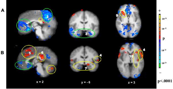
 Figure 3. Hemodynamic response in deqi (52 runs/37 subjects) versus deqi mixed with sharp pain (52 runs/29 subjects) during acupuncture stimulation at right LI4, ST36 and LV3, The prominent deactivation of the MPFC (1), MPC (2) and MTL (3) seen with deqi absent pain (Fig A) was attenuated in the presence of pain (Fig B). With pain, activation of the sensorimotor and association cortices (4) became more prominent, and a subset of the limbic and paralimbic regions such as the midC/SMA (5), postC_BA23 (6), Amy (7), and cerebellar vermis (8) became activated. The right anterior insula (9) was markedly activated in deqi while smaller areas of anterior and posterior insula (10) were activated bilaterally in pain. P<0.0001.
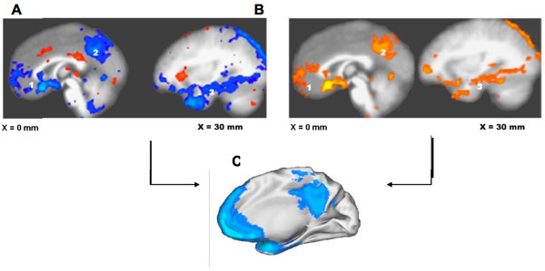
 Figure 4. Overlap of limbic-paralimbic-neocortical network (LPNN) in acupuncture with the default mode network (DMN); The BOLD response of the LPNN by GLM analysis (Fig.A) and by model-free Fuzzy Cluster analysis (Fig B) during acupuncture stimulation at LI4, ST36 and LV3 show virtually identical patterns with the DMN (Fig.C, adapted from Shulman et al, 1997). Clusters of regions with coherent signal decreases appear in the medial prefrontal cortex (1), postero-medial cortex (2) and temporal lobe (3). p<0.0001.
Discussion
The employment of proper techniques, the experience of the acupuncturist and a large sample size to permit separation from confounding effects of inadvertent noxious stimulation are important in the acquisition and interpretation of acupuncture fMRI data. Our studies of several classical acupoints indicate that acupuncture modulates the limbic network, an important intrinsic regulatory system of the human brain. The network shows significant overlap with the default mode of the resting brain. These associated autonomic nervous function changes can be demonstrated by physiological monitoring.
Acknowledgments
The work was supported by the NIH/National Center for Complementary and Alternative Medicine (R21AT00978) (1-P01-002048-01) (K01-AT-002166-01), the National Center for Research Resources (P41RR14075), the Mental Illness and Neuroscience Discovery Institute (MIND) and the Brain Project Grant (NS 34189).
References
- Hui KKS, Liu J, Marina O, Napadow V, Haselgrove C, Kwong KK, Kennedy DN, Makris N. The integrated response of the human cerebro-cerebellar and limbic systems to acupuncture stimulation at ST 36 as evidenced by fMRI. Neuroimage. 2005;27(3):479–496. doi: 10.1016/j.neuroimage.2005.04.037. [DOI] [PubMed] [Google Scholar]
- Hui KKS, Marina O, Claunch JD, Nixon EE, Fang J, Liu J, Li M, Napadow V, Vangel M, Makris N, Chan ST, Kwong KK, Rosen BR. Acupuncture mobilizes the brain's default mode and its anti-correlated network in healthy subjects. Brain Res. 2009;1287:84–103. doi: 10.1016/j.brainres.2009.06.061. [DOI] [PMC free article] [PubMed] [Google Scholar]
- Napadow V, Dhond RP, Purdon P, Kettner N, Makris N, Kwong KK, Hui KK. Correlating acupuncture fMRI in the human brainstem with heart rate variability. Conf Proc IEEE Eng Med Biol Soc. 2005;5:4496–4499. doi: 10.1109/IEMBS.2005.1615466. [DOI] [PubMed] [Google Scholar]
- Shulman GL, Corbetta M, Buckner RL, Raichle ME, Fiez JA, Miezin FM, Petersen SE. Top-down modulation of early sensory cortex. Cereb. Cortex. 1997;7(3):193–206. doi: 10.1093/cercor/7.3.193. [DOI] [PubMed] [Google Scholar]


