ABSTRACT
In the last decade 3D and live 3D ultrasound or the so called 4D (3D/4D) in examination of the fetal heart evolved very rapidly with the development of the new technique called Spatiotemporal Image Corelation – STIC, which enables the aquisition of a volume data concomitent with the beating heart. It appears that 3D/4D ultrasound in fetal echocardiography may make an important contribution to the diagnosis of congenital heart disease, to interdisciplinary management, to parental counseling and to medical personal training.
Keywords: fetal echocardiography, 3D/4D, spatiotemporal image correlation
INTRODUCTION
Three dimensional (3D) and four dimensional (4D) applications in the scanning of fetal heart has largely developed in the last ten years. No other organ or system has this progress so evident that as in the fetal cardiovascular system. Congenital heart disease (CHD) is the most common group of malformations in neonates, occuring in 8 per 1000 live births (1). In 2006 the International Society of Ultrasound in Obstetrics and Gynecology (ISUOG) published practice guidelines for the screening of CHD during the second trimester of pregnancy and outlined two levels for the screening of low risk fetuses for heart anomalies. The first level is the basic 4-chamber view and the second level is the extended basic scan which includes examination of the arterial outflow tracts (2). Also the term "fetal echocardiogram" refers to a detailed sonographic evaluation of the fetal heart which is performed by a specialist in prenatal diagnosis of CHD.
There are several imaging modalities that can be used to evaluate the fetal heart anomalies, from M-mode techniques, to color Doppler and to the use of the new 3D/4D ultrasonography. The new technological development allows a real-time 3D/4D of the examination of the fetal heart (3). These ultrasound techniques can have a major contribution to the understanding of normal and abnormal fetal heart and can also extend the benefits of the prenatal cardiac screening. With the introduction of the so called "virtual planes" to fetal cardiac examination, we are able to obtain views of the fetal heart that are not generally accesible with the use of standard 2D examination (4). ❑
3D/4D TECHNIQUES IN FETAL CARDIAC EXAMINATION
There are three steps that must be followed when we are using 3D/4D fetal echocardiography: volume aquisition, volume display and volume manipulation.
Volume acquisition
The volume acquisition can be done either static 3D, either on-line 4D (direct volume scan), either spatiotemporal image correlation-STIC- (off-line 4D which is an indirect volume scan).
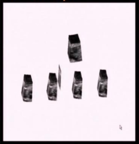
Spatio-temporal image correlation acquisition (STIC) is an automated volume acquisition with the array of the transducer by performing a slow single sweep, recording a single 3D data consisting of many 2D frames one behind the other. The volume of interest (VOI) is set for a period of time between 7.5 sec to 30 sec (usualy 10 sec), and a sweep angle between 20 to 40° (usually 25°). After this, the system processes the volume data and detect systolic peak which are used to calculate fetal heart rate. The systolic peaks define the heart cycle. The resultant 40 consecutive volumes represents a reconstructed complete heart cycle. One can extract from that volume acquisition any convenient plane we want, which means that from a single volume we can get the image of 4-chamber view, or 5-chamber view, or three vessels etc (5). In other words, the acquisition of a volume is realized in 2 steps: in the first step data are aquired by a single automatic volume sweep and in the second step the system analyzes the data in their spatial and temporal domains and processes a 4D sequence (Figure 1). STIC can be used with gray scale, or color Doppler, or power Doppler, or B-flow fetal echocardiography (6).
Figure 1. STIC – Acquisition of fetal heart.
The static 3D represents stored still images as a volume data set and we don’t have neither heart rate and neither motion. This technique is not used for assessing events connected with the heart cycle, or to myocardial wall or valve movement, but can be used to appreciate size and relationship of cardiac structures. The static 3D is used with uniform power Doppler or B-flow.
On-line 4D or direct volume scan, can be obtained only by using a 2D transducer with rapid acquisition of 20-30 volumes or with a matrix 3D transducer.
Volume display
After acquisition we can have the volume display in three different imaging modalities: gray-scale, color Doppler, or power Doppler. Each of these modalities can display 2 possible formats: multiplanar or volume-rendered.
The multiplanar format is the first system that allows simultaneous evaluation of several 2D planes. The rendered format can be displayed in the following applications: gray, color and glassbody (7).
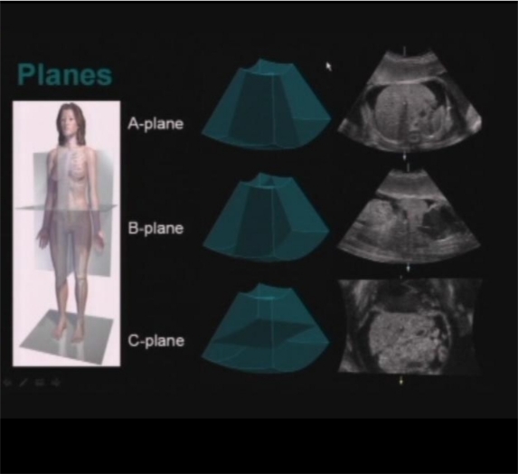
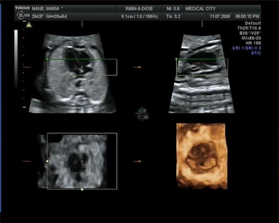
In multiplanar reconstruction mode the screen is divided in four frames: A (upper left), B (upper right), C (lower left) and the fourth (lower right) is the rendered image. Each of the three frames represents one of the three orthogonal planes of the volume. The reference dot represents the intersection of the three planes (Figure 2). Moving the reference dot the operator manipulates the volume to display any plane within the volume. The operator can scroll through the volume and can obtain sequentially each of the five classic five planes of fetal echocardiography (7,8). The multiplanar analysis is reliable in assessing the different cardiac planes, in screening studies and in evaluating different anomalies (9,10). Color and power Doppler can be added to gray scale STIC technology, which can asses the hemodynamic changes during the cycle (11).
Figure 2. Multiplanar reconstruction.
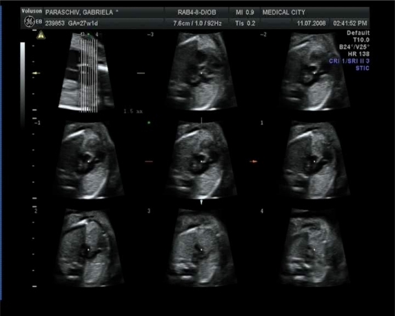
Besides the three plane of multiplanar mode, we can use also the tomographic mode in which we obtain a lot of parallel transverse views from a 3D/4D volume data set (Figure 3).
Figure 3. Tomographic ultrasound imagine.
Volume manipulation
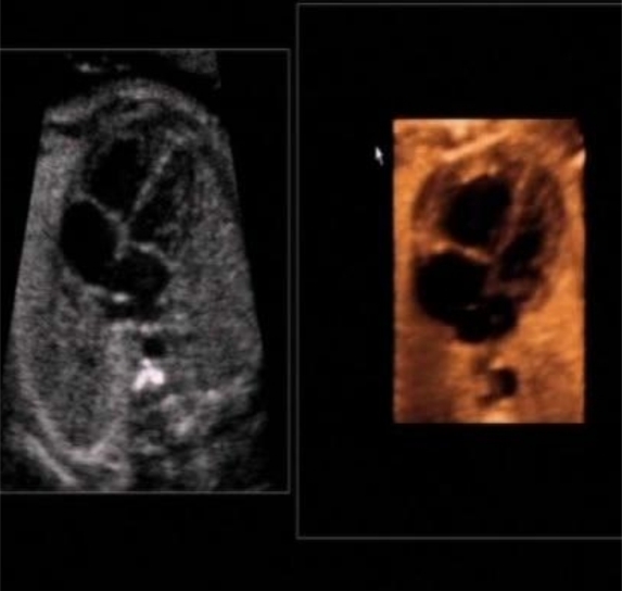
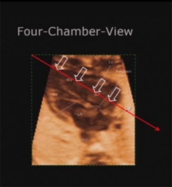
Rendering surface mode is a mode of analysis of the aquired volume. The operator places the rendering box around the region of interest (ROI) within the volume in order to show a slice of that volume (Figure 4).
Figure 4. Rendered image of 4-chamber view.
When we have in the A frame a good four chamber view one possibility is that the operator can place the rendering box around the interventricular septum, and in the D frame we will see the rendered image of the interventricular septum the so called "en face " view of the septum (9). We can determine if we want to see the septum from right to left or from left to right, depending where we place the green line (the active line or the rendered view direction) of the rendered box (Figure 5).
Figure 5. Rendered image of IVS.
Usually for rendering to be considered as "successful" the lateral view of the interventricular plane should display the entire septum and the foramen ovale, seen from the left ventricle. In another view "the base of the heart" (or the common atrioventricular plane-CAV plane), the rendered view direction is placed in the atria at the level of the valves and permit to see the "en face" view of both atrioventricular valves and semilunar valves, which is very helpful for diagnosing atrioventricular septal defect (12). So, for this plane to be considered as "successful" the rendered image had to clearly display all four cardiac annuli (Figure 6).
Figure 6. Rendered image of CAV plane.
Rendering of the volume data with color alone or the combination of gray-scale and color (the so called glass body mode) can give us information concerning the arrangement of the great vessels. The 3D power Doppler is directionless and is most effectively associated with 3D static scanning. 3D power Doppler reconstruct the blood vessels in the volume of interest (VOI) zone. 3D power Doppler reconstructs the vascular tree of the fetal abdomen and thorax (13), relieving the operator of the necessity to reconstruct a mental image of the anomalous vessel from a series of 2D planes. High definition power flow Doppler is the newest development in color Doppler applications and uses high resolution and a small sample volume to produce images with two colour directional information. It gives us information about vessels with very low velocity and has the advantage of showing flow direction. So, it is used specially for very small vessels. It can be used with static 3D, with STIC and with glass body mode to produce high resolution image of vascular tree.
The rendering mode-transparent minimum is a display in which the heart and blood vessels are seen in a transparent projection, so that the structures that are anechoic are visualized in a 3D projection of black-appearing vessels.
The rendering mode-inversion display mode represents the minimum mode rendering information which is simply inverted, thus presenting the hipoechoic structures as echogenic solid and eliminating into black most of the surrounding tissue information (that is the fluid filled spaces such as the cardiac chambers now appear white while the myocardium has disappeared). In fetal echocardiography it can be applied to create digital casts of the cardiac chambers and vessels (14).
Advantages of 3D/4D examination of fetal heart
The main advantages of the examination 3D/4D versus 2D of the fetal heart are:
Introduction of the virtual plane gives us views of the fetal heart not generally accessible with 2D approach.
Storage of volumes of cardiac anomalies or of the normal heart for review of the findings or for a second opinion.
For fetal heart screening.
The time used for scanning is shorter for 3D examination (3-4 minutes).
There are a few disadvantages of the examination 3D/4D of fetal heart:
We need a very good ultrasound machine.
We need a special training to obtain 3D volume data sets (how to choose the appropriate slice thickness within the volume, the region of interest).
Pitfalls of 3D/4D echocardiography
Fetal cardiac ultrasound using 3D/4D has some artifacts that are specific to 3D/4D acquisition and post-processing.
The quality of a STIC acquisition can be negatively modified by fetal body movement, by breathing mouvements, hiccups. The acquisition is improved when the fetus is quiet, and when we are using the shortest scan time. In the multiplanar display, in the B plane we can see the artifacts generate by fetal movement even though in the A-plane the acquisition is good. It is important to remember that the quality of the initial acquisition will affect all other stages of fetal heart evaluation.
The original angle at which the scan is performed is very important because can influence the quality of the planes aquired. Usually it is recommended that the sweep angle corresponds to the number of weeks of gestation (15).
Shadows generate a specific problem for 3D/4D acquisition, because when we start scanning from the 2D plane acoustic shadows may not be apparent, but they may be present within the aquired volume block in 3D/4D. So, the fetal position with the spine up creates some problem of acquisition and the acquisition is easiest when the spine of the fetus is not in ventral position.
When we are using 3D rendering we can applicate some adjusting designed to smooth the image. These adjustings can lead to loss of data from the original scan, so when we are using 3D rendering we must use always in the same time with the A-plane 2D image for comparison.
In evaluating congenital heart disease Meyer-Wittkopf evaluated 3D cardiac volume sets and 2D diagnosed cardiac lesions and compared the views of the heart in both modalities. They concluded that 3D added some advantages in diagnosis (16). Also most recently Benacerraf compared the acquisition and analysis for 2D and 3D examination of fetal anatomy scanning and concluded that 3D examination is superior in the accuracy of the diagnosis (17). The data obtained with 3D/4D fetal heart examination open new avenues for introducing fetal echocardiography programs to distant or poorly served medical areas (18). ❑
CONCLUSION
The STIC technique acquisition used in fetal echocardiography has the great advantage that allows obtaining imaging of a beating fetal heart and from a volume data set it is possible to obtain any plane within the fetal heart at anytime during the cardiac cycle.
Volume data display can be achieved in planes (single, multiplanar, tomographic), as a rendered 3D volume in surface, glass-body, minimum, or inversion modes.
Clinically 3D/4D fetal cardiac scanning can be used to store volumes of cardiac anomalies, as a tool for screening examinations and also in teaching courses (19,20). So, this technology has reached the stage when it is reproducible and added a real value in screening accuracy, but also it can be used in research to measure the volume of cardiac chambers and develop an approach to demonstrate cardiac anomalies. ❑
References
- 1.Garne E, Stoll C, Clementi M. Evaluation of prenatal diagnosis of congenital heart disease by ultrasound: experience from 20 European countries. Ultras Obstetr Gynec. 2001;17:386–391. doi: 10.1046/j.1469-0705.2001.00385.x. [DOI] [PubMed] [Google Scholar]
- 2.International Society of Ultrasound in Obstetrics and Gynecology – Cardiac scanning guidelines of the fetus: guidelines for performing the basic and extended basic cardiac scan Ultras Obstetr Gynec 2006. 27 107 113 [DOI] [PubMed] [Google Scholar]
- 3.Chaoui R, Hoffman J, Heling K. Three dimensional (3D) and 4D color fetal Doppler echocardiography using STIC correlation. Ultras Obstetr Gynec. 2004;23:535–545. doi: 10.1002/uog.1075. [DOI] [PubMed] [Google Scholar]
- 4.Yagel S, Weissman A, Rotstein Z. Congenital heart defects: natural course and in utero development. Circulation. 1997;96:550–555. doi: 10.1161/01.cir.96.2.550. [DOI] [PubMed] [Google Scholar]
- 5.Goncalves L, Lee W, Espinoza J, et al. Examination of the fetal heart by four dimensional ultrasound with STIC. Ultra Obstetr Gynec. 2006;27:336–348. doi: 10.1002/uog.2724. [DOI] [PubMed] [Google Scholar]
- 6.Goncalves l, Lee W, Espinoza J. Four dimensional fetal echocardiography with STIC a sistematic study of standard cardiac views assesed by different observers. Ultras Obstetr Gynec. 2003;22:50–53. doi: 10.1080/14767050500127765. [DOI] [PubMed] [Google Scholar]
- 7.De Vore G, Falkensammer P, Slansky M, et al. Spatiotemporal image correlation new technology for evaluation of fetal heart. Ultras Obstetr Gynec. 2003;22:380–387. doi: 10.1002/uog.217. [DOI] [PubMed] [Google Scholar]
- 8.Yagel S, Cohen S, Achiron R. Examination of the fetal heart by five short axis views A proposed screening method for comprehensive cardiac evaluations. Ultras Obstetr Gynec. 2001;17:367–369. doi: 10.1046/j.1469-0705.2001.00414.x. [DOI] [PubMed] [Google Scholar]
- 9.Yagel S, Benachi A, Bonner D, et al. Rendering in fetal cardiac scanning: the intracardiac septa and the coronal atrioventricular valves planes. Ultras Obstetr Gynec. 2006;28:266–274. doi: 10.1002/uog.2843. [DOI] [PubMed] [Google Scholar]
- 10.DeVore G, Polanco B, Sklansky M, et al. The spin technique a new method for examination of the fetal outflow tracts using three-dimensional ultrasound. Ultras Obstetr Gynec. 2004;24:72–82. doi: 10.1002/uog.1085. [DOI] [PubMed] [Google Scholar]
- 11.Chaoui R, Heling K. New developments in fetal heart scanning; three and four dimensional fetal echocardiography. Semin Fetal Neonatal Med. 2005;10:567–577. doi: 10.1016/j.siny.2005.08.008. [DOI] [PubMed] [Google Scholar]
- 12.Ionescu C, Gheorghiu D, Davitoiu B, et al. Vizualizarea planului valvular atrioventricular la fetii cu defect septal atrioventricular complet folosind tehnica STIC. Gineco.ro. 2009;Vol. 5:210–214. [Google Scholar]
- 13.Chaoui R, Kalache K, Hartnung J. Application of three dimensional power Doppler ultrsound in prenatal diagnosis. Ultras Obstetr Gynec. 2001;17:22–29. doi: 10.1046/j.1469-0705.2001.00305.x. [DOI] [PubMed] [Google Scholar]
- 14.Goncalves L, Espinoza J, Lee W. Three dimensional and 4D reconstruction of the aortic and ductal arches using inversion mode: a new rendering algorithm for vizualization of fluid filled anatomical structures. Ultras Obstetr Gynec. 2004;24:696–698. doi: 10.1002/uog.1754. [DOI] [PubMed] [Google Scholar]
- 15.Abuhamad A, Falkensammer P, Zhao Y. Automated sonography: defining the spatial relationship of standard diagnostic fetal cardiac planes in the second trimester of pregnancy. J Ultras Med. 2007:501–507. doi: 10.7863/jum.2007.26.4.501. [DOI] [PubMed] [Google Scholar]
- 16.Wittkopf Meyer, Cooper S, Vaughan J, et al. Three dimensional echocardiographic analysis of congenital heart disease in the fetus –comparaison with 2D cross sectional section. Ultras Obstetr Gynec. 2001;17:485–492. doi: 10.1046/j.1469-0705.2001.00429.x. [DOI] [PubMed] [Google Scholar]
- 17.Benacerraf B, Shipp T, Bromley B. Three dimensional ultrasound of the fetus: volume imaging. Radiology. 2006;238:988–996. doi: 10.1148/radiol.2383050636. [DOI] [PubMed] [Google Scholar]
- 18.Michailidis G, Simpson J, Karidas C. Detailed three dimensional fetal echocardiography facilitated by an internet link. Ultras Obstetr Gynec. 2001;18:325–328. doi: 10.1046/j.0960-7692.2001.00520.x. [DOI] [PubMed] [Google Scholar]
- 19.Uittenbogaard B, Haak C, Spreeuwenberg M, et al. A systematic analysis of the feasibility of four dimensional ultrasound imaging using STIC in routine fetal echocardiography. Ultras Obstetr Gynec. 2008;31:625–632. doi: 10.1002/uog.5351. [DOI] [PubMed] [Google Scholar]
- 20.Ionescu D, Gheorghiu D, Pacu I, et al. Aplicatia TUI-STIC poate fi introdusa in screeningul prenatal? Lucrare publicata la Congresul al 7-lea de Medicina Perinatala. 2007 Oct;:11–12. [Google Scholar]


