

Abstract
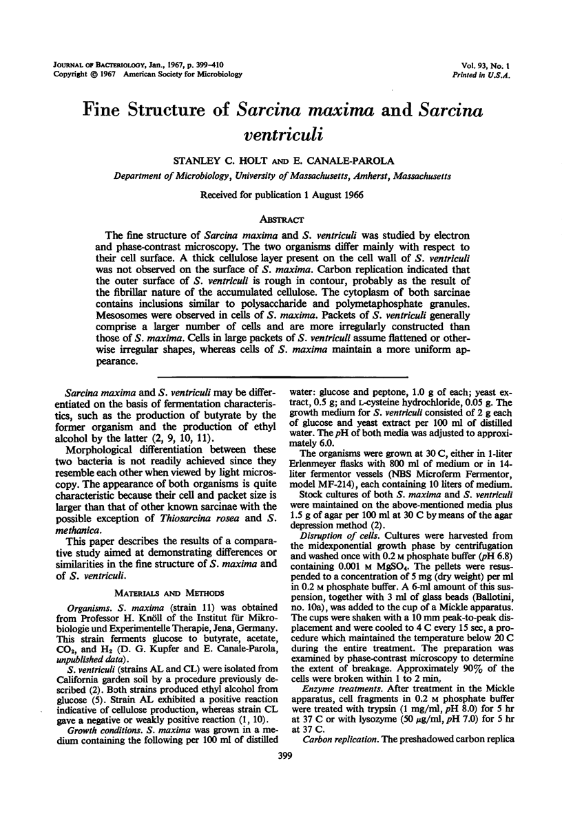
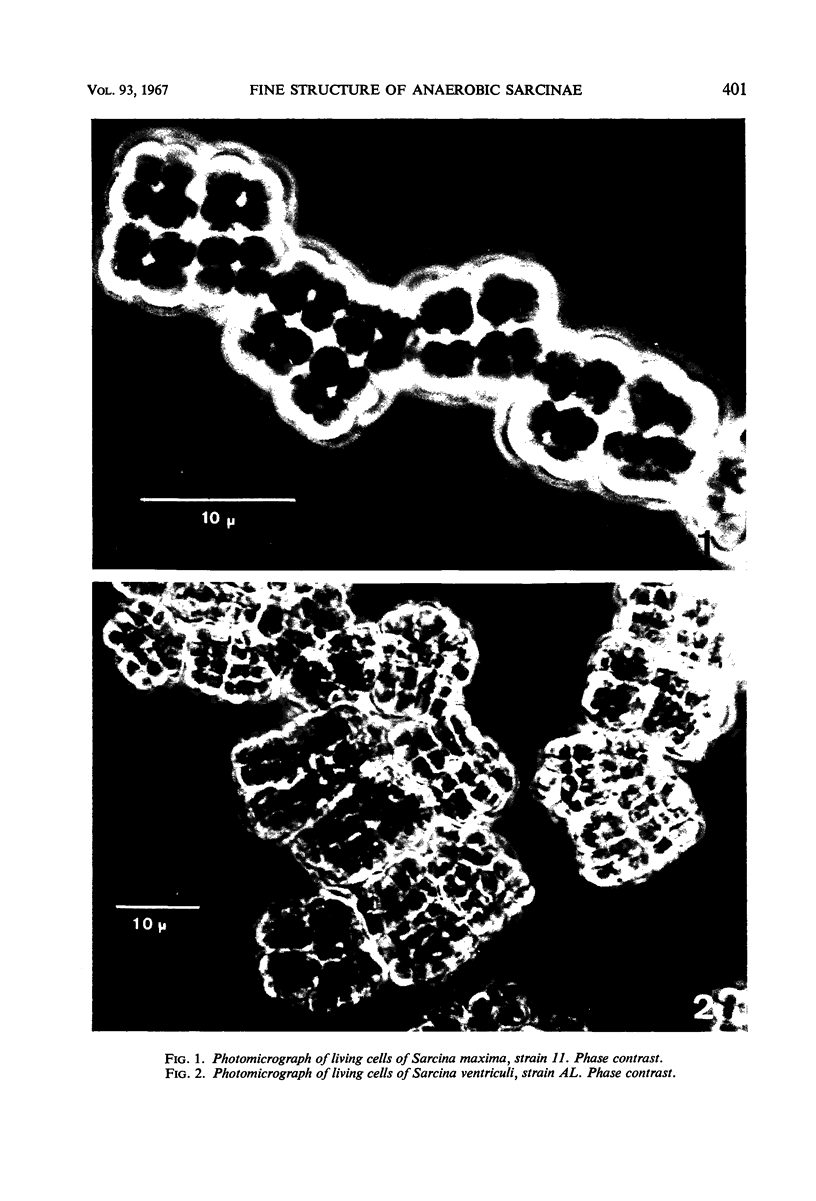
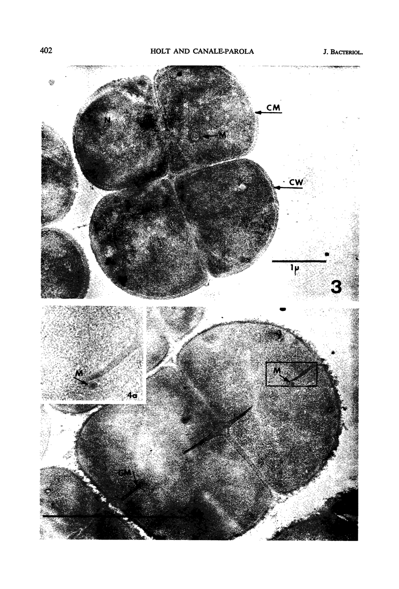
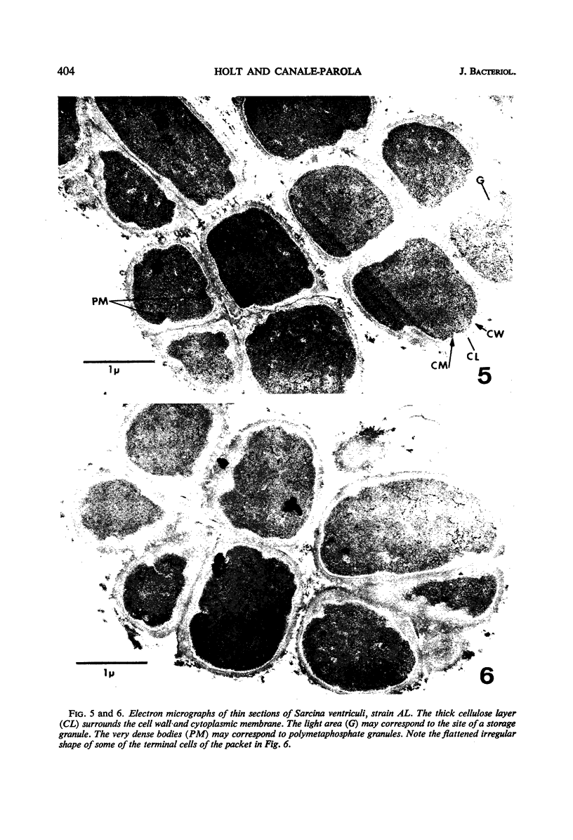
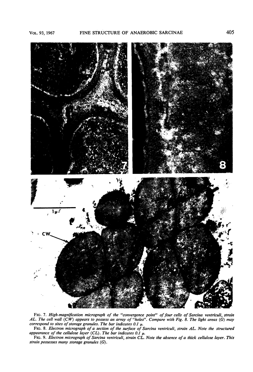
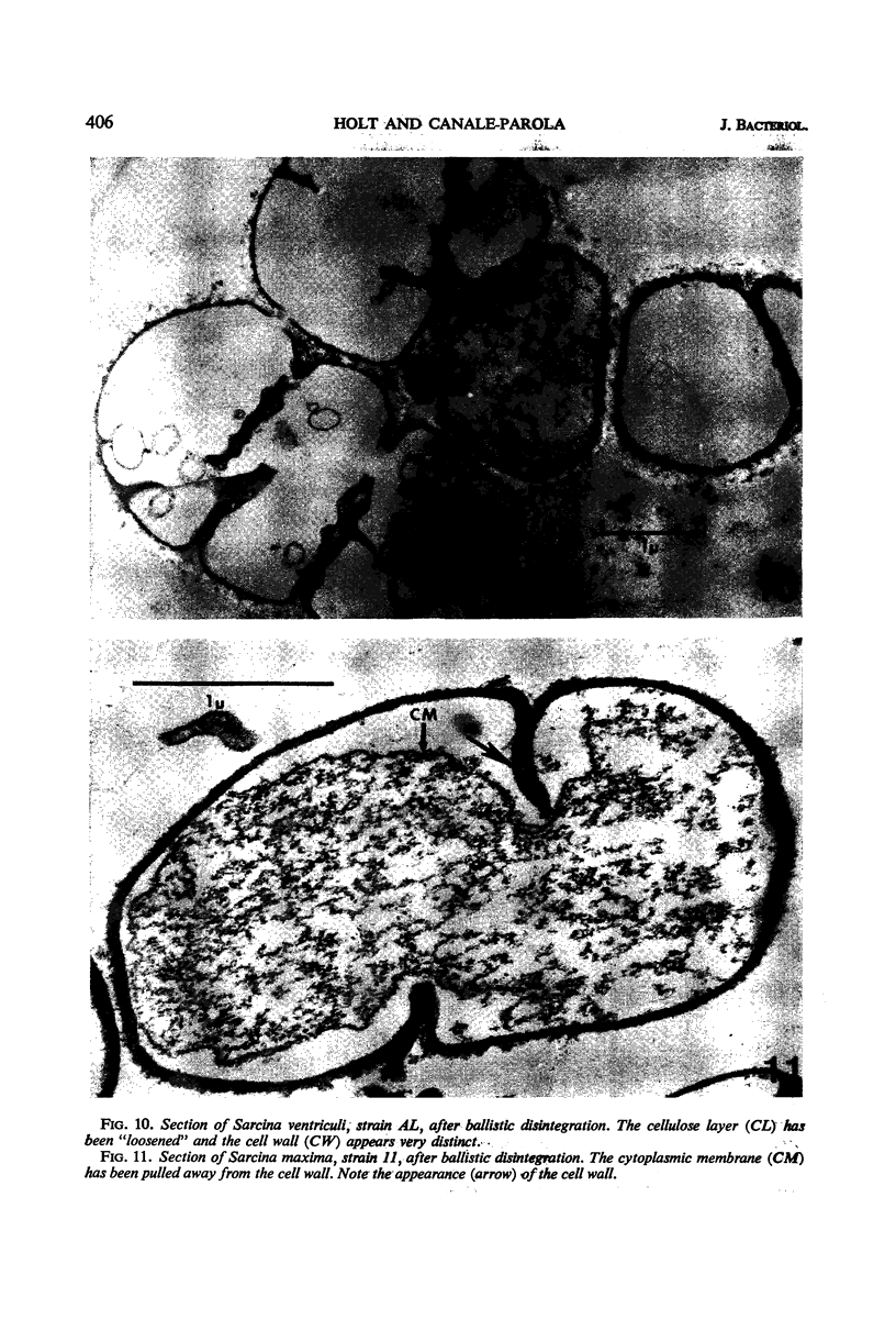
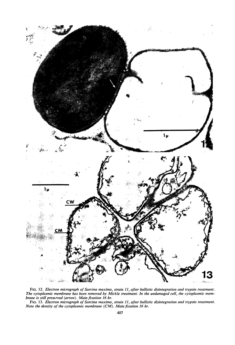
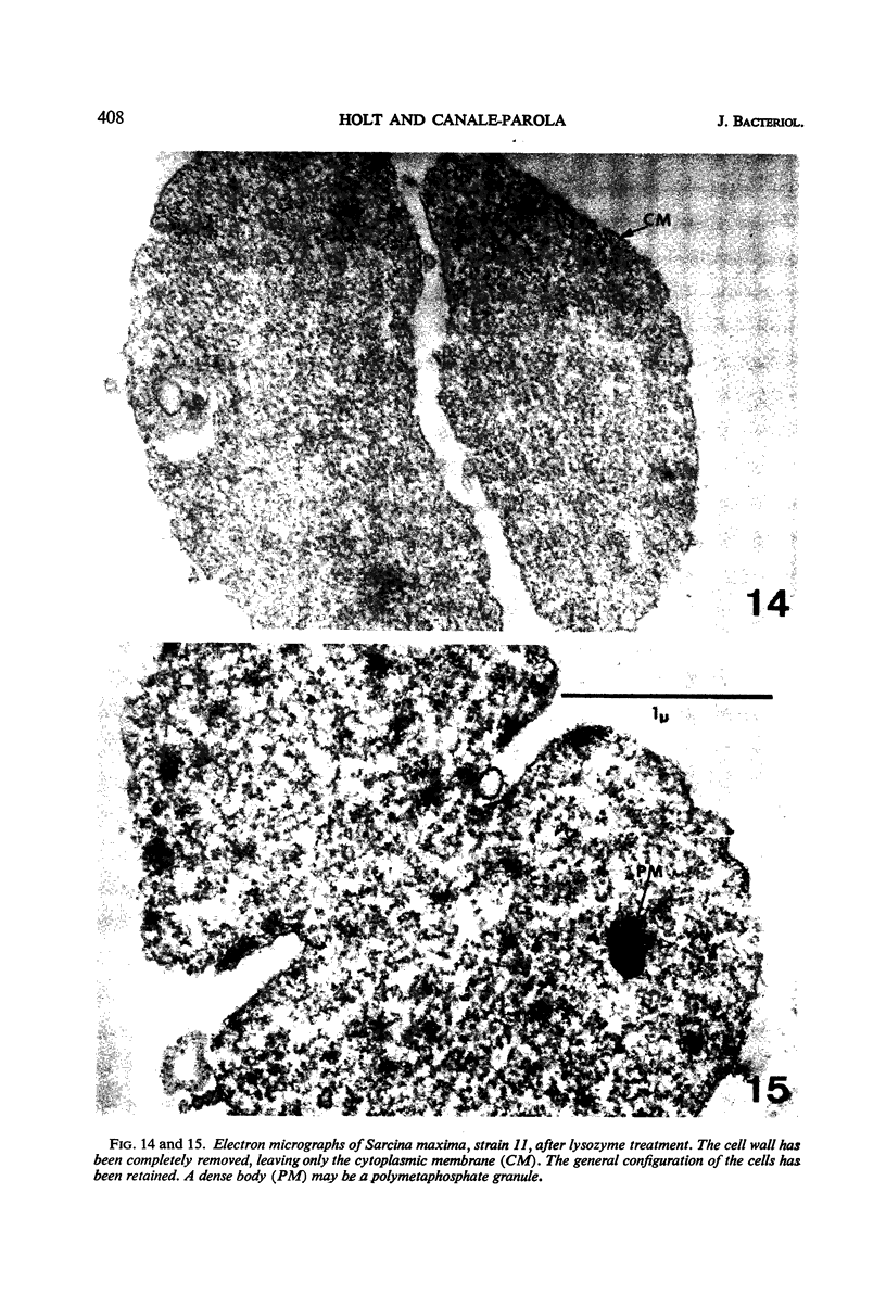
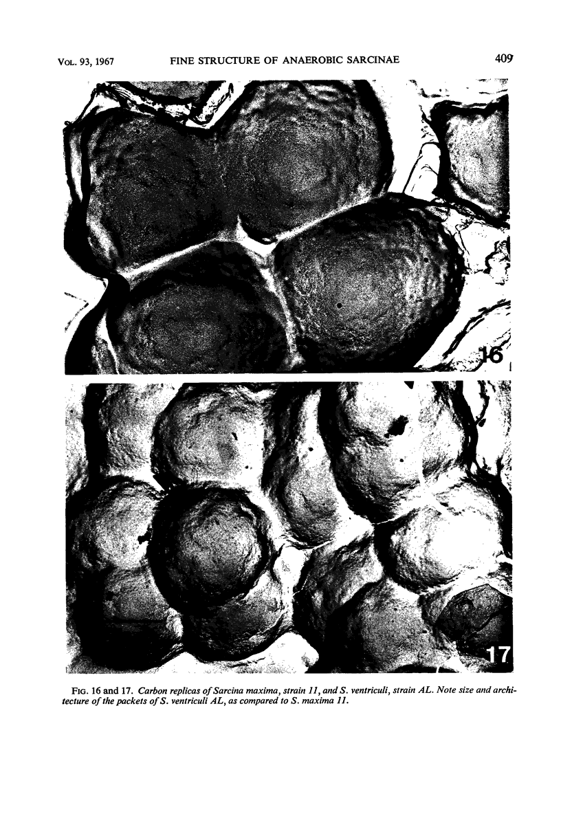
The fine structure of Sarcina maxima and S. ventriculi was studied by electron and phase-contrast microscopy. The two organisms differ mainly with respect to their cell surface. A thick cellulose layer present on the cell wall of S. ventriculi was not observed on the surface of S. maxima. Carbon replication indicated that the outer surface of S. ventriculi is rough in contour, probably as the result of the fibrillar nature of the accumulated cellulose. The cytoplasm of both sarcinae contains inclusions similar to polysaccharide and polymetaphosphate granules. Mesosomes were observed in cells of S. maxima. Packets of S. ventriculi generally comprise a larger number of cells and are more irregularly constructed than those of S. maxima. Cells in large packets of S. ventriculi assume flattened or otherwise irregular shapes, whereas cells of S. maxima maintain a more uniform appearance.
Full text
PDF
Images in this article
Selected References
These references are in PubMed. This may not be the complete list of references from this article.
- AUBERT J. P., MILHAUD G., VAN NIEL C. B. Etude de la glycolyse de Zymosarcina ventriculi. Ann Inst Pasteur (Paris) 1956 Sep;91(3):363–368. [PubMed] [Google Scholar]
- CANALE-PAROLA E., BORASKY R., WOLFE R. S. Studies on Sarcina ventriculi. III. Localization of cellulose. J Bacteriol. 1961 Feb;81:311–318. doi: 10.1128/jb.81.2.311-318.1961. [DOI] [PMC free article] [PubMed] [Google Scholar]
- CANALE-PAROLA E., WOLFE R. S. Studies on Sarcina ventriculi. I. Stock culture method. J Bacteriol. 1960 Jun;79:857–859. doi: 10.1128/jb.79.6.857-859.1960. [DOI] [PMC free article] [PubMed] [Google Scholar]
- DE BOER W. E., SPIT B. J. A NEW TYPE OF BACTERIAL CELL WALL STRUCTURE REVEALED BY REPLICA TECHNIQUE. Antonie Van Leeuwenhoek. 1964;30:239–248. doi: 10.1007/BF02046729. [DOI] [PubMed] [Google Scholar]
- KARNOVSKY M. J. Simple methods for "staining with lead" at high pH in electron microscopy. J Biophys Biochem Cytol. 1961 Dec;11:729–732. doi: 10.1083/jcb.11.3.729. [DOI] [PMC free article] [PubMed] [Google Scholar]
- KELLENBERGER E., RYTER A., SECHAUD J. Electron microscope study of DNA-containing plasms. II. Vegetative and mature phage DNA as compared with normal bacterial nucleoids in different physiological states. J Biophys Biochem Cytol. 1958 Nov 25;4(6):671–678. doi: 10.1083/jcb.4.6.671. [DOI] [PMC free article] [PubMed] [Google Scholar]
- LUFT J. H. Improvements in epoxy resin embedding methods. J Biophys Biochem Cytol. 1961 Feb;9:409–414. doi: 10.1083/jcb.9.2.409. [DOI] [PMC free article] [PubMed] [Google Scholar]
- Thornley M. J., Horne R. W., Glauert A. M. The fine structure of Micrococcus radiodurans. Arch Mikrobiol. 1965 Jul 20;51(3):267–289. doi: 10.1007/BF00408143. [DOI] [PubMed] [Google Scholar]