



Abstract
B cells are subjected to selection at multiple checkpoints during their development. The selection of antibody heavy chains is difficult to study because of the large diversity of the CDR3. In order to study the selection of individual antibody heavy chain variable region genes (VH), we performed CDR3 spectratyping of 75–300 rearrangements per individual VH in C57BL6/J mice. We measured the fraction of rearrangements that were in-frame (IF fraction) in B cell DNA. We demonstrate that individual VH genes have different IF fractions, ranging from 10% to 90%, and that these IF fractions are reproducible in different mice. For most VHs, the IF fraction in pro-B cells approximated 33% and then shifted to the nearly final (mature) B cell value by the cycling pre-B cell stage. The frequency of high IF VH usage increased in cycling pre-B cells compared to pro-B cells, whereas this did not occur for low IF VHs. The IF fraction did not shift as much in BCR-expressing B cells and was minimally affected by light chain usage for most VH. High IF clan II/III VHs share more positively charged CDR2 sequences, whereas high IF clan I J558 CDR2 sequences are diverse. These data indicate that individual VHs are subjected to differential selection, that VH IF fraction is mainly established through pre-BCR mediated selection, that it may operate differently in clan I vs. II/III VHs, and that it has a lasting influence on the antibody repertoire.
Introduction
Immunoglobulin loci of developing B cells undergo a series of DNA rearrangements (V(D)J recombination) that culminate in the assembly of antibody heavy and light chain variable regions (1–2). A large and diverse repertoire of antibodies is created by the recombination of multiple V, D and J segments; variability at the junctions between these gene segments; and numerous combinatorial possibilities for heavy + light chain pairing. But this diversity comes at a cost: non-productive (out of frame) rearrangements occur frequently and, among the rearrangements that are productive (in-frame, IF), many of the resulting antibodies are autoreactive (3).
The manner in which the primary antibody repertoire is purged of autoreactive receptors is fundamental to the understanding of self-tolerance. It is typically assumed that the major stage of bone marrow B cell development where censoring of the primary antibody repertoire takes place is at the Pre-B to naive B cell transition. At this stage, B cells with autoreactive IgM antibodies can edit their antibody receptor specificity by undergoing further light chain rearrangement (reviewed in (4)) or undergo clonal deletion. It seems logical for editing of autoreactivity to occur after the full antibody (heavy + light chain) has been formed, but several lines of evidence suggest that antibody heavy chains are also subjected to specificity-based selection during early B cell development.
For example, antibody heavy chains are thought to undergo “structural selection” at the pro-B to pre-B cell transition. Consistent with this model, heavy chains that pair well with the surrogate light chain result in a pre-BCR that is capable of signaling via associated Igα and Igβ domains (5), resulting in down-regulation of the V(D)J recombinase and IL-7-dependent proliferation in large, cycling pre-B cells (Hardy bone marrow (BM) fraction C’, hereafter Fr. C’ (6–8)). Ten Boekel, Rolink and Melchers showed that approximately half of antibody heavy chains were able to pair well with the surrogate light chain and promote B cell development (9). But why some heavy chains pair well with surrogate light chain and others pair poorly is incompletely understood, in part because early experiments did not distinguish between selection based on CDR3 (which generally derives a minority of its sequence from the VH segment) vs. selection elsewhere in the VH. Furthermore, the mechanism of pre-BCR signaling is unclear, e.g., is it ligand-dependent? Compelling data indicate that galectin 1, secreted by bone marrow stromal cells, binds to the λ5 unique region of the pre-BCR and influences pre-B cell differentiation and proliferation (10–11). On the other hand, the crystal structure of the human pre-BCR Fab, along with other biochemical data, suggest that the long tails of the surrogate light chains could mediate the self-assembly of multimers (5, 12–13). Furthermore, recent data implicate a role for N-linked glycosylation within the H chain constant region in surrogate light chain binding and pre-BCR crosslinking (14).
Other recent evidence suggests that antibody heavy chains in developing pre-B cells are subjected to distinctive selection, particularly with regard to autoreactivity: surrogate light chain knock-out mice develop spontaneous autoimmunity, including the production of anti-nuclear antibodies (15). The heavy chains of antibodies from surrogate light chain knock-out mice are enriched for positively charged arginine residues in the CDR3, resembling the canonical sequences of anti-DNA antibodies (15–16). In the absence of λ5, these heavy chains were expressed on the cell surface, bound to nuclear antigens such as DNA, and permitted B cell proliferation. In contrast, in the presence of surrogate light chain, freshly isolated anti-DNA pro-B cells were reduced in frequency after a few days in culture, suggestive of negative selection. Additionally, two groups have demonstrated that heavy chains with CDR3s that mimic or are derived from known autoantibodies promote surrogate light chain independent B cell maturation and proliferation (17–18).
Work from other laboratories supports the selection of DH reading frames, CDR3 length and amino acid content during early B cell development (3, 9, 18–23). Earlier studies of antibody heavy chain selection also revealed a progressive shift in the frequency of VH usage, particularly within the 7183 family, from 3’ (D-proximal) in the earliest stage B cells to more 5’ VH usage in more mature B cell subsets. This shift in VH usage is recapitulated during ontogeny, with fetal B cell repertoires exhibiting increased 3’ VH gene usage (24–27). However, the analysis of selection based upon VH frequency is problematic because multiple factors, including the frequency of rearrangement, can independently influence the observed VH gene usage (28). Furthermore, the frequency of VH rearrangement is not necessarily proportional to whether the VH is positively or negatively selected. A case in point is the most 3’ VH, 81X, which is frequently but often non-productively rearranged (29). The analysis of CDR3 sequences of certain VH genes, including 81X, VH12 and Q52 (9, 22–23, 30–31), clearly documents specific changes in the repertoire that occur during the pre-BCR selection step. These and other studies have led to the prevailing view that the CDR3, which contributes the lion’s share of H chain diversity (32), is the critical region for H chain selection (for example, (33–34)). In point of fact, however, the selective consequences of VH sequences outside the CDR3 are almost entirely unknown. Only a handful of studies, focused on specific VHs such as 81X, VH11 and VH12, have documented selection of amino acids outside of the CDR3 (23, 30, 35).
A more systematic study of VH selection would enhance our understanding of tolerance mechanisms that apply specifically to antibody heavy chains. For example, VH replacement is a mechanism of antibody heavy chain editing that exchanges a VH gene in an existing VDJ rearrangement with a new VH gene by invasion into a cryptic heptamer near the 3’ end of the VH (36–38). As such, VH replacement does not remove the existing CDR3, but rather adds to it and swaps out the VH gene. If the CDR3 is the dominant factor in heavy chain selection, this receptor editing mechanism should be ineffective. Understanding the developmental timing of VH selection would also provide better insights into how the pre-BCR vs. the BCR are involved in autoantibody regulation (15).
Accordingly, we have applied a more general approach to the issue of H chain selection by measuring the fraction of in-frame rearrangements (IF fractions) of twenty different VH genes in genomic DNA of B cells from C57BL/6J (hereafter B6) mice. In the B6 spleen, we found that different VH genes had different IF fractions, ranging from 10% to 90%. Most of the clan I VH genes, including all but one member of the J558 family that were evaluated, had a high IF fraction. In a more detailed analysis of BM B cell subsets, most VHs exhibited an IF fraction of approximately 33% at the pro-B cell stage and the IF fraction increased substantially for high IF VHs by the large cycling pre-B cell stage. These findings suggest that pre-BCR driven selection influences the VH IF fraction. In support of this hypothesis, the IF fraction appears to be positively correlated with an increase in rearrangement frequency as cells progress from the pro-B to the pre-B stage. In contrast to the large changes in the VH IF fraction at the pro-B to pre-B cell transition, changes in BCR-expressing B cell subsets tended to be more subtle and, for most heavy chains, light chain usage did not significantly alter the IF fraction. Taken together, these findings suggest that the major shift in the IF fraction occurs at the pro-B to pre-B cell transition and involves pre-BCR rather than BCR-based selection.
Materials and Methods
Mice
B6 mice were purchased from Jackson Laboratories (Bar Harbor, ME) and maintained at the University of Pennsylvania, School of Medicine under an Institutional Animal Care and Use Committee-approved protocol. Vκ8 light chain knock-in mice were maintained at the University of Chicago under an IACUC-approved protocol.
Flow cytometry
Cell suspensions from 3–4 month old B6 mice were prepared from femurs, tibias, and spleens in FACS buffer (PBS, 0.5% BSA, 0.01% NaN3, 1 mM EDTA) after red blood cell lysis (ACK Lysing Buffer; BioWhittaker, Walkersville, MD). All antibodies were purchased from eBioscience (San Diego, CA), BD Biosciences (San Jose, CA) and BioLegend (San Diego, CA). The following antibody-fluorophore combinations were used to resolve BM subsets: FITC anti–heat-stable antigen (30F1), PE anti-CD43 (S7), PE-Cy7 anti-IgM (II/41), PE-Cy5.5 anti-CD19 (1D3), APC anti-CD93 (AA4.1), APC-AF750 anti-B220 (RA3-6B2), Pacific Blue anti-IgD (11–26). BM B cell fractions are defined as follows, based upon the scheme by Hardy (7): Fr.B-C (B220+CD19+CD43+IgM−IgD−CD24dimCD93+), Fr.C’ (B220+CD19+CD43+IgM−IgD−CD24briCD93+), Fr.D (B220+CD43−IgM−IgD−CD93+), Fr.E is B220+CD43−IgM+IgD−CD93+ and Fr.F is B220+CD43−IgMdim/+IgD+CD93−. The following antibodies were used to resolve splenic B cell subsets: FITC anti-IgM (II/41), PE anti-CD21 (7G6), APC anti-CD93 (AA4.1) and APC-AF750 anti-B220 (RA3-6B2). Splenic B cell subsets were defined as follows: Transitional (B220+CD93+), Follicular (B220+CD93−IgMdimCD21dim) and Marginal zone (B220+CD93−IgMbriCD21bri). For all sorts, dead cells were first eliminated by DAPI staining and cell doublets/aggregates by pulse width gating. The purities of the Fr. B-C sorted cell populations were 98–99%. FACS analysis and sorting were performed on the LSR II and FACSAria cytometers, respectively (BD Bioscience, San Jose, CA) in the University of Pennsylvania Flow Cytometry core facility. Flow cytometry data were analyzed using FlowJo software (version7.5.5, Treestar Inc., Ashland, OR). For sorting of λ+ B cells, splenic B cells were stained with APC anti-λ (RML-42), FITC anti-κ (187.1), APC-AF750 anti-B220 and PE-Cy5.5 anti-CD19.
CDR3 Spectratyping
CDR3 spectratyping was performed to evaluate the VH IF fractions. Genomic DNA was isolated from either spleens or sorted cells, purified according to the manufacturer’s directions using PureGene (Qiagen, Valencia CA), and amplified using the VH primers in Supplemental Table 1 and a labeled reverse primer in JH2 (5’-FAM-CTG TGA GAG TGG TGC CTT G-3’). All primers were synthesized by Integrated DNA Technologies (Coralville, IN). Each 20 μL PCR contained 5-50 ng genomic DNA, 1x PCR buffer (AmpliTaq GoldTM 10x PCR buffer I with 15 mM MgCl2 (Roche Applied Sciences, Indianapolis, IN)), 0.2 mM of each dNTP (Promega, Madison, WI), 0.6 μM of each primer, and 1 U of DNA polymerase (AmpliTaq Gold (Roche Applied Sciences)). The cycling conditions were: 10 min at 94°C, followed by 40 cycles of 94°C for 30 s, Ta (Supplemental Table 1) for 30 s, and 72°C for 30 s, followed by 20 min at 72°C. To minimize notching artifacts due to non-uniform adenosine addition by the Taq polymerase, some VH primers were modified by “PIGtailing” (incorporating the sequence GTTTCTT at the 5’ end, see italicized sequences in Supplemental Table 1). This modification resulted in nearly 100% adenylation of the 3’ end of the FAM-labeled opposite strand, consistent with what has been described previously (39). 2 μL of PCR products were resolved by capillary electrophoresis on an ABI 3100 analyzer (Applied Biosciences Inc., Foster City, CA).
CDR3 length analysis
Capillary electropherograms were generated and analyzed using ABI Genotyper 3.7 or ABI Peak Scanner Software, version 1.0. Peak sizes were interpolated using a ROX ladder “DS30” (Applied Biosystems) alone or combined with our homemade ladder (sizes of the homemade ladder are: 195, 198, 201, 204, 207, 210, 213, 216, 219 bp). The IF fraction was determined by counting the number of peaks that are IF and dividing by the total number of peaks (IF+OF). The CDR3 lengths were calculated, beginning with the conserved cysteine (Cys) residue at the 3’ end of the VH (position 96) and ending with the last conserved glycine (Gly) in the middle of JH2.
CDR3 peak count metadata
In Fig. 2B, the numbers of peaks and IF fraction (the latter in percent) for each VH are: J558.88 n=92 76%, J558.85 n=86 84%, J558.72 n=124 85%, 3609.11 n=152 81%, J558.47 n=55 42%, VH15 n=84 29%, VH10 n=91 69%, 3609.1 n=109 42%, J606.1 n=178 81%, VH12 n=218 21%, 3609N.2 n=371 36%, S107.3/4 n=107 82%, VH11 n=109 27%, SM7.1/2/4 n=77 88%, S107.1 n=78 15%, Q52.8/5 n=63 38%, 7183.9 n=68 75%, 7183.4 n=100 60%, Q52.2 n=86 45%, 7183.2 n=93 13%. In Fig. 3A, the numbers of peaks and IF fraction analyzed for each subset are: J606.1, Fr. B-C n=83 39%, Fr. C’ n=231 87%; VH10, Fr.B-C n=84 30%, Fr.C’ n=69 57%; VHJ558.47 Fr. B-C n=56 38%, Fr. C’ n=22 64%; VH3609N.2 Fr. B-C n=48 35%, Fr. C’ n=31 35%; VH7183.2 Fr. B-C n= 69 32%, Fr. C’ n=55 29%. In Fig. 3B the numbers of peaks analyzed for each subset are: J606.1, Fr. B-C n=83, Fr. C’ n=231; VHJ558.85 Fr. B-C n=42, Fr. C’ n=88; VH3609N.2 Fr. B-C n=47, Fr. C’ n=21; VHJ558.47 Fr. B-C n=18, Fr. C’ n=4; VH7183.2 Fr. B-C n= 69, Fr. C’ n=55. The number of peaks in Fig. 3B are lower for some of the VH than in Fig. 3A because in Fig. 3B all of the measurements were performed on highly diluted genomic DNA samples for optimal quantification of rearrangement frequency. In Fig. 4A, the numbers of rearrangements and the IF fractions analyzed for each subset and each VH are: J606.1, Fr.B-C n=83 39%, Fr.C’ n=231 87%, Fr.D n=72 89%, Fr.E n=74 88%, Fr.F n=43 91%, Trans n=118 86%, FO n=123 82%, MZ n=273 87%; 3609N.2, Fr.B-C n=48 35%, Fr.C’ n=31 35%, Fr.D n=92 46%, Fr.E n=76 39%, Fr.F n=61 46%, Trans n=47 33%, FO n=68 31%, MZ n=61 38%; VH10, Fr.B-C n=84 30%, Fr.C’ n=69 57%, Fr.D n=53 59%, Fr.E n=50 56%, Fr.F n=54 54%, Trans n=29 66%, FO n=19 68%, MZ n=75 68%. In Fig. 4B the numbers of rearrangements and their IF fractions are 7183.2-Vκ8 n=64 14%, VH15-Vκ8 n=61 25%, 3609N.2-Vκ8 n=129 25%, J558.47-Vκ8 n=64 39%, 3609.1-Vκ8 n=109 32%, VH10-Vκ8 n=35 69%, J558.88-Vκ8 n=49 84%, 3609.11-Vκ8 n=51 78%, J606.1-Vκ8 n=100 77%, J558.72-Vκ8 n=77 82%, SM7.1/2/4-Vκ8 n=50 90%; 7183.2-λ n=40 13%, VH12-λ n=27 15%, 3609N.2-λ n=29 28%, VH10-λ n=98 82%, 3609.11-λ n=55 89%, J606.1-λ n=31 77%, J558.85-λ n=77 91%, J558.72-λ n=79 85%, SM7.1/2/4-λ n=17 94%. For the VHs in this comparative analysis, the κ peak numbers and IF fractions are the same as in Fig. 2.
Figure 2. VHs differ in their IF fractions.

A. Mouse-to-mouse reproducibility of the VH IF fraction. VHIF fraction analysis was performed on spleen DNA from 3-month-old B6 mice (n=2) for VH J606.1 (white bars) and 3609N.2 (grey bars). The numbers of J606.1- JH2 rearrangements and their corresponding IF fractions (the latter in percent) were: mouse #1, 124 peaks, 80%; mouse #2, 194 peaks, 81%. For 3609N.2-JH2: mouse #1, 290 peaks, 36%; mouse #2, 293 peaks, 33%. The IF fractions calculated for individual mice are plotted for each VH. The difference in the average IF fraction between the two VHs is significant (p≪1 x 10⁻¹⁰ by cumulative binomial probability). B. Survey of the IF fraction in 20 different VHs. VH IF fractions were measured in genomic DNA from 3-month-old B6 mice spleen DNA (n=2). VH IF fractions are arranged in the order of their location in the germline locus, with 5’ VHs on the left. The numbers of rearrangements and the IF fractions analyzed for each VH are given in the Materials and Methods section. Some of the primers bind to more than one VH (see Supplemental Table 1 for primer information).
Figure 3. Establishment of the VH IF fraction occurs early during B cell development.
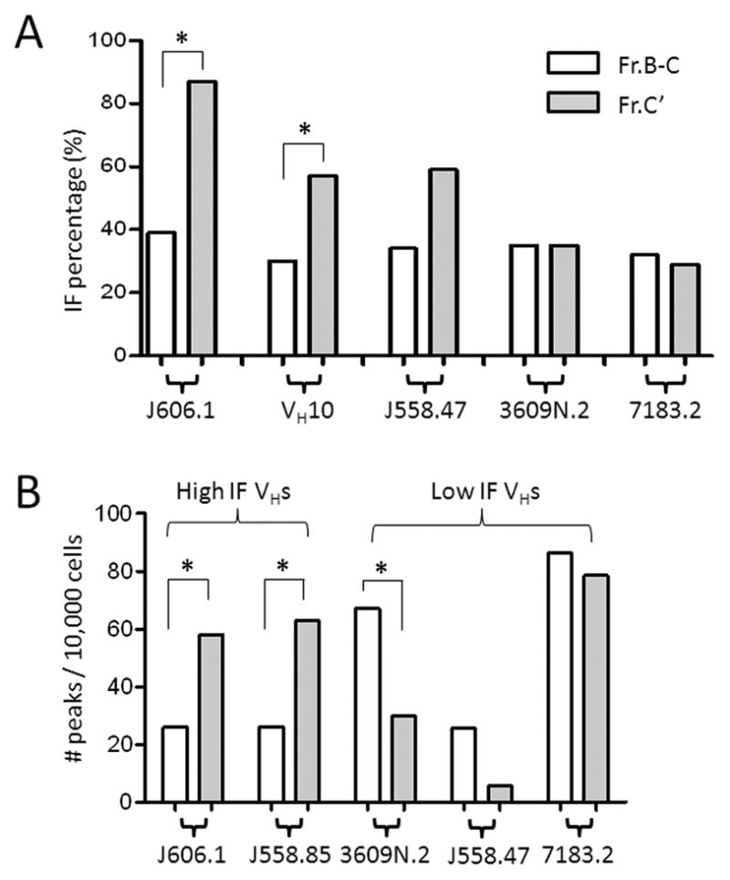
A. VH IF analysis in bone marrow pro-B (Fr. B-C) and cycling pre-B (Fr. C’) cells. IF fractions (given in percent) for J606.1-JH2, VH10-JH2, J558.47-JH2, 3609N.2-JH2 and 7183.2-JH2 rearrangements are plotted for each B cell subset. White bars denote Fr. B-C and gray bars represent Fr. C’. Asterisks denote IF fractions that differ significantly between Fr. B-C and Fr. C’ (p<0.05 by Binomial probability). B. VH peak number analysis in Fr. B-C and Fr. C’. Plotted are the frequencies of VHJ606.1-JH2, J558.85-JH2, 3609N.2-JH2, J558.47-JH2 and 7183.2-JH2 rearrangements per 10,000 sorted B cells from Fr. B-C versus Fr. C’. The total raw peak numbers are given in the Materials and Methods section. Asterisks denote peak numbers that differ significantly between Fr. B-C and C’ (p<0.05 by Chi square test). Data for both panels in the figure are pooled from sorted cells of 3-month-old B6 (n=4–8 mice).
Figure 4. Minimal changes in the VH IF fraction in BCR-expressing B cells.

A. VH IF analysis in B cell subsets in bone marrow and spleen. IF fractions (given in percent) for J606.1-JH2, 3609N.2-JH2 and VH10-JH2 rearrangements are plotted for each B cell subset. The same data for Fr. B-C and C’ from Fig. 3A are re-plotted here for comparison. The numbers of rearrangements and the IF fractions analyzed for each subset and each VH are given in Materials and Methods. Asterisks denote IF fractions that differ significantly between the indicated subsets (p<0.05 by Binomial probability). B. The VH IF fraction is not significantly altered by light chain usage for most VHs. The VH IF fraction is compared among splenocytes (mostly κ+, filled circles, same data as in Fig. 2B), λ+ B cells (open squares, n=2 mice) and Vκ8+ splenocytes (open triangles, n= 2 mice). The IF fractions (percentages) of different VH-JH2 rearrangements are plotted in order of increasing VH IF fraction, based upon the unsorted splenocyte data. Only VH10-JH2 rearrangements display a statistically significant difference in the IF fraction in the different light chain expressing populations (black arrow, p<0.05 by the Mann-Whitney test). The numbers of rearrangements and their IF fractions are given in Materials and Methods.
Cloning of PCR products and sequencing
PCR products were cloned into pCR2.1 TOPO per the manufacturer’s directions (Invitrogen, Carlsbad, CA). Randomly selected clones were sequenced at the University of Pennsylvania DNA sequencing facility, and analyzed using Ig-BLAST (40).
Protein data analysis
The following crystal structure data were obtained from the protein database (PDB): 2H32 (human pre-BCR Fab, (12)), 3GHB (human VH3 anti-HIV Fab, (41)) and 1KC5 (anti-hepatitis virus peptide Fab, (42)). All of the amino acid sequence alignments, 3-dimensional structure visualization and isoelectric point (pI) calculations were performed using Geneious software (Geneious v4.5, available from http://www.geneious.com/.) B6 germline VH sequences (43) were analyzed for the CDR2 pI using Geneious software.
Statistical analysis and modeling
The overall IF fraction can be estimated using a simple model of rearrangement, as described in Fig. 1. Other mathematical models are described in the legend to Supplemental Fig. 2. In order to determine if two different VHs have a statistically significant difference in their IF fractions in spleen DNA, we used binomial probabilities. Statistical comparisons of median CDR3 lengths and CDR2 pI correlations with the IF fraction were performed with a 2-tailed Mann-Whitney test. Statistical significance of the rearrangement frequencies of low (IF fraction <50%) vs. high (>70%) IF VHs in pro-B vs. pre-B cells was analyzed using a Chi square test. P-values of less than 0.05 were considered statistically significant.
Figure 1. Measurement and theories of the VH IF fraction.

A. Schematic of spectratyping PCR. Shown is a simplified H chain locus with two VHs (J606.1 and 7183.4), two DHs, two JH gene segments and one CH. The primers (arrows) specifically amplify rearrangements that involve the J606.1 VH, either DH and JH2. The reverse primer in JH2 has a fluorescent tag (star) that is used to analyze product sizes by capillary electrophoresis. Boxes denote exons, lines represent introns, and dashed lines show which gene segments participated in a VHJ606.1-DH-JH2 rearrangement. B. Spectratypes of VHJ606.1 - JH2 rearrangements. Spleen DNA from 3-month-old B6 mice was amplified using a VH-specific J606.1 primer and a FAM-labeled JH2 primer. PCR products from five individual reactions are displayed as spectratypes. In each spectratype, the y-axis represents the fluorescence intensity and x-axis gives the product sizes in nucleotides (nt). Out of frame peaks (OF) are marked with asterisks. C. Calculation of the VH IF fraction. The in-frame (IF) fraction of a single VH amongst a population of B cells can be envisioned as a ratio of the number of productive (VHDJH+) rearrangements to the total number of productive and non-productive rearrangements (VHDJH+ + VHDJH−) of that particular VH in a population of B cells. B cells expressing a given VH can have one of two genotypes, VHDJH+/DJH or VHDJH+/VHDJH−. D. Model of VH IF fraction under conditions of uniform selection. According to a simplified model of H chain gene rearrangement, B cells undergo a maximum of two rearrangement attempts per cell and rearrangement promptly ceases once a productive rearrangement has occurred (27). With estimated B cell genotype frequencies of 0.6 for VDJ+/DJ and 0.4 for VDJ+/VDJ-, the overall IF fraction (VDJ+/(VDJ+ + VDJ−), according to this model, is (0.6 + 0.4)/(0.6 + 0.4 + 0.4) = 0.71. E. Model of VH IF fraction under conditions of differential VH selection. Under conditions of differential VH selection, VHs will have different IF fractions. According to this model, if a VH has a high IF fraction, then B cells that express a functional H chain rearrangement (green cells) will be more frequent than B cells that harbor the same H chain as a nonfunctional rearrangement (red cells). The converse occurs if a VH has a low IF fraction.
Results
Using CDR3 spectratyping, it is possible to survey the diversity of a single set of VH and JH gene rearrangements that include any of the DH genes (Fig. 1A). When many antibody gene rearrangements are amplified from a peripheral B cell population, the CDR3 spectratype typically assumes a Gaussian distribution with peaks that appear at 3 nucleotide intervals, due to the over-representation of in-frame (IF) rearrangements. But when single VH-JH primer sets are used at limiting amounts of input DNA, it is evident that some of the rearrangements are out of frame (Fig. 1B, asterisks). To define a rearrangement as having an IF length, the number of nucleotides in the CDR3 that spans the 3’ end of the VH, through the DH gene segment and into the 5’ end of the JH gene segment must be an integral multiple of three (Fig. 1C). All rearrangements that fail this test of size are, by definition, out of frame (OF). However, not all sequences that are of the correct IF length are productive because they could harbor stop codons. Thus, the measured IF fraction, [IF/(IF+OF)] x 100%, is a maximal estimate of the true IF fraction.
To obtain an accurate measurement of the frequencies of individual rearrangements by CDR3 spectratyping, B cell genomic DNA was diluted to very low concentrations so that it was unlikely that more than one rearrangement corresponded to a peak of a given size. The strategy was to perform several independent amplifications of the same DNA sample and count the numbers of IF vs. OF sized peaks to determine the overall IF fraction. To prove that single PCR amplifications were detecting only one rearrangement per peak, amplicons of a given size were cloned and sequenced from single reactions. In each case only one sequence of a given size was repeatedly recovered per reaction, as expected if each peak in a single reaction corresponds to one and only one rearrangement (Supplemental Fig. 1). We also performed computer simulations to estimate the probability of obtaining more than one rearrangement per peak (Supplemental Fig. 2A). This problem is analogous to estimating the likelihood of two people in a group having the same birthday, and the “collision probability” can be modeled using different underlying distributions of rearrangement data. Assuming a normal distribution with a standard deviation of 10 peaks, a range of 60 rearrangement lengths, and 3 peaks per PCR (which resemble our typical reaction conditions), the collision probability is approximately 10%.
The IF fraction differs in different VH genes throughout the H chain locus
If VH genes are not subjected to distinctive selective pressures, the expected IF fraction is approximately 70% (Fig. 1D). This assumes a simple model with instant and complete feedback inhibition of rearrangement once an IF rearrangement is produced and a maximum of two rearrangement attempts (27). Alternatively, if a particular VH were favored, one would expect more of the B cells to express that VH as an IF rearrangement than as an OF rearrangement (Fig. 1E). Conversely, if a VH were counter-selected, more B cells should harbor that VH as an OF rearrangement than as an IF rearrangement (Fig. 1E).
To determine if different VHs have different IF fractions, we first analyzed the IF fractions of two VHs (J606.1 and 3609N.2) which are closely positioned in the middle of the germline H chain locus in B6 mice. The measured IF fractions of J606.1 and 3609N.2 rearrangements were 80% and 34%, respectively. These findings were reproducible in two individual B6 mice (Fig. 2A). Differences in the IF fraction can be modeled using binomial probabilities (see Materials and Methods), and the difference between J606.1 and 3609N.2 is highly significant (n>200 rearrangements per group, p≪1x10⁻¹⁰). To further validate these findings, we performed DNA sequence analysis. Nine of eleven (~80%) sequences cloned from J606.1-JH2 and 3 of 8 (~40%) from 3609N-JH2 had IF VH rearrangements, consistent with the spectratyping results (data not shown).
In order to determine if other VHs also differ significantly in their IF fractions, we next examined the IF fractions amongst other VH families, including the most D-proximal VH (7183.2, equivalent to 81X in BALB/c), the most D-distal VH (J558.88), some B-1 lineage-associated VHs (VH11, VH12, Q52, S107), some that are overused in anti-nuclear antibodies (ANAs) (J558.85 (VMU3.2), J558.72 (VH124), 7183.9) and one that is overused in non-ANAs (SM7 (V130), (44)). (The VH nomenclature for B6 is applied to all VH names (43) and DH names (45) in this paper.) The measured IF fractions of different VHs are displayed in Fig. 2B. At 75 peaks per VH, a difference of 10% or more in the IF fraction is statistically significant, based upon cumulative binomial probabilities (p<0.05). By this criterion, several of the VHs varied significantly in their IF fractions from one another, with a range of IF fractions of 10% to 90% (Fig. 2B). Hereafter we refer to the VH IF fraction as being “high” if it is >70%, “intermediate” (50–70%) or “low” (<50%), based upon measurements in splenic B cells. 70% is the cut-off predicted by the standard rearrangement model (Fig. 1D), whereas any IF fraction below 50% requires some form of counter-selection. (Even if all B cells underwent complete V(D)J rearrangement on both alleles, the minimum VH IF fraction in the absence of counter-selection would be 50%.) To determine if the VH IF fraction is reproducible in different B6 mice, the data for each VH were analyzed separately for each mouse and compared between mice (for all VHs having more than 20 peaks per mouse). This analysis revealed that the average difference in the IF fraction between different mice was 6% which was not statistically significant (data not shown).
We analyzed the same data set with respect to CDR3 length (Supplemental Fig. 3A). To compare CDR3 lengths between different VHs, we defined the CDR3 as beginning with the conserved cysteine (Cys) residue at the 3’ end of the VH (position 96) and ending with the last conserved glycine (Gly) in the middle of JH2. This definition of the CDR3 length allowed for accurate comparison of VH sequence lengths when there is extensive nibbling of the JH. The following VH have additional nucleotides at the end of framework 3 (CAR): 3609.11 (4 nt), 3609.1 (6 nt), VH12 (6 nt), VH11 (2 nt), S107.1 (4 nt), Q52.8/5 (2 nt); therefore their mean CDR3 lengths are longer. There is no statistically significant correlation between the mean CDR3 length and the VHIF fraction, if one stratifies the data based upon VHs (r2=0.03).
The major shift in the VH IF fraction occurs at the pro-B to pre-B cell transition
We considered two possible explanations for differences in the IF fraction amongst rearrangements of different VHs. The first was that successful rearrangement of different VHs occurred with different probabilities. The second possibility was that all VHs rearranged randomly (e.g. each started off with an IF fraction of 33%) and were subsequently subjected to differential selection. To distinguish between these alternatives, we analyzed the IF fraction at different stages of B cell development, using sorted B cell subsets from the bone marrow of B6 mice (Fig. 3A and see Materials and Methods for sort parameters). We analyzed the IF fraction of a high IF (J606.1), one intermediate IF (VH10) and three low IF (J558.47, 3609N.2 and 7183.2) VH in BM pro-B cells. All five pro-B IF fractions are approximately 33%. Therefore, these data disfavor biased recombination as an explanation for the different VH IF fractions.
To evaluate the role of the pre-BCR in shaping the VH IF fraction, we next analyzed the IF fraction in cycling pre-B cells (Fr. C’). Consistent with earlier reports of H chain selection being tied to pre-BCR expression (9), the IF fraction increased for the high and intermediate IF VH (p<0.05). Conversely, none of the low IF VHs exhibited a statistically significant increase in the IF fraction at the cycling pre-B cell stage.
The frequency of high IF VH rearrangements increases in cycling pre-B cells compared to pro-B cells
To determine if the VH IF fraction is linked to expansion at the cycling pre-B cell stage, we compared the frequency of VH rearrangements in Fr. B-C vs. Fr. C’ (Fig. 3B). If the VH IF fraction is linked to proliferation in Fr. C’, then B cells expressing a low IF VH would be expected to undergo fewer rounds of cell division than B cells expressing a high IF VH. Consistent with this prediction, the number of J606.1 and J558.85 (both high IF VH) rearrangements increased in Fr. C’ compared to Fr. B-C (p<0.05), whereas the number of 3609N.2, J558.47 and 7183.2 rearrangements were either lower or equivalent in Fr. B-C and Fr. C’. These data are consistent with the hypothesis that B cells expressing high IF VHs undergo more rounds of cell division at the cycling pre-B cell stage than B cells expressing low IF VHs.
Minor shifts in the VH IF fraction occur in BCR-expressing B cell subsets
To determine if BCR mediated selection also influences the VH IF fraction, we analyzed a high IF VH (J606.1), an intermediate IF VH (VH10) and a low IF VH (3609N.2) at different stages of development (Fig. 4A). For J606.1 and 3609N.2, there were no statistically significant changes in the IF fraction in BCR-expressing B cell subsets. Inthe case of VH10, however, there were smaller but statistically significant changes that occurred beyond the cycling pre-B cell stage. To further evaluate the influence of the BCR on the VH IF fraction, we measured the VH IF fraction for several different VH genes in populations of splenic B cells that differed with respect to their BCR light chains (Fig. 4B). Unsorted B6 splenocytes express mostly kappa light chains, whereas lambda B cells and Vκ8 B cells express more restricted light chain repertoires. When the VH repertoires of these different light chain expressing B cell populations were compared, only VH10 rearrangements exhibited a statistically significant shift in the IF fraction in the λ-expressing B cells compared to Vκ8 or kappa expressing B cells (Fig. 4B). When the average CDR3 lengths were compared, VH10, J606.1, J558.85 and J558.72 rearrangements differed significantly between the κ-expressing splenocytes vs. the more light chain restricted B cell populations (Supplemental Fig. 3C). Taken together, these data indicate that the major shift in the IF fraction occurs between Fr. B-C and Fr. C’. Furthermore, the stability of the VH IF fraction in BCR-expressing developmental stages and populations with different light chains suggests that the VH IF fraction is relatively insensitive to BCR-mediated selection.
Sequence comparison and structural modeling of VHs
The preceding experiments indicate that the VH IF fraction is determined mainly by selection at the pro-B to pre-B cell transition and may be linked to pre-B cell proliferation in cycling pre-B cells. To search for a mechanism for this selective process, we compared the amino acid sequences of high vs. low IF VHs. We began by aligning all of the VH amino acid sequences and looking for motifs that were shared amongst the high IF VHs. Individual VHs can be grouped on the basis of their framework sequences into larger families called clans. Clan I contains the large D-distal J558 family, as well as SM7 and VH15. Clans II and III contain all of the remaining VH families.
We noticed that nearly all of the clan I VH genes had a high IF fraction. When we used a degenerate primer to amplify several members of the J558 VH family on B6 splenocytes, the spectratypes yielded a telltale pattern of peaks oscillating at 3 nucleotide intervals, with virtually no peaks in between, consistent with a very high overall IF fraction for the entire J558 family (data not shown). The only low-IF J558 family member that we identified was J558.47, which had a promoter that lacked a TATA box, unlike other J558 family members (43). The only other clan I member with a low IF fraction was VH15, which had a very different amino acid sequence from all other clan I (or clan II/III) family members. When we surveyed the CDR2 sequences of different J558 family members, we observed considerable variation in the isoelectric point (pI) and hydrophobicity, but all of them appeared to have high IF fractions when analyzed in individual spectratyping studies with VH-specific primers (data not shown). Thus nearly all of the J558 family members that were surveyed appear to have high IF VHs, despite a diversity of CDR2 sequences. As expected, based upon these data, there was no statistically significant correlation between the CDR2 pI and the VH IF fraction amongst clan I VHs (data not shown). In contrast to clan I, the IF fractions of clan II/III VHs were far more variable. Also of note, high IF clan II/III VHs tended to have a high frequency of positively charged residues-- specifically, the pIs in the β-strand region within their CDR2s were high (Fig. 5).
Figure 5. Modeling of charged motifs in clan II/III VH CDR2.

A. Isoelectric point (pI) analysis of the CDR2 in high versus low IF clan II/III VHs. Plotted is the mean CDR2 pI +/− standard deviation for each VH group. Each dot corresponds to a single VH sequence. Only the non-conserved 5’ portion of the CDR2 sequence is used for this analysis (see boxed regions in Fig. 5B). The high IF group is defined as having an IF fraction of greater than or equal to 50% and low IF is less than 50%. The difference between the high IF vs. low IF clan II/II CDR2 median pI is significant (p<0.01 by Mann-Whitney test). B. Alignment of high IF vs. low IF germline VH amino acid sequences. The amino acid sequences are based on the germline sequences for B6 (from (43)). The CDR2 is indicated by a horizontal line and the consensus sequence is given on top of each alignment. The motifs used for isoelectric point analysis are enclosed in boxes.
Discussion
The data presented herein demonstrate that individual VH are subjected to distinctive selection. Each VH has a characteristic fraction of in-frame rearrangements, and that IF fraction is reproducible in different mice. The IF fractions of twenty different VHs range from 10% to 90%. For the VHs studied in detail, the IF fraction is determined at the pro-B to pre-B cell transition, implicating the pre-BCR in VH selection.
How can the range of IF fractions in different VH be explained? We propose that B cells expressing low IF VHs undergo fewer rounds of pre-B cell division than B cells expressing high IF VHs. The observed range of cell divisions at the pre-B stage is 4-6 (13), which could readily accommodate the observed range of IF fractions (Supplemental Fig. 2B and 2C). This model predicts that pre-B cells that have undergone more rounds of division will be more likely to express high IF VHs and is supported by our analysis of peak number frequencies in Fr. B-C vs. C’, which increase in Fr. C’ for a high IF VH but not for a low IF VH. This model also predicts that the IF fractions of high vs. low IF VHs will diverge with increasing numbers of pre-B cell divisions (the IF fraction of high IF VHs will get higher and low IF VHs will get lower).
Individual VHs seem to either have a high or a low IF fraction. There were comparatively few VHs that had intermediate IF fractions. The simplest explanation for this seemingly bimodal distribution is that high IF VHs pair well with surrogate L chain and/or signal well through Igα and Igβ. VHs reported to function poorly in pre-BCR signaling or expression, including 7183.2 (81X), VH11, and VH12, all had low IF fractions. Furthermore, many of the low IF VHs that we surveyed had non-canonical promoter sequences. For example, the only J558 family member with a low IF fraction, J558.47, has a promoter that lacks a TATA box (43). All other members of the J558 family that we studied had high IF fractions and the J558 family is known to exhibit the highest level of transcription of any of the mouse VH families studied (46). 7183.2 lacks two Pu.1 binding sites and an initiator sequence that are shared by most of the other 7183 family members (43). Similarly, Q52.8, 3609N.2 and 3609.1 have promoter sequences that either lack elements or exhibit altered spacing of elements compared to other VHs in their families with higher IF fractions (43). These promoter elements may be weak, potentially resulting in lower RNA transcript abundance and, ultimately perhaps, lower levels of H chain protein expression. If the amount of H chain protein is rate-limiting for pre-BCR assembly, higher levels of H chain proteins could result in increased pre-BCR signaling, driving more rounds of pre-B cell division.
An important unresolved question is whether or not the VH IF fraction is correlated with autoreactivity. In this connection, it is intriguing that most high IF clan II/III VHs have high isoelectric points in CDR2. It is possible that autoreactive or multireactive high pI clan II or III VHs are positively selected at the pre-BCR checkpoint. Positive selection of autoreactive H chains has been suggested for B-1 and fetal B cells, which tend to be enriched for the expression of certain clan II/III VHs (47–49). The enrichment of autoreactive or multireactive natural (auto)antibody specificities in the B-1 repertoire may represent an innate system of defense against commonly encountered pathogens (48, 50–51). Perhaps these positively charged residues mediate binding to a negatively charged selecting ligand or alter the overall pre-BCR structure in such a way that binding is possible (10, 52–53). Another possibility is that positively charged amino acids in the CDR2 of clan II/III high IF VH interact with negatively charged residues in the VpreB tail. The murine VpreB tail contains both positively and negatively charged amino acids, it is possible that the VpreB tail forms an amphipathic helix in which one face exhibits mostly positively charged residues whereas the other face exhibits mostly negatively charged residues.
In contrast to clan II/III VHs, clan I VHs exhibit a diversity of CDR2 amino acid sequences with varying pIs. Despite the variations in the CDR2 pI, nearly all clan I VHs have high IF fractions. This could mean that sequences outside of CDR2 that are conserved between different J558 family members are conferring a selective advantage upon clan I VHs. Or, perhaps J558 VHs undergo selection for charge motifs that can at least partially cancel one another out. For example, J558 VHs with negatively charged CDR2 sequences could have more positively charged CDR3 sequences and vice versa. Such a model of charge distribution amongst the CDRs is reminiscent of the earlier observation that antibodies with anti-DNA H chains (which usually have high pIs due to the presence of arginine residues (16)) often have L chains with low pIs (54). Similarly, the selection for antibodies that contain both positively charged and negatively charged CDRs typifies what Mohan and colleagues have described as “hardwiring of autoreactive motifs” into the antibody repertoire (55). In particular, the presence of a negatively charged Asp residue at H50 can veto DNA binding by positively charged amino acids in the CDRs. By selecting for H chains with positively charged or negatively charged CDRs or, as we argue here, potentially both, one creates the opportunity for single antibodies to bind to multiple and varied antigens, including autoantigens (56). We focused on CDR2 because it was the most variable germline-encoded sequence in the VH, but it is entirely possible that sequences outside of CDR2 are important for ligand binding or surrogate light chain interaction or both. For example, within the high IF clan I VHs, there are hydrophobic residues at positions 20, 51 and an arginine residue at position 40, which are not found in some of the low IF clan I VHs.
Alternatively, rather than positive selection of high IF VHs by binding to self- antigen, low IF VHs could be subject to negative selection, either by VH replacement or apoptosis. With regard to VH replacement, it is interesting that most of the D-distal VH genes (most members of the J558 family) have high IF fractions. By virtue of their position in the H chain locus, clan I VHs would be the ones most likely to replace the more D-proximal low IF clan II/III VHs. As discussed above, it is possible that structural features of clan I VHs make them more suitable as high IF VHs. Alternatively, a high IF fraction of distal VH genes could be explained by the timing of different VH rearrangements; D-proximal VH genes usually rearrange first and by the time a distal VH gene has rearranged, perhaps the window of opportunity for further rearrangement is more limited, causing fewer B cells with non-productive distal VH rearrangements to survive. With regard to apoptosis, an analysis in mice with Bcl-xL transgenes revealed an increased frequency of pro-B cells and an accumulation of non-productive rearrangements as well as rearrangements involving D reading frame 2 (57). These data suggest that B cells have a limited time window in which H chain rearrangements are permitted prior to death (they have a high crash factor). In a subsequent study, the same group showed that self-reactive Bcl-xL transgenic B cells were able to escape death but were regulated by other pathways that enforced self-tolerance, namely anergy and receptor editing (58).
Curiously, we were able to repeatedly recover two sequences from independent PCR amplifications VH J558.85-JH2 rearrangements in pro-B cells (Supplemental Fig. 1C). This finding is perplexing as pro-B cells are not known to proliferate. These sequences were not observed in water or fibroblast DNA, but we cannot unequivocally rule out the possibility of PCR contamination. We did not observe clonal expansions of any other VH rearrangement in pro-B cells. The mechanism by which these sequences arise is unclear, but we considered the possibility of homology mediated joining, given the findings of Chukwuocha, Nadel and Feeney, who observed that a VH in the S107 family exhibited an elevated frequency of IF rearrangements in cytoplasmic immunoglobulin negative pre-B cells from newborn mice (59). However, in the case of the rearrangements observed herein, they were observed in adult mice and there is no obvious sequence homology between J558.85 and DH DSP2.9 or an alteration in the RSS of J558.85 that readily explains these findings.
Perhaps the most intriguing property of the VH IF fraction is that it is nearly entirely established by the cycling pre-B cell stage: for most VHs the IF fraction did not shift nearly as much beyond the cycling pre-B cell stage as it did before it. Furthermore, the IF fraction of most VHs does not differ significantly in λ vs. κ-expressing B cells. The lack of large differences in IF fraction in more mature B cell subsets suggests that the repertoire of H chains is either so dominant and/or so large that many different rearrangements of a given H chain can preserve the IF fraction, even when there is considerable narrowing of the repertoire. Narrowing of the repertoire beyond the naive B cell stage is strongly supported by the reduced level of light chain diversity in mature recirculating compared to naive bone marrow B cells in a J558 heavy chain transgenic mouse model (60). Despite narrowing with regard to particular H+L chain pairs, the antibody repertoire can nevertheless be extremely diverse. The elegant study of Owen and Klinman demonstrated massive clonotypic diversity in the IgM response to phosphorylcholine, despite this response being restricted to only a few VH (61). Thus, the H chain repertoire is established early during B cell development yielding the IF fraction or selection set-point for each VH. From a nearly limitless diversity of H chains, antibody specificities are subsequently narrowed to a more “useful” array of protective specificities in more mature B cell stages, based upon H+L chain pairing and further fine-tuning of antigen specificity via somatic mutation.
Overall, our data suggest that the H chain IF fraction can be used as a measure of H chain selection at the pro-B to pre-B cell transition. An altered IF fraction could point to an early B cell tolerance checkpoint defect or to altered early B cell development. B cell tolerance checkpoint defects have been observed in animal models and human subjects with lupus and type 1 diabetes, as early as the L chain rearrangement stage in pre-B cells (62–63). The H chain IF fraction potentially surveys an even earlier stage of B cell development and selection. It is intriguing that certain VHs such as VH4-34, VH3-23, VH1-69, VH3-30 and others are over-represented in humans with autoimmune disease or in polyreactive antibodies (64–69). It will be interesting to determine if the VH IF fraction for these and other VHs is different in individuals with autoimmunity, potentially providing insights into the stage of B cell development where immune tolerance is defective in individual patients with autoimmunity. A measure of early B cell tolerance or development, such as the VH IF fraction, may be helpful in targeting therapy for autoimmunity to the relevant B cell subset(s). In addition, an abnormal IF fraction of a single germline VH could reveal a more specific change in the antibody repertoire, such as clonal expansion in response to a particular (auto)antigen (70).
Supplementary Material
Acknowledgments
We thank the University of Pennsylvania flow cytometry core facility and the molecular genotyping facilities at the Hospital of the University of Pennsylvania and the Department of Genetics core laboratory at the University of Pennsylvania for skilled technical assistance. We thank Siyuan Hu for his expert help with illustrations. We also gratefully acknowledge Michael Denny, Avinash Bhandoola, Michael Cancro, Robert Wilson, Olga Kalinina, Sara Smith, Lee Herzenberg, Robert F. Thomas Jr. and Denise Gay for helpful discussions.
non-standard abbreviations
- IF fraction
fraction of rearrangements that are in-frame
- IF
in-frame
- OF
out of frame
- VH
antibody H chain variable region gene
- Fr. B-C
bone marrow pro-B cells
- Fr. C’
bone marrow cycling pre-B cells
Footnotes
This work was supported by NIH grants R01 DE017590, RO1AR34156, and 5R01GM020964-34.
References
- 1.Tonegawa S. Somatic generation of antibody diversity. Nature. 1983;302:575–581. doi: 10.1038/302575a0. [DOI] [PubMed] [Google Scholar]
- 2.Sakano H, Kurosawa Y, Weigert M, Tonegawa S. Identification and nucleotide sequence of a diversity DNA segment (D) of immunoglobulin heavy-chain genes. Nature. 1981;290:562–565. doi: 10.1038/290562a0. [DOI] [PubMed] [Google Scholar]
- 3.Wardemann H, Yurasov S, Schaefer A, Young JW, Meffre E, Nussenzweig MC. Predominant autoantibody production by early human B cell precursors. Science. 2003;301:1374–1377. doi: 10.1126/science.1086907. [DOI] [PubMed] [Google Scholar]
- 4.Nemazee D. Receptor editing in lymphocyte development and central tolerance. Nat Rev Immunol. 2006;6:728–740. doi: 10.1038/nri1939. [DOI] [PubMed] [Google Scholar]
- 5.Melchers F. The pre-B-cell receptor: selector of fitting immunoglobulin heavy chains for the B-cell repertoire. Nat Rev Immunol. 2005;5:578–584. doi: 10.1038/nri1649. [DOI] [PubMed] [Google Scholar]
- 6.Herzog S, Reth M, Jumaa H. Regulation of B-cell proliferation and differentiation by pre-B-cell receptor signalling. Nat Rev Immunol. 2009;9:195–205. doi: 10.1038/nri2491. [DOI] [PubMed] [Google Scholar]
- 7.Hardy RR, Carmack CE, Shinton SA, Kemp JD, Hayakawa K. Resolution and characterization of pro-B and pre-pro-B cell stages in normal mouse bone marrow. J Exp Med. 1991;173:1213–1225. doi: 10.1084/jem.173.5.1213. [DOI] [PMC free article] [PubMed] [Google Scholar]
- 8.Mårtensson IL, Rolink A, Melchers F, Mundt C, Licence S, Shimizu T. The pre-B cell receptor and its role in proliferation and Ig heavy chain allelic exclusion. Sem Immunol. 2002;14:335–342. doi: 10.1016/s1044-5323(02)00066-0. [DOI] [PubMed] [Google Scholar]
- 9.Boekel Et, Melchers F, Rolink AG. Changes in the VH gene repertoire of developing precursor B lymphocytes in mouse bone marrow mediated by the pre-B cell receptor. Immunity. 1997;7:357–368. doi: 10.1016/s1074-7613(00)80357-x. [DOI] [PubMed] [Google Scholar]
- 10.Gauthier L, Rossi B, Roux F, Termine E, Schiff C. Galectin-1 is a stromal cell ligand of the pre-B cell receptor (BCR) implicated in synapse formation between pre-B and stromal cells and in pre-BCR triggering. Proc Natl Acad Sci U S A. 2002;99:13014–13019. doi: 10.1073/pnas.202323999. [DOI] [PMC free article] [PubMed] [Google Scholar]
- 11.Espeli M, Mancini SJC, Breton C, Poirier F, Schiff C. Impaired B cell development at the pre-BII cell stage in galectin-1 deficient mice due to inefficient pre-BII-stromal cell interactions. Blood. 2009;113:5878–5886. doi: 10.1182/blood-2009-01-198465. [DOI] [PubMed] [Google Scholar]
- 12.Bankovich AJ, Raunser S, Juo ZS, Walz T, Davis MM, Garcia KC. Structural insight into pre-B cell receptor function. Science. 2007;316:291–294. doi: 10.1126/science.1139412. [DOI] [PubMed] [Google Scholar]
- 13.Vettermann C, Herrmann K, Jäck HM. Powered by pairing: The surrogate light chain amplifies immunoglobulin heavy chain signaling and preselects the antibody repertoire. Sem Immunol. 2006;18:44–55. doi: 10.1016/j.smim.2006.01.001. [DOI] [PubMed] [Google Scholar]
- 14.Ubelhart R, Bach MP, Eschbach C, Wossning T, Reth M, Jumaa H. N-linked glycosylation selectively regulates autonomous precursor BCR function. Nat Immunol. 11:759–765. doi: 10.1038/ni.1903. [DOI] [PubMed] [Google Scholar]
- 15.Keenan RA, De Riva A, Corleis B, Hepburn L, Licence S, Winkler TH, Martensson I-L. Censoring of autoreactive B cell development by the pre-B cell receptor. Science. 2008;321:696–699. doi: 10.1126/science.1157533. [DOI] [PubMed] [Google Scholar]
- 16.Radic MZ, Mackle J, Erikson J, Mol C, Anderson WF, Weigert M. Residues that mediate DNA binding of autoimmune antibodies. J Immunol. 1993;150:4966–4977. [PubMed] [Google Scholar]
- 17.Köhler F, Hug E, Eschbach C, Meixlsperger S, Hobeika E, Kofer J, Wardemann H, Jumaa H. Autoreactive B cell receptors mimic autonomous pre-B cell receptor signaling and induce proliferation of early B Cells. Immunity. 2008;29:912–921. doi: 10.1016/j.immuni.2008.10.013. [DOI] [PubMed] [Google Scholar]
- 18.Minegishi Y, Conley ME. Negative selection at the pre-BCR checkpoint elicited by human mu heavy chains with unusual CDR3 regions. Immunity. 2001;14:631–641. doi: 10.1016/s1074-7613(01)00131-5. [DOI] [PubMed] [Google Scholar]
- 19.Gu H, Kitamura D, Rajewsky K. B cell development regulated by gene rearrangement: arrest of maturation by membrane-bound D mu protein and selection of DH element reading frames. Cell. 1991;65:47–54. doi: 10.1016/0092-8674(91)90406-o. [DOI] [PubMed] [Google Scholar]
- 20.Zemlin M, Schelonka RL, Ippolito GC, Zemlin C, Zhuang Y, Gartland GL, Nitschke L, Pelkonen J, Rajewsky K, Schroeder HW., Jr Regulation of repertoire development through genetic control of DH reading frame preference. J Immunol. 2008;181:8416–8424. doi: 10.4049/jimmunol.181.12.8416. [DOI] [PMC free article] [PubMed] [Google Scholar]
- 21.Ivanov II, Schelonka RL, Zhuang Y, Gartland GL, Zemlin M, Schroeder HW., Jr Development of the expressed Ig CDR-H3 repertoire is marked by focusing of constraints in length, amino acid use, and charge that are first established in early B cell progenitors. J Immunol. 2005;174:7773–7780. doi: 10.4049/jimmunol.174.12.7773. [DOI] [PubMed] [Google Scholar]
- 22.Martin DA, Bradl H, Collins TJ, Roth E, Jack HM, Wu GE. Selection of Ig mu heavy chains by complementarity-determining region 3 length and amino acid composition. J Immunol. 2003;171:4663–4671. doi: 10.4049/jimmunol.171.9.4663. [DOI] [PubMed] [Google Scholar]
- 23.Wang H, Ye J, Arnold LW, McCray SK, Clarke SH. A VH12 transgenic mouse exhibits defects in pre-B cell development and is unable to make IgM+ B cells. J Immunol. 2001;167:1254–1262. doi: 10.4049/jimmunol.167.3.1254. [DOI] [PubMed] [Google Scholar]
- 24.Riley SC, Connors SJ, Klinman NR, Ogata RT. Preferential expression of variable region heavy chain gene segments by predominant 2,4-dinitrophenyl-specific BALB/c neonatal antibody clonotypes. Proc Natl Acad Sci U S A. 1986;83:2589–2593. doi: 10.1073/pnas.83.8.2589. [DOI] [PMC free article] [PubMed] [Google Scholar]
- 25.Sheehan KM, Mainville CA, Willert S, Brodeur PH. The utilization of individual VH exons in the primary repertoire of adult BALB/c mice. J Immunol. 1993;151:5364–5375. [PubMed] [Google Scholar]
- 26.Marshall AJ, Paige CJ, Wu GE. V(H) repertoire maturation during B cell development in vitro: differential selection of Ig heavy chains by fetal and adult B cell progenitors. J Immunol. 1997;158:4282–4291. [PubMed] [Google Scholar]
- 27.Alt FW, Yancopoulos GD, Blackwell TK, Wood C, Thomas E, Boss M, Coffman R, Rosenberg N, Tonegawa S, Baltimore D. Ordered rearrangement of immunoglobulin heavy chain variable region segments. EMBO J. 1984;3:1209–1219. doi: 10.1002/j.1460-2075.1984.tb01955.x. [DOI] [PMC free article] [PubMed] [Google Scholar]
- 28.Feeney AJ, Goebel P, Espinoza CR. Many levels of control of V gene rearrangement frequency. Immunol Rev. 2004;200:44–56. doi: 10.1111/j.0105-2896.2004.00163.x. [DOI] [PubMed] [Google Scholar]
- 29.Decker DJ, Boyle NE, Klinman NR. Predominance of nonproductive rearrangements of VH81X gene segments evidences a dependence of B cell clonal maturation on the structure of nascent H chains. J Immunol. 1991;147:1406–1411. [PubMed] [Google Scholar]
- 30.Decker DJ, Kline GH, Hayden TA, Zaharevitz SN, Klinman NR. Heavy chain V gene-specific elimination of B cells during the pre-B cell to B cell transition. J Immunol. 1995;154:4924–4935. [PubMed] [Google Scholar]
- 31.Hayden TA, Riegert P, Kline GH. Detection of Functional VH81X Heavy Chains in adult mice with an assessment of complementarity-determining region 3 diversity and capacity to form pre-B cell receptor. J Immunol. 2002;169:1970–1977. doi: 10.4049/jimmunol.169.4.1970. [DOI] [PubMed] [Google Scholar]
- 32.Davis MM. The evolutionary and structural ‘logic’ of antigen receptor diversity. Sem Immunol. 2004;16:239–243. doi: 10.1016/j.smim.2004.08.003. [DOI] [PubMed] [Google Scholar]
- 33.von Boehmer H, Melchers F. Checkpoints in lymphocyte development and autoimmune disease. Nat Immunol. 11:14–20. doi: 10.1038/ni.1794. [DOI] [PubMed] [Google Scholar]
- 34.Ippolito GC, Pelkonen J, Nitschke L, Rajewsky K, Schroeder HW., Jr Antibody repertoire in a mouse with a simplified D(H) locus: the D-limited mouse. Ann N Y Acad Sci. 2003;987:262–265. doi: 10.1111/j.1749-6632.2003.tb06058.x. [DOI] [PubMed] [Google Scholar]
- 35.Wasserman R, Li YS, Shinton SA, Carmack CE, Manser T, Wiest DL, Hayakawa K, Hardy RR. A novel mechanism for B cell repertoire maturation based on response by B cell precursors to pre-B receptor assembly. J Exp Med. 1998;187:259–264. doi: 10.1084/jem.187.2.259. [DOI] [PMC free article] [PubMed] [Google Scholar]
- 36.Kleinfield R, Hardy RR, Tarlinton D, Dangl J, Herzenberg LA, Weigert M. Recombination between an expressed immunoglobulin heavy-chain gene and a germline variable gene segment in a Ly 1+ B-cell lymphoma. Nature. 1986;322:843–846. doi: 10.1038/322843a0. [DOI] [PubMed] [Google Scholar]
- 37.Reth M, Gehrmann P, Petrac E, Wiese P. A novel VH to VHDJH joining mechanism in heavy-chain-negative (null) pre-B cells results in heavy-chain production. Nature. 1986;322:840–842. doi: 10.1038/322840a0. [DOI] [PubMed] [Google Scholar]
- 38.Chen C, Nagy Z, Luning Prak E, Weigert M. Immunoglobulin heavy chain gene replacement: a mechanism of receptor editing. Immunity. 1995;3:747–756. doi: 10.1016/1074-7613(95)90064-0. [DOI] [PubMed] [Google Scholar]
- 39.Brownstein MJ, Carpten D, Smith JR. Modulation of non-templated nucleotide addition by Taq DNA polymerase: primer modifications that facilitate genotyping. BioTechniques. 1996;20:1004–1010. doi: 10.2144/96206st01. [DOI] [PubMed] [Google Scholar]
- 40.Altschul SF, Gish W, Miller W, Myers EW, Lipman DJ. Basic local alignment search tool. J Mol Biol. 1990;215:403–410. doi: 10.1016/S0022-2836(05)80360-2. [DOI] [PubMed] [Google Scholar]
- 41.Burke V, Williams C, Sukumaran M, Kim SS, Li H, Wang XH, Gorny MK, Zolla-Pazner S, Kong XP. Structural basis of the cross-reactivity of genetically related human anti-HIV-1 mAbs: implications for design of V3-based immunogens. Structure. 2009;17:1538–1546. doi: 10.1016/j.str.2009.09.012. [DOI] [PMC free article] [PubMed] [Google Scholar]
- 42.Nair DT, Singh K, Siddiqui Z, Nayak BP, Rao KV, Salunke DM. Epitope recognition by diverse antibodies suggests conformational convergence in an antibody response. J Immunol. 2002;168:2371–2382. doi: 10.4049/jimmunol.168.5.2371. [DOI] [PubMed] [Google Scholar]
- 43.Johnston CM, Wood AL, Bolland DJ, Corcoran AE. Complete sequence assembly and characterization of the C57BL/6 mouse Ig heavy chain V region. J Immunol. 2006;176:4221–4234. doi: 10.4049/jimmunol.176.7.4221. [DOI] [PubMed] [Google Scholar]
- 44.Sedrak P, Hsu K, Mohan C. Molecular signatures of anti-nuclear antibodies contribution of heavy chain framework residues. Mol Immunol. 2003;40:491–499. doi: 10.1016/s0161-5890(03)00223-2. [DOI] [PubMed] [Google Scholar]
- 45.Ye J. The immunoglobulin IGHD gene locus in C57BL/6 mice. Immunogenetics. 2004;56:399–404. doi: 10.1007/s00251-004-0712-z. [DOI] [PubMed] [Google Scholar]
- 46.Buchanan KL, Smith EA, Dou S, Corcoran LM, Webb CF. Family-specific differences in transcription efficiency of Ig heavy chain promoters. J Immunol. 1997;159:1247–1254. [PubMed] [Google Scholar]
- 47.Hardy RR. B-1 B cells: development, selection, natural autoantibody and leukemia. Curr Opin Immunol. 2006;18:547–555. doi: 10.1016/j.coi.2006.07.010. [DOI] [PubMed] [Google Scholar]
- 48.Chen C, Stenzel-Poore MP, Rittenberg MB. Natural auto- and polyreactive antibodies differing from antigen-induced antibodies in the H chain CDR3. J Immunol. 1991;147:2359–2367. [PubMed] [Google Scholar]
- 49.Duan B, Morel L. Role of B-1a cells in autoimmunity. Autoimmun Rev. 2006;5:403–408. doi: 10.1016/j.autrev.2005.10.007. [DOI] [PubMed] [Google Scholar]
- 50.Seidl KJ, Wilshire JA, MacKenzie JD, Kantor AB, Herzenberg LA, Herzenberg LA. Predominant VH genes expressed in innate antibodies are associated with distinctive antigen-binding sites. Proc Natl Acad Sci U S A. 1999;96:2262–2267. doi: 10.1073/pnas.96.5.2262. [DOI] [PMC free article] [PubMed] [Google Scholar]
- 51.Baumgarth N, Tung JW, Herzenberg LA. Inherent specificities in natural antibodies: a key to immune defense against pathogen invasion. Springer Semin Immun. 2005;26:347–362. doi: 10.1007/s00281-004-0182-2. [DOI] [PubMed] [Google Scholar]
- 52.Bradl H, Wittmann J, Milius D, Vettermann C, Jack HM. Interaction of murine precursor B cell receptor with stroma cells is controlled by the unique tail of λ5 and stroma cell-associated heparan sulfate. J Immunol. 2003;171:2338–2348. doi: 10.4049/jimmunol.171.5.2338. [DOI] [PubMed] [Google Scholar]
- 53.Cary S, Krishnan M, Marion TN, Silverman GJ. The murine clan VH III related 7183, J606 and S107 and DNA4 families commonly encode for binding to a bacterial B cell superantigen. Mol Immunol. 1999;36:769–776. doi: 10.1016/s0161-5890(99)00085-1. [DOI] [PubMed] [Google Scholar]
- 54.Li H, Jiang Y, Prak EL, Radic M, Weigert M. Editors and editing of anti-DNA receptors. Immunity. 2001;15:947–957. doi: 10.1016/s1074-7613(01)00251-5. [DOI] [PubMed] [Google Scholar]
- 55.Chang S, Yang L, Moon YM, Cho YG, Min SY, Kim TJ, Kim YJ, Patrick W, Kim HY, Mohan C. Anti-nuclear antibody reactivity in lupus may be partly hard-wired into the primary B-cell repertoire. Mol Immunol. 2009;46:3420–3426. doi: 10.1016/j.molimm.2009.07.014. [DOI] [PMC free article] [PubMed] [Google Scholar]
- 56.Doyle CM, Han J, Weigert MG, Luning Prak ET. Consequences of receptor editing at the lambda locus: multireactivity and light chain secretion. Proc Natl Acad Sci U S A. 2006;103:11264–11269. doi: 10.1073/pnas.0604053103. [DOI] [PMC free article] [PubMed] [Google Scholar]
- 57.Fang W, Mueller DL, Pennell CA, Rivard JJ, Li YS, Hardy RR, Schlissel MS, Behrens TW. Frequent aberrant immunoglobulin gene rearrangements in pro-B cells revealed by a bcl-xL transgene. Immunity. 1996;4:291–299. doi: 10.1016/s1074-7613(00)80437-9. [DOI] [PubMed] [Google Scholar]
- 58.Fang W, Weintraub BC, Dunlap B, Garside P, Pape KA, Jenkins MK, Goodnow CC, Mueller DL, Behrens TW. Self-reactive B lymphocytes overexpressing Bcl-xL escape negative selection and are tolerized by clonal anergy and receptor editing. Immunity. 1998;9:35–45. doi: 10.1016/s1074-7613(00)80586-5. [DOI] [PubMed] [Google Scholar]
- 59.Chukwuocha RU, Nadel B, Feeney AJ. Analysis of homology-directed recombination in VDJ junctions from cytoplasmic Ig- pre-B cells of newborn mice. J Immunol. 1995;154:1246–1255. [PubMed] [Google Scholar]
- 60.Levine MH, Haberman AM, Sant’Angelo DB, Hannum LG, Cancro MP, Janeway CA, Jr, Shlomchik MJ. A B-cell receptor-specific selection step governs immature to mature B cell differentiation. Proc Natl Acad Sci U S A. 2000;97:2743–2748. doi: 10.1073/pnas.050552997. [DOI] [PMC free article] [PubMed] [Google Scholar]
- 61.Owen JA, Sigal NH, Klinman NR. Heterogeneity of the BALB/c IgM anti-phosphorylcholine antibody response. Nature. 1982;295:347–348. doi: 10.1038/295347a0. [DOI] [PubMed] [Google Scholar]
- 62.Lamoureux JL, Watson LC, Cherrier M, Skog P, Nemazee D, Feeney AJ. Reduced receptor editing in lupus-prone MRL/lpr mice. J Exp Med. 2007;204:2853–2864. doi: 10.1084/jem.20071268. [DOI] [PMC free article] [PubMed] [Google Scholar]
- 63.Panigrahi AK, Goodman NG, Eisenberg RA, Rickels MR, Naji A, Luning Prak ET. RS rearrangement frequency as a marker of receptor editing in lupus and type 1 diabetes. J Exp Med. 2008;205:2985–2994. doi: 10.1084/jem.20082053. [DOI] [PMC free article] [PubMed] [Google Scholar]
- 64.Haynes BF, Fleming J, St Clair EW, Katinger H, Stiegler G, Kunert R, Robinson J, Scearce RM, Plonk K, Staats HF, Ortel TL, Liao HX, Alam SM. Cardiolipin polyspecific autoreactivity in two broadly neutralizing HIV-1 antibodies. Science. 2005;308:1906–1908. doi: 10.1126/science.1111781. [DOI] [PubMed] [Google Scholar]
- 65.Payne AS, Ishii K, Kacir S, Lin C, Li H, Hanakawa Y, Tsunoda K, Amagai M, Stanley JR, Siegel DL. Genetic and functional characterization of human pemphigus vulgaris monoclonal autoantibodies isolated by phage display. J Clin Invest. 2005;115:888–899. doi: 10.1172/JCI24185. [DOI] [PMC free article] [PubMed] [Google Scholar]
- 66.Roark JH, Bussel JB, Cines DB, Siegel DL. Genetic analysis of autoantibodies in idiopathic thrombocytopenic purpura reveals evidence of clonal expansion and somatic mutation. Blood. 2002;100:1388–1398. [PubMed] [Google Scholar]
- 67.Pugh-Bernard AE, Silverman GJ, Cappione AJ, Villano ME, Ryan DH, Insel RA, Sanz I. Regulation of inherently autoreactive VH4-34 B cells in the maintenance of human B cell tolerance. J Clin Invest. 2001;108:1061–1070. doi: 10.1172/JCI12462. [DOI] [PMC free article] [PubMed] [Google Scholar]
- 68.Lecerf JM, Chen Y, Richalet-Secordel P, Wang X, Stollar BD. Autoreactivity of human VH domains from cDNA libraries: analysis with a bacterial expression system. J Immunol. 1998;161:1274–1283. [PubMed] [Google Scholar]
- 69.Sanz I, Dang H, Takei M, Talal N, Capra JD. VH sequence of a human anti-Sm autoantibody. Evidence that autoantibodies can be unmutated copies of germline genes. J Immunol. 1989;142:883–887. [PubMed] [Google Scholar]
- 70.Manjarrez-Orduno N, Quach TD, Sanz I. B cells and immunological tolerance. J Invest Dermatol. 2009;129:278–288. doi: 10.1038/jid.2008.240. [DOI] [PMC free article] [PubMed] [Google Scholar]
Associated Data
This section collects any data citations, data availability statements, or supplementary materials included in this article.