

Abstract
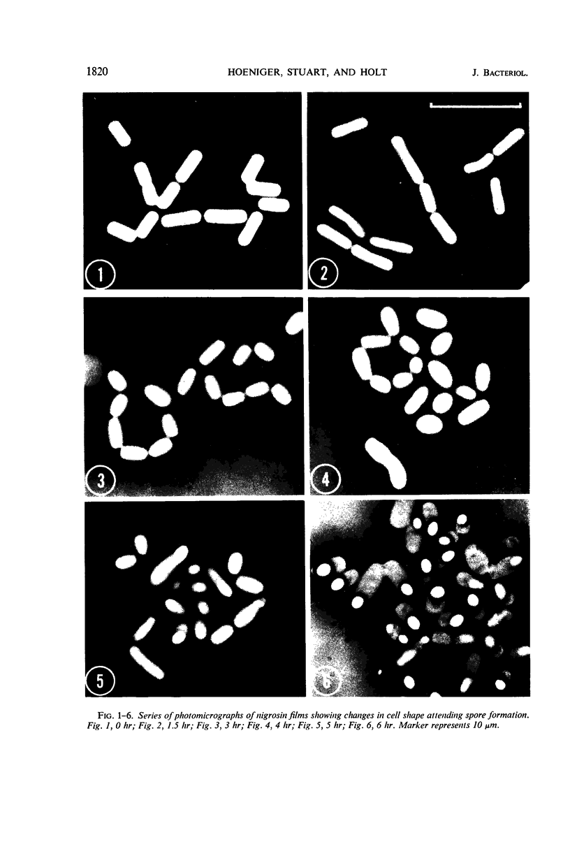
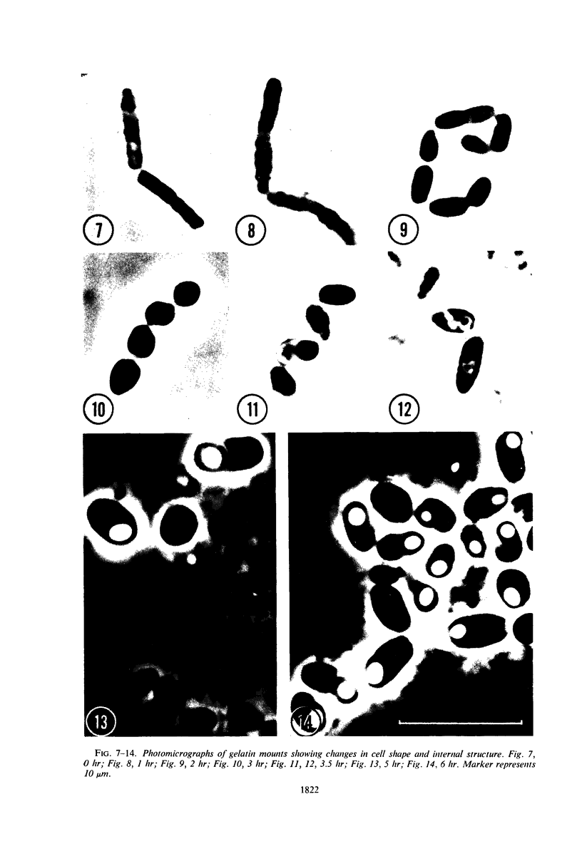
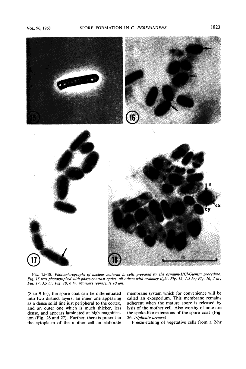
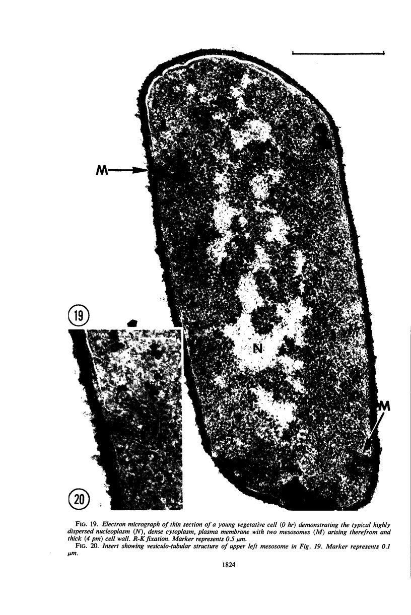
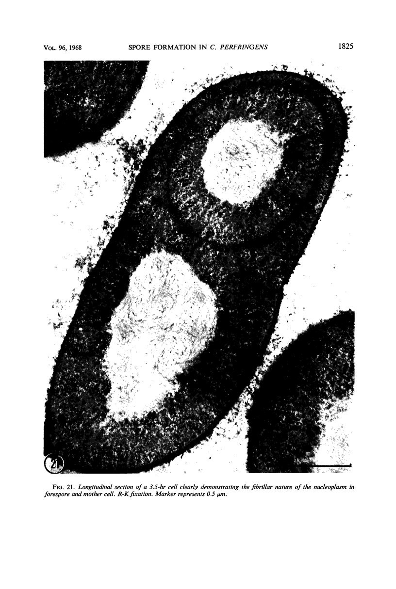
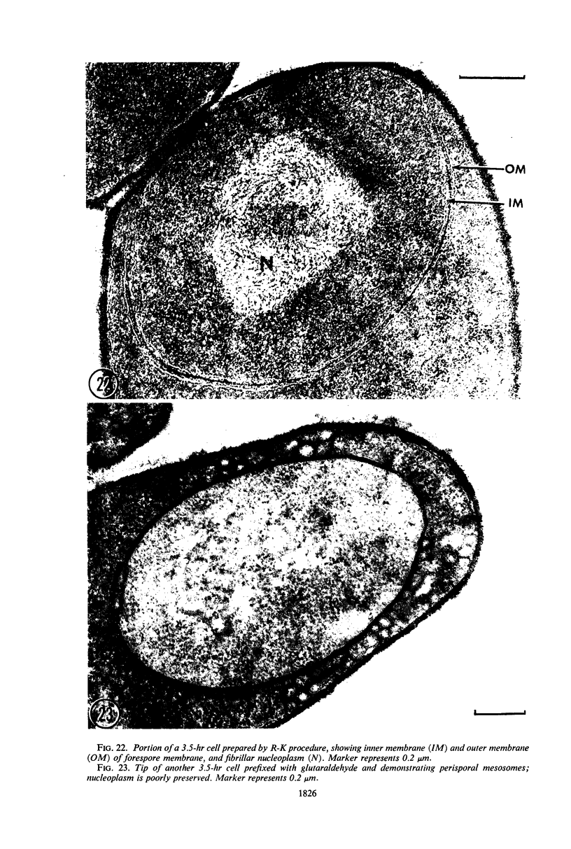
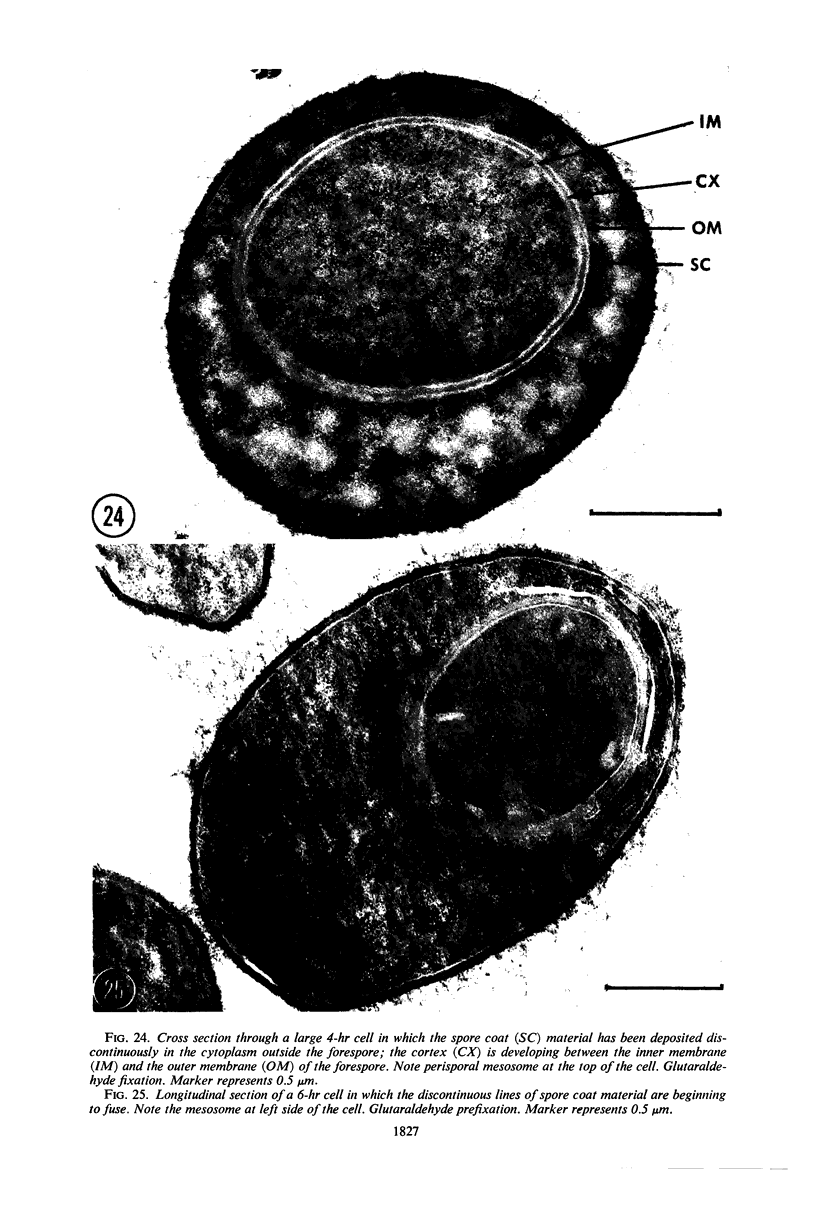
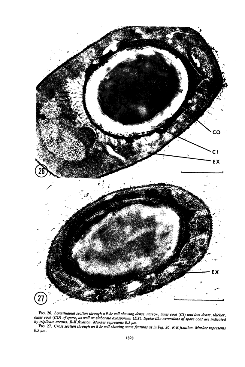
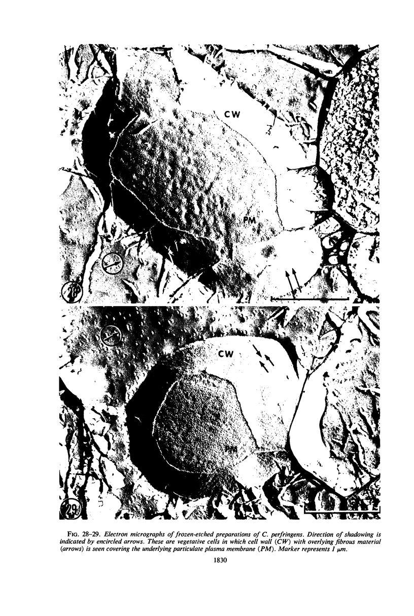
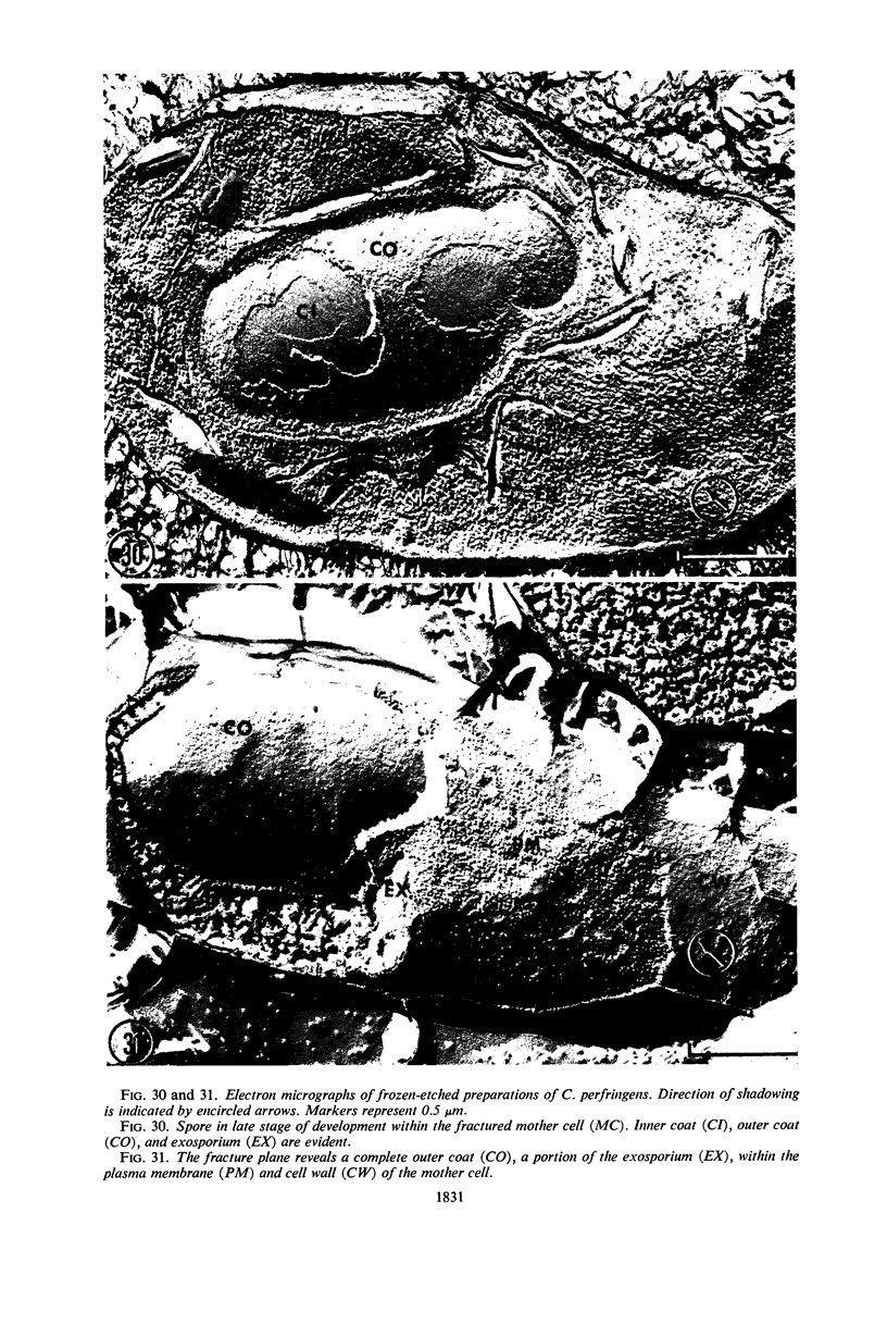
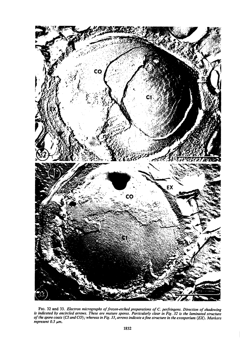
The sequential morphological events in spore formation by Clostridium perfringens type D were observed in Ellner's medium where 80 to 100% of the cells formed spores. Gross structural changes were studied with the light microscope under phase-contrast, and in fixed cells by the use of both nigrosin and Giemsa preparations. Fine structure was examined with the electron microscope in both thin sections and frozen-etched preparations. During the first 3 hr of incubation, the original rod-shaped cells became ellipsoid to ovoid in shape; by 5 to 6 hr, subterminal spores had developed within these enlarged cells. The fine structural sequence was in most respects identical to that in other Bacillaceae, although some stages were illustrated with particular clarity. A unique feature was the development of a convoluted, membranous exosporium which adhered to the outer surface of the two coats and had an unusual fine structure resembling a rectangular array of subunits.
Full text
PDF
Images in this article
Selected References
These references are in PubMed. This may not be the complete list of references from this article.
- ALBRYCHT H., TREMBOWLER P. Wpłlywniektórych czynników w podłloźu na zarodnikowanie laseczek z grupy Cl. perfringens. Med Dosw Mikrobiol. 1959;11(1):13–18. [PubMed] [Google Scholar]
- ANGELOTTI R., HALL H. E., FOTER M. J., LEWIS K. H. Quantitation of Clostridium perfringens in foods. Appl Microbiol. 1962 May;10:193–199. doi: 10.1128/am.10.3.193-199.1962. [DOI] [PMC free article] [PubMed] [Google Scholar]
- Bayen H., Frehel C., Ryter A., Sebald M. Etude cytologique de la sporulation chez Clostridium histolyticum. Souche sporogène et mutants de sporulation. Ann Inst Pasteur (Paris) 1967 Aug;113(2):163–173. [PubMed] [Google Scholar]
- ELLNER P. D. A medium promoting rapid quantitative sporulation in Clostridium perfringens. J Bacteriol. 1956 Apr;71(4):495–496. doi: 10.1128/jb.71.4.495-496.1956. [DOI] [PMC free article] [PubMed] [Google Scholar]
- Ellar D. J., Lundgren D. G. Fine structure of sporulation in Bacillus cereus grown in a chemically defined medium. J Bacteriol. 1966 Dec;92(6):1748–1764. doi: 10.1128/jb.92.6.1748-1764.1966. [DOI] [PMC free article] [PubMed] [Google Scholar]
- FITZ-JAMES P. C. Participation of the cytoplasmic membrane in the growth and spore fromation of bacilli. J Biophys Biochem Cytol. 1960 Oct;8:507–528. doi: 10.1083/jcb.8.2.507. [DOI] [PMC free article] [PubMed] [Google Scholar]
- Fitz-James P. C. MORPHOLOGY OF SPORE DEVELOPMENT IN CLOSTRIDIUM PECTINOVORUM. J Bacteriol. 1962 Jul;84(1):104–114. doi: 10.1128/jb.84.1.104-114.1962. [DOI] [PMC free article] [PubMed] [Google Scholar]
- Groom R. A., Strong D. H. Sporulation of Clostridium perfringens (welchii) in four laboratory media. J Appl Bacteriol. 1966 Aug;29(2):308–318. doi: 10.1111/j.1365-2672.1966.tb03481.x. [DOI] [PubMed] [Google Scholar]
- HALL H. E., ANGELOTTI R., LEWIS K. H., FOTER M. J. CHARACTERISTICS OF CLOSTRIDIUM PERFRINGENS STRAINS ASSOCIATED WITH FOOD AND FOOD-BORNE DISEASE. J Bacteriol. 1963 May;85:1094–1103. doi: 10.1128/jb.85.5.1094-1103.1963. [DOI] [PMC free article] [PubMed] [Google Scholar]
- HASHIMOTO T., NAYLOR H. B. Studies of the fine structure of microorganisms. II. Electron microscopic studies on sporulation of Clostridium sporogenes. J Bacteriol. 1958 Jun;75(6):647–653. doi: 10.1128/jb.75.6.647-653.1958. [DOI] [PMC free article] [PubMed] [Google Scholar]
- Hodgkiss W., Ordal Z. J., Cann D. C. The morphology and ultrastructure of the spore and exosporium of some Clostridium species. J Gen Microbiol. 1967 May;47(2):213–225. doi: 10.1099/00221287-47-2-213. [DOI] [PubMed] [Google Scholar]
- Holt S. C., Trüper H. G., Takács B. J. Fine structure of Ectothiorhodospira mobilis strain 8113 thylakoids: chemical fixation and freeze-etching studies. Arch Mikrobiol. 1968;62(2):111–128. doi: 10.1007/BF00410398. [DOI] [PubMed] [Google Scholar]
- KRASILNIKOV N. A., DUDA V. I., SOKOLOV A. A. VYROSTY NA POVERKHNOSTI SPOR ANAEROBNYKH BAKTERI I IZ RODA COLSTRIDIUM. Mikrobiologiia. 1964 May-Jun;33:454–458. [PubMed] [Google Scholar]
- Kim C. H., Cheney R., Woodburn M. Sporulation of Clostridium perfringens in a modified medium and selected foods. Appl Microbiol. 1967 Jul;15(4):871–876. doi: 10.1128/am.15.4.871-876.1967. [DOI] [PMC free article] [PubMed] [Google Scholar]
- MASON D. J., POWELSON D. M. Nuclear division as observed in live bacteria by a new technique. J Bacteriol. 1956 Apr;71(4):474–479. doi: 10.1128/jb.71.4.474-479.1956. [DOI] [PMC free article] [PubMed] [Google Scholar]
- MOOR H. DIE GEFRIER-FIXATION LEBENDER ZELLEN UND IHRE ANWENDUNG IN DER ELEKTRONENMIKROSKOPIE. Z Zellforsch Mikrosk Anat. 1964 Apr 28;62:546–580. [PubMed] [Google Scholar]
- OHYE D. F., MURRELL W. G. Formation and structure of the spore of Bacillus coagulans. J Cell Biol. 1962 Jul;14:111–123. doi: 10.1083/jcb.14.1.111. [DOI] [PMC free article] [PubMed] [Google Scholar]
- PIVNICK H., HAUSCHILD A. H., GORENSTEIN B., HABEEB A. F. EFFECT OF CONTROLLED PH ON TOXINOGENESIS BY CLOSTRIDIUM PERFRINGENS TYPE D. Can J Microbiol. 1965 Feb;11:45–55. doi: 10.1139/m65-007. [DOI] [PubMed] [Google Scholar]
- Pope L., Yolton D. P., Rode L. J. Appendages of Clostridium bifermentans spores. J Bacteriol. 1967 Oct;94(4):1206–1215. doi: 10.1128/jb.94.4.1206-1215.1967. [DOI] [PMC free article] [PubMed] [Google Scholar]
- Propst A., Möse J. R. Sporulation und Germination beim Clostridium butyricum M 55. Elektronenmikroskopische Untersuchung. Zentralbl Bakteriol Orig. 1966 Nov;201(3):373–395. [PubMed] [Google Scholar]
- REYNOLDS E. S. The use of lead citrate at high pH as an electron-opaque stain in electron microscopy. J Cell Biol. 1963 Apr;17:208–212. doi: 10.1083/jcb.17.1.208. [DOI] [PMC free article] [PubMed] [Google Scholar]
- RYTER A. ETUDE MORPHOLOGIQUE DE LA SPORULATION DE BACILLUS SUBTILIS. Ann Inst Pasteur (Paris) 1965 Jan;108:40–60. [PubMed] [Google Scholar]
- RYTER A., JACOB F. ETUDE AU MICROSCOPE 'ELECTRONIQUE DE LA LIAISON ENTRE NOYAU ET M'ESOSOME CHEZ BACILLUS SUBTILIS. Ann Inst Pasteur (Paris) 1964 Sep;107:384–400. [PubMed] [Google Scholar]
- RYTER A., KELLENBERGER E., BIRCHANDERSEN A., MAALOE O. Etude au microscope électronique de plasmas contenant de l'acide désoxyribonucliéique. I. Les nucléoides des bactéries en croissance active. Z Naturforsch B. 1958 Sep;13B(9):597–605. [PubMed] [Google Scholar]
- Remsen C. C. Fine structure of the mesosome and nucleoid in frozen-etched Bacillus subtilis. Arch Mikrobiol. 1968;61(1):40–47. doi: 10.1007/BF00704290. [DOI] [PubMed] [Google Scholar]
- Remsen C. C. The fine structure of frozen-etched Bacillus cereus spores. Arch Mikrobiol. 1966 Sep 8;54(3):266–275. doi: 10.1007/BF00408999. [DOI] [PubMed] [Google Scholar]
- Robinow C. F., Marak J. A fiber apparatus in the nucleus of the yeast cell. J Cell Biol. 1966 Apr;29(1):129–151. doi: 10.1083/jcb.29.1.129. [DOI] [PMC free article] [PubMed] [Google Scholar]
- Rode L. J., Crawford M. A., Williams M. G. Clostridium spores with ribbon-like appendages. J Bacteriol. 1967 Mar;93(3):1160–1173. doi: 10.1128/jb.93.3.1160-1173.1967. [DOI] [PMC free article] [PubMed] [Google Scholar]
- SCHNEIDER M. D., GRECZ N., ANELLIS A. Sporulation of Clostridium botulinum types A, B, and E, Clostridium perfringens, and putrefactive anaerobe 3679 in dialysis sacs. J Bacteriol. 1963 Jan;85:126–133. doi: 10.1128/jb.85.1.126-133.1963. [DOI] [PMC free article] [PubMed] [Google Scholar]
- SMITH A. G., ELLNER P. D. Cytological observations on the sporulation process of Clostridium perfringens. J Bacteriol. 1957 Jan;73(1):1–7. doi: 10.1002/path.1700730102. [DOI] [PMC free article] [PubMed] [Google Scholar]
- TAKAGI A., KAWATA T., YAMAMOTO S. Electron microscope studies on ultrathin sections of spores of the Clostridium group, with special reference to the sporulation and germination process. J Bacteriol. 1960 Jul;80:37–46. doi: 10.1128/jb.80.1.37-46.1960. [DOI] [PMC free article] [PubMed] [Google Scholar]
- TAKAGI A., KAWATA T., YAMAMOTO S., KUBO T., OKITA S. Electron microscopic studies on ultrathin sections of spores of Clostridium tetani and Clostridium histolyticum, with special reference to sporulation and spore germination process. Jpn J Microbiol. 1960 Apr;4:137–155. doi: 10.1111/j.1348-0421.1960.tb00162.x. [DOI] [PubMed] [Google Scholar]
- Takagi A., Nakamura K., Ueda M. Electron microscope studies of the intracytoplasmic membrane system in Clostridium tetani and Clostridium botulinum. Jpn J Microbiol. 1965 Sep;9(3):131–143. doi: 10.1111/j.1348-0421.1965.tb00282.x. [DOI] [PubMed] [Google Scholar]
- Walker P. D., Thomson R. O., Baillie A. Fine structure of clostridia with special reference to the location of antigens and enzymes. J Appl Bacteriol. 1967 Dec;30(3):444–449. doi: 10.1111/j.1365-2672.1967.tb00322.x. [DOI] [PubMed] [Google Scholar]
- Yolton D. P., Pope L., Williams M. G., Rode L. J. Further electron microscope characterization of spore appendages of Clostridium bifermentans. J Bacteriol. 1968 Jan;95(1):231–238. doi: 10.1128/jb.95.1.231-238.1968. [DOI] [PMC free article] [PubMed] [Google Scholar]
- van Iterson W. Symposium on the fine structure and replication of bacteria and their parts. II. Bacterial cytoplasm. Bacteriol Rev. 1965 Sep;29(3):299–325. doi: 10.1128/br.29.3.299-325.1965. [DOI] [PMC free article] [PubMed] [Google Scholar]