Abstract
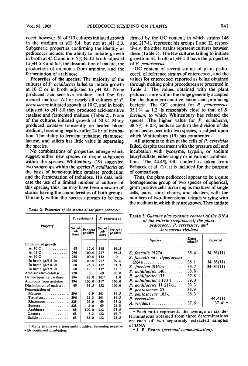
The pediococci residing on plants resemble the lactobacilli, but they differ from the streptococci in their limited distribution and low population level on plants. They are a subgroup within the genus Pediococcus which grow freely in neutral media and require neither NaCl nor CO2. They are most readily recognized by the ability to initiate growth in liquid media, acidified to pH 5.0, which contain 1.5% sodium acetate. In stained preparations the cells occur singly and in pairs, short chains, and clusters. The occurrence of two-dimensional tetrads may be rare; this varies with the individual culture and with the culture medium. The terminal pH in 2% glucose broth varies from 3.6 to 4.3. Ability to initiate growth at 45 C, production of ammonia from arginine, dissimilation of malate, and fermentation of arabinose are confirmatory characteristics. The subgroup contains only two quite similar, but differentiable, species. P. acidilactici initiates growth at 50 C and produces catalase on heated blood medium but does not produce acid-sensitive catalase; a majority of the strains fail to initiate growth at 10 C and many fail to ferment maltose and lactose. P. pentosaceus initiates growth at 10 C but not at 50 C and produces acid-sensitive catalase; catalase production on heated blood medium is transient; a majority of the cultures ferment maltose, salicin, and trehalose. No carbohydrate serves reliably to differentiate between the species. The guanine plus cytosine ratio of P. pentosaceus deoxyribonucleic acid (DNA) was determined to be 35.1 ± 1.2 and that of P. acidilactici DNA is 38.5 ± 0.8.
Full text
PDF
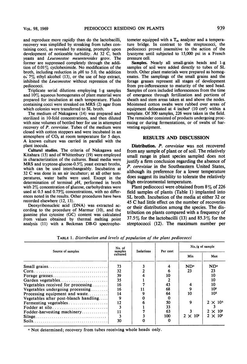
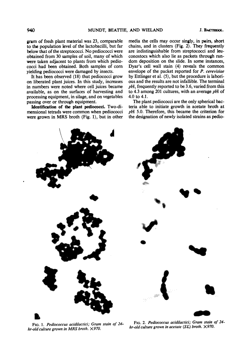
Images in this article
Selected References
These references are in PubMed. This may not be the complete list of references from this article.
- DEIBEL R. H., NIVEN C. F., Jr Comparative study of Gaffkya homari, Aerococcus viridans, tetrad-forming cocci from meat curing brines, and the genus Pediococcus. J Bacteriol. 1960 Feb;79:175–180. doi: 10.1128/jb.79.2.175-180.1960. [DOI] [PMC free article] [PubMed] [Google Scholar]
- Dyar M. T. A Cell Wall Stain Employing a Cationic Surface-active Agent as a Mordant. J Bacteriol. 1947 Apr;53(4):498–498. doi: 10.1128/jb.53.4.498-498.1947. [DOI] [PMC free article] [PubMed] [Google Scholar]
- Ettlinger L., Herrmann C., Staehelin A. Morphologie und Teilungsmodus von Pediococcus cerevisiae. Pathol Microbiol (Basel) 1965;28(4):561–566. [PubMed] [Google Scholar]
- FELTON E. A., EVANS J. B., NIVEN C. F., Jr Production of catalase by the pediococci. J Bacteriol. 1953 Apr;65(4):481–482. doi: 10.1128/jb.65.4.481-482.1953. [DOI] [PMC free article] [PubMed] [Google Scholar]
- Kerbaugh M. A., Evans J. B. Aerococcus viridans in the hospital environment. Appl Microbiol. 1968 Mar;16(3):519–523. doi: 10.1128/am.16.3.519-523.1968. [DOI] [PMC free article] [PubMed] [Google Scholar]
- MARMUR J., DOTY P. Determination of the base composition of deoxyribonucleic acid from its thermal denaturation temperature. J Mol Biol. 1962 Jul;5:109–118. doi: 10.1016/s0022-2836(62)80066-7. [DOI] [PubMed] [Google Scholar]
- Mundt J. O., Graham W. F., McCarty I. E. Spherical Lactic Acid-producing Bacteria of Southern-grown Raw and Processed Vegetables. Appl Microbiol. 1967 Nov;15(6):1303–1308. doi: 10.1128/am.15.6.1303-1308.1967. [DOI] [PMC free article] [PubMed] [Google Scholar]
- Mundt J. O., Hammer J. L. Lactobacilli on plants. Appl Microbiol. 1968 Sep;16(9):1326–1330. doi: 10.1128/am.16.9.1326-1330.1968. [DOI] [PMC free article] [PubMed] [Google Scholar]


