Abstract
This article describes and compares the fat pad clearance procedure developed by DeOme KB et al.1 and the sparing procedure developed by Brill B et al.2, followed by the mammary epithelial transplant procedure. The mammary transplant procedure is widely used by mammary biologists because it takes advantage of the fact that significant development of the mammary epithelium doesn't occur until after puberty. At 3 weeks of age, growth of the mammary epithelial tree is confined to the vicinity of the nipple and the fat pad is largely devoid of mammary epithelium, but by 7 weeks of age the epithelial ductal tree extends throughout the entire fat pad. Therefore, if this small portion of the fat pad containing epithelium, the region between the nipple and the lymph node, is removed at 3 weeks of age, the endogenous epithelium will never populate the mammary fat pad and the fat pad is described as "cleared". At this time, mammary epithelium from another source can be transplanted in the cleared fat pad where it has the potential to extend mammary ductal trees through out the fat pad. This procedure has been utilized in many experimental models including the examination of tumor phenotype in transgenic mammary epithelial tissue without the confounding effects of genotype on the entire animal3, in the identification of mammary stem cells by transplanting cells in limited dilution4,5, determining if hyperplastic nodules proceed to mammary tumors6, and to assess the effect of prior hormone exposure on the behavior of the mammary epithelium7,8.
Three week old host mice are anesthetized, cleaned and restrained on a surgical stage. A mid-sagittal incision is made through the skin, but not the peritoneum, extending from the pubis to the sternum. Oblique cuts are made through the skin from the mid-sagittal incision across the pelvis toward each leg. The skin is pulled away from the peritoneum to expose the 4th inguinal mammary gland. The fat pad is cleared by removing the fat pad tissue anterior to the lymph node. Epithelium fragments or epithelial cells are transplanted into the remaining cleared fat pad and the mouse is closed.
Protocol
1. Preparation of Transplant Epithelium
Fresh or frozen donor epithelial tissue can be transplanted into the cleared fat pad, but prior preparation of frozen donor epithelium is more convenient with a minimal decrease in transplantation efficiency (>85% efficiency). The protocol for the preparation of frozen donor epithelium is as follows:
Before preparing donor transplant epithelium, prepare freeze media for epithelium and aliquot about 1ml into cryovials. This can be stored at 4°C for up to 6 months until needed. Freeze media contains DMEM/F12 media buffered with HEPES and NaHCO3, 10% fetal bovine serum (FBS) and 10% dimethyl sulphoxide (DMSO).
Harvest mammary epithelium from a recently euthanized female mouse.
Wash the mouse with 70% EtOH.
Make a mid-sagittal cut through the body wall extending from the pubis to the sternum without piercing the serous membranes of the ventral body cavity. Make an oblique cut from the initial incision at the pubis toward each hind leg (Figure 1).
Peel the body wall away from the serous membranes to expose the 5th inguinal mammary gland attached to the fascia on the underside of the body wall. The best way to achieve this is by grasping the serous membranes carefully with the curved portion of a pair of curved forceps, being careful not to pierce the ventral body cavity. Simultaneously, grasp the cut edge of the integument with straight serrated forceps and pull the body wall away from the serous membranes.
Identify the lymph node within the mammary gland. The lymph node is located at the intersection of three prominent blood vessels in the mammary gland and can be felt as a dense nodule within the fat pad (Figure 2). With the tip of a pair of eye scissors, nip the adipose tissue surrounding the node. With a fore finger on the epidermal side of the skin, apply pressure to raise the node out of the fat pad and pinch it off with a pair of curved forceps. Discard the lymph node.
While holding the margins of the skin, grasp the mammary fat pad with the curved edge of a pair of curved forceps and gradually peel the fat pad away from the body wall. Follow the direction of the fat pad towards the dorsal surface of the mouse as you remove the entire mammary gland in one piece.
Put the mammary gland on a sterile plastic cutting block. Add a small amount of media, about 200 μl, to the surface of the gland to keep it moist. Mince the gland into 1mm pieces with a fresh razor blade. Divide the minced gland into four portions and distribute among four cryovials of freeze media with forceps. Put the cryovials in a Nalgene "Mr. Frosty cell freezer which has 250ml of isopropyl alcohol in the outer compartment. Put the Nalgene freezer in a -70°C freezer for 24 hours. Store frozen vials of mammary fragments in liquid nitrogen until required for transplantation. Each cryovial will contain enough mammary gland fragments to transplant into the cleared fat pads of 4 to 8 host mice.
2. Preparation of Transplant Cells
Our lab has transplanted different cell types, including immortalized mammary epithelial cell lines, mammary tumor cell lines or primary mammary cells into a mouse cleared fat pad. The protocol for the preparation of Comma-D cells for transplantation is as follows:
Comma-D cells are cultured in regular MECL media containing DMEM:F12 supplemented with 2% Adult Bovine Serum (ABS), mEGF, Insulin, Antibiotic/Antimycotic (AB/AM).
Harvest Comma-D cells when the cells are 85%-90% confluent.
Centrifuge the cell suspension at 1200rpm for 5 minutes to pellet the cells. Resuspend the cells in fresh MECL media and adjust the cell concentration to 5 x 10⁶/ml.
For the transplantation of primary mammary cells, the primary mammary epithelial cells are isolated from mouse 5th mammary glands through mechanical and enzymatic dissociation. Cells are resuspended in DMEM:F12 media and adjusted to the desired concentration, usually 5 x 10⁶/ml.
3. Standard Procedure for Clearing the Mouse Mammary Fat Pad
Anesthetize the three week old host mouse. Select mice that are <12 g to ensure that mammary ductal growth has not been initiated. We use 200mg/kg of body weight of 2,2,2-tribromoethanol.
After the mouse is anesthetized, firmly restrain the mouse, ventral side up with limbs outstretched, on a surgical stage so that the abdomen is taunt. Prior to surgery, administer analgesics; either bupivacaine (<1mg/kg) or buprenorphine (0.05mg/kg), subcutaneously.
Grasp the skin with forceps a few millimeters above the pelvis and lift it up from the abdomen. Nip the skin with scissors and to make a mid-sagittal cut through the body wall extending from the pubis to the sternum. Do not pierce the serous membranes of the ventral body cavity. Do not make your initial incision too close to the urethra so that wound clips will not interfere with the mouse's ability to urinate.
From the initial incision, make oblique cuts through the skin from the mid-line across the pelvis towards each hind leg.
Use the curved area of the curved forceps to gently grasp the serous membranes. Use tissue forceps to grasp the cut margin of the skin and pull the body wall away from the serous membranes and expose the 5th inguinal mammary gland located on the fascia on the underside of the body wall.
Cauterize the nipple, node and two arteries leading to the node (Figure 2)
Separate the fourth and fifth glands. Cauterize the margin of the fifth gland to prevent ingrowth from the 6th gland.
Divide the fat pad of the fourth mammary gland by cutting across the fat pad with scissors just ventral (side towards nipple) of the node. Use curved forceps to firmly grasp the ventral portion of the fat pad and pull it away from the body wall in one piece. Press the ventral portion of the fat pad between two slides and put in a glass histology staining dish containing Carnoy's fixative. This tissue will be fixed and stained later with carmine alum solution to check for cleared margins (Figure 3). Clean the fascia around the region of the removed portion of the fat pad to remove all traces of the mammary gland with the curved forceps.
4. Alternative "Sparing Procedure for Clearing the Fat Pad
After one has become competent with the standard procedure for clearing the mouse mammary fat pad, they may prefer to attempt the less invasive sparing procedure developed by Brill et al. (2008)2.
Prepare the mouse as for the standard procedure.
Grasp the nipple of the fourth inguinal mammary gland and cut a 3-5 mm circumference circle around the nipple with the tip of scissors.
Pull the nipple away from the body to detach the cut portion of skin and draw the fourth inguinal mammary gland out through the hole.
Identify the node. Use the cautery to cauterize the node and the 6th mammary gland through the hole.
Cut and detach the most superficial portion of the 5th mammary gland just before the node. The nipple, circular area of skin and portion of the mammary fat pad containing epithelium are removed in one piece.
5. Tissue/Cell Transplantation
For transplanting epithelium fragments, defrost a cryovial of epithelium and transfer the epithelial fragments to a Petri dish containing fresh media to dilute out the DMSO. Make a small hole or pocket in the cleared fat pad with scissor tip for the new epithelium. Put a 1mm piece of donor epithelium into the pocket with needle forceps. Hold the pocket closed with tissue forceps as you withdraw the needle forceps so the tissue remains in place.
For transplanting cells, inject 50,000 cells in 10 μl of media into each fat pad using a 50μl Hamilton syringe with a 22s gauge needle.
6. Closing/Revival of Mouse
Arrange the skin for closing. Grasp opposing margins of skin, hold them together with forceps, lift them away from the serous membranes of the ventral body cavity and close with wound clips. Typically, it is necessary to use two clips to close the mid-sagittal incision and one clip for each oblique incision for the standard procedure. Avoid obstructing the urethra. It may be necessary to close gaps in the skin at the intersection of the mid-sagittal and oblique incisions with a suture. The sparing procedure will only require one wound clip for each side.
. Place the mouse in her cage and keep the cage under a heating lamp until the mouse has revived.
The wound clips should be removed after 2 weeks.
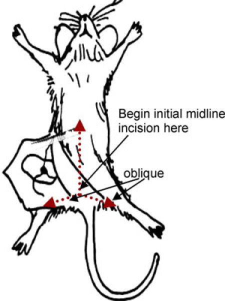 Figure 1. Demonstration of incision pattern.
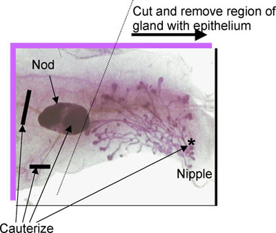 Figure 2. Location of lymph node, blood vessels, nipple and endogenous epithelium. Cauterize at locations indicated.
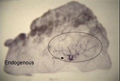 Figure 3. Whole mount demonstrates that endogenous epithelium is restricted to the area within the circle and therefore margins are "clear'.
7. Representative Results
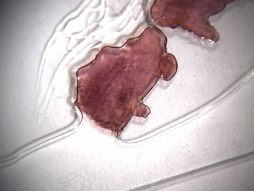 Figure 4. Whole mount of removed fat pad from 3 week old mouse showing cleared margins.
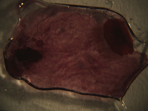 Figure 5. Whole mount from a 10 week old mouse showing fat pad cleared of endogenous mammary epithelium.
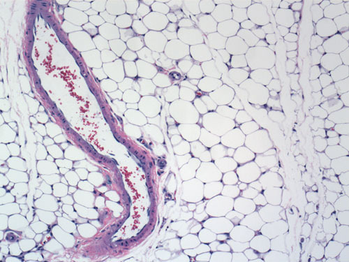 Figure 6. Hematoxylin and eosin stained section of a cleared fat pad from a 10 week old mouse.
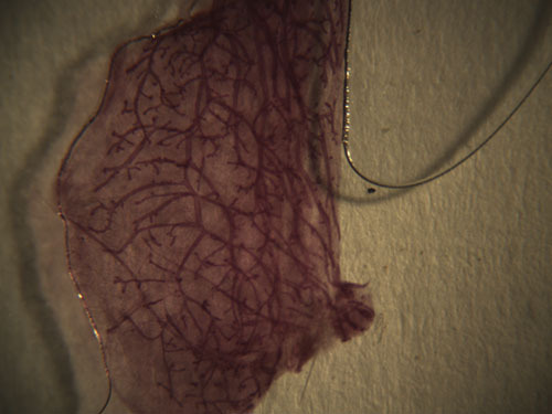 Figure 7. Whole mount from a 10 week old mouse showing normal mammary ductal tree developed from transplanted epithelium.
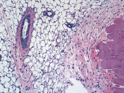 Figure 8. Hematoxylin and eosin stained section of mammary ducts developed from transplanted epithelium.
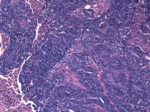 Figure 9. Hematoxylin and eosin stained section of a mammary tumor developed from transplanted epithelium.
Discussion
In this report, we present two alternative methods for clearing the mouse mammary fat pad in preparation for mammary epithelial transplants. The standard procedure has been in use since its development in 1959 by DeOme et al.1 While more invasive, the standard method is the preferred method for training the novice because the structure and orientation of the mammary gland are readily viewed. However, the sparing procedure may be preferred by the more experienced surgeon because of several advantages2. Most importantly, the sparing procedure is less invasive, requires less time and can be performed using a brief period of inhalation anesthesia. This reduces the recovery period for the mouse. The sparing procedure can also be performed on younger mice (<21 days) ensuring successful clearance of epithelium. In addition, there is a reduction of tissue adhesion after surgery which may be advantageous if experiments require repeat surgery to remove primary tumors, yet allow for continued long-term observation of the mouse for metastasis.
The transplant portion of the procedure takes advantage of the fact that full development of the mammary gland does not occur until after puberty. Prior removal of the small portion of the fat pad containing the endogenous epithelium "clears" the remaining fat pad of the young mouse and eliminates the potential for the development of epithelial ductal trees. Experimental epithelium can be transplanted into the cleared fat pad of the host mouse and assessment of the resulting outgrowths can be made without the confounding effects of the endogenous host epithelium. Therefore, this procedure is useful for assessing the capacity of mammary epithelial cells to develop in vivo.
References
- DeOme KB, Faulkin LJ, Jr, Bern HA, Blair PB. Development of mammary tumors from hyperplastic alveolar nodules transplanted into gland-free mammary fat pads of female C3H mice. Cancer Res. 1959;19:515–520. [PubMed] [Google Scholar]
- Brill B, Boecher N, Groner B, Shemanko CS. A sparing procedure to clear the mouse mammary fat pad of epithelial components for transplantation analysis. Laboratory Animals. 2008;42:104–110. doi: 10.1258/la.2007.06003e. [DOI] [PubMed] [Google Scholar]
- Jerry DJ, Kittrell FS, Kuperwasser C, Laucirica R, Dickinson ES, Bonilla PJ, Butel JS, Medina D. A mammary-specific model demonstrates the role of the p53 tumor suppressor gene in tumor development. Oncogene. 2000;19:1052–1058. doi: 10.1038/sj.onc.1203270. [DOI] [PubMed] [Google Scholar]
- Shackleton M, Vaillant F, Simpson KJ, Stingl J, Smyth GK, Asselin-Labat M-L, Wu L, Lindman GJ, Visvader JE. Generation of a functional mammary gland from a single stem cell. Nature. 2006;439:84–88. doi: 10.1038/nature04372. [DOI] [PubMed] [Google Scholar]
- Stingl J, Eirew P, Ricketson I, Shackleton M, Vaillant F, Choi D, Li HI, Eaves CJ. Purification and unique properties of mammary epithelial stem cells. Nature. 2006;439:993–997. doi: 10.1038/nature04496. [DOI] [PubMed] [Google Scholar]
- Medina D. Preneoplastic Lesions in Murine Mammary Cancer. Cancer Res. 1976;36:2589–2595. [PubMed] [Google Scholar]
- Wagner K-U, Boulanger CA, Henry Sgagias, Hennighausen M, Smith GHL. An adjunct mammary epithelial cell population in parous females: its role in functional adaptation and tissue renewal. Development. 2002;129:1377–1386. doi: 10.1242/dev.129.6.1377. [DOI] [PubMed] [Google Scholar]
- Dunphy KA, Blackburn AC, Haoheng Y, O Connell LR, Jerry DJ. Estrogen and progesterone induce persistent increases in p53-dependent apoptosis and suppress mammary tumors in BALB/c-Trp53+/- mice. Breast Cancer Res. 2008;10(3):R43–R43. doi: 10.1186/bcr2094. [DOI] [PMC free article] [PubMed] [Google Scholar]


