Abstract
Optical projection tomography (OPT) imaging is a powerful tool for three-dimensional imaging of gene and protein distribution patterns in biomedical specimens. We have previously demonstrated the possibility, by this technique, to extract information of the spatial and quantitative distribution of the islets of Langerhans in the intact mouse pancreas. In order to further increase the sensitivity of OPT imaging for this type of assessment, we have developed a protocol implementing a computational statistical approach: contrast limited adaptive histogram equalization (CLAHE). We demonstrate that this protocol significantly increases the sensitivity of OPT imaging for islet detection, helps preserve islet morphology and diminish subjectivity in thresholding for tomographic reconstruction. When applied to studies of the pancreas from healthy C57BL/6 mice, our data reveal that, at least in this strain, the pancreas harbors substantially more islets than has previously been reported. Further, we provide evidence that the gastric, duodenal and splenic lobes of the pancreas display dramatic differences in total and relative islet and β-cell mass distribution. This includes a 75% higher islet density in the gastric lobe as compared to the splenic lobe and a higher relative volume of insulin producing cells in the duodenal lobe as compared to the other lobes. Altogether, our data show that CLAHE substantially improves OPT based assessments of the islets of Langerhans and that lobular origin must be taken into careful consideration in quantitative and spatial assessments of the pancreas.
Key words: pancreas, OPT, islets, contrast normalization, β-cell mass
Introduction
The mouse pancreas consists of the splenic, duodenal and gastric lobes. The latter, which is associated with the splenic lobe from which it is developed, is conserved in a number of rodent species and is suggested to correspond to the auricle or “ear of the pancreas” in humans.1 The possibility to accurately measure β-cell mass (BCM) and islet number is a prerequisite for interpretation of results in many areas of diabetes research. In particular, the possibility to study diabetes disease dynamics and/or the effect of genetic mutations on islet/β-cell development often depend on such assessments. Pioneering morphometric studies of BCM in mice were performed already 50 years ago2,3 and until this day assessments of BCM have to a large extent relied on stereological sampling techniques. OPT4 which is essentially the optical equivalent of x-ray tomography,5 enables high-resolution 3D visualization of protein expression patterns in small biological specimens by the detection of antibody markers. Previously, we described an OPT-based protocol that enables extraction of parameters such as BCM, islet number and 3D islet co-ordinates, throughout the volume of the intact pancreas.6 As such, OPT imaging has been successfully utilized e.g., in studies of BCM dynamics during T1D disease progression7 and for evaluating the impact on BCM by targeted gene ablation.8 In this report we attempt to further increase the sensitivity of OPT for islet/BCM assessments by incorporating a computational statistical approach; CLAHE9 to the tomographic projection data. CLAHE was originally applied to medical imaging as an improvement to adaptive histogram localization (AHE).10–12 CLAHE and AHE operate by equalizing the histogram of individual non-overlapping blocks across the digital image area (see Materials and Methods, Sup. Fig. 1 and Sup. Video 1) but in contrast to AHE, CLAHE constrain the image contrast in order to avoid the appearance of saturated pixels and the amplification of background noise. Utilizing this technique, we here investigate the lobular distribution of islets/BCM in mice and reveal significant heterogeneities between the lobular compartments of the pancreas.
Results
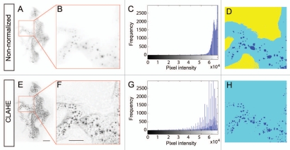
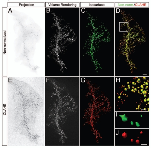
By applying the CLAHE script to the individual projection images before tomographic reconstruction we could increase the relative ratio between insulin-labelled islets (from here on referred to as islets) with weak signal intensity and non-labelled cells without interference from quasi-saturated areas (Fig. 1). This procedure conveyed several advantages for the tomographic reconstruction process. Most importantly, it enabled the detection of islets exhibiting very weak signals intensities, which would normally be “thresholded out” during the tomographic reconstruction process to preserve the morphology of islets exhibiting stronger signal intensities. To illustrate this, projections of identical data sets with and without normalization is shown together with reconstructed volume and iso-surface renderings (Fig. 2). In comparison both to our previous OPT-based protocol7 and reported point counting morphometric analyses13 our data suggest that the developed approach (implementing CLAHE) allows for the visualization of a considerably larger amount of islets. When applied to the entire pancreas (all three lobes) from 8-week female C57Bl/6 mice, the CLAHE based protocol revealed an average islet count of 4,483 (SEM ± 216, n = 5). This figure is significantly higher (≈80%) than what has been previously described by point counting morphometric assessments of the same strain at the same age.13 It should be noted however that differences in how an islet is defined (to be counted as an islet) differs between the approaches (see Material and Methods and ref. 13), which may to some extent contribute to this discrepancy. To exclude the possibility that CLAHE induced artefacts that contributed to our figures, pancreata were stained with secondary antibody only, scanned and subjected to CLAHE before reconstruction. We could hereby not detect any objects/volumes introduced by the contrast normalisation routine (Sup. Fig. 2). Furthermore, in post scanning morphometric assessments (see Material and Methods) performed on tissue sections, all individual islets detected by the CLAHE processed OPT data could be accounted for (data not shown). Given that we in our protocol exclude objects smaller than 10 voxels (to prevent including artificial objects by general noise, see Materials and Methods) it should be noted that in these analyses we could detect additional small “islets” consisting of a few insulin positive cells. Together, our results show that CLAHE significantly increases the sensitivity of OPT for islet detection, decreases subjectivity in islet segmentation and as a consequence improves preservation of islet morphology (Fig. 2D and H–J). As such, we predict that the CLAHE approach also increases the accuracy in volumetric assessments of islet β-cell volumes.
Figure 1.
CLAHE facilitates OPT-based assessments of islets and BCM. (A–H) Illustration of the effect of CLAHE (E–H) as compared to non-normalized tomographic projection data (A–D). (A and B) Original projection image. (E and F) Images showing effect of CLAHE when applied to (A). (C and G) Intensity histograms of the patches shown in (B and F) respectively. (D and H) Segmentation of the patches shown in (B and F) respectively. Scale bar in (E) is 2 mm in (A and E). Scale bar in (F) is 1 mm in (B and F).
Figure 2.
Adaptive contrast normalization facilitates detection of islets by OPT imaging. (A–J) OPT images of splenic pancreas from eight-week C57Bl/6 mice labelled for insulin showing non-normalized images (A–C) and CLAHE processed images (E–G) of OPT projection data (A and E), resultant volume-renderings (B and F) and iso-surface reconstructions (C and G). (D) Overlay of the non-normalized data in ((C), pseudo colored green) and the CLAHE processed data ((G), pseudo colored red). CLAHE processing enables detection of a larger number of insulin labelled islets (red only). (H) high magnification image corresponding to inset in (D). (I and J) Representative high magnification iso-surface reconstructions (of high intensity islets) illustrating the effect on islet morphology with non-normalized (I, green) versus CLAHE processed data (J, red). Scale bar in (D) is 1,000 µm in (A–G). Scale bar in (H) is 500 µm. Scale bar in (I) is 500 µm in (I and J).
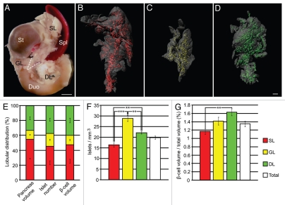
The relative distribution of islets/BCM between the pancreatic lobes has previously not been reported. In particular, no such accounts are available utilizing a high-resolution approach that is independent on the extrapolation of two-dimensional data. To address this issue, we subjected insulin stained splenic, duodenal and gastric lobes from healthy eight-week C57Bl/6 mice to OPT imaging implementing the CLAHE protocol. These analyses revealed striking heterogeneities between the three lobes of the pancreas with regard to both islet and BCM distribution (Fig. 3). Notably, the duodenal lobe displayed a 20% higher relative islet count (islets per pancreas volume) and a 40% higher BCM (β-cell volume per pancreas volume) compared with the splenic lobe (Fig. 3F and G). An even more dramatic difference in islet count was observed between the gastric and the splenic lobe. The gastric lobe, which constitutes ≈12% of the total pancreatic volume, strikingly harbors close to 75% more islets per pancreas volume than does the splenic lobe (Fig. 3F). The average size of these islets, which contribute to ≈17% of the total islet count, is however smaller than those of the splenic and duodenal lobes and contribute to ≈12% of the overall BCM (Fig. 3E). Collectively these data reveal that the three lobes of the pancreas display significant heterogeneities regarding their relative distribution of islets and BCM.
Figure 3.
The lobular compartments of the pancreas display significant differences in relative β-cell mass and islet densities. (A) Photomicrograph of a gut segment including the stomach, duodenum, spleen and pancreas from a C57Bl/6 mouse at eight weeks. For comparative OPT assessments, the lobular compartments were separated based on morphologic features and the relationship to embryological origin (indicated by broken lines). (B–D) Representative iso-surface reconstructions of the islet β-cell volumes in the splenic (B, red), gastric (C, yellow) and duodenal (D, green) lobes of the pancreas from C57Bl/6 mice at eight weeks. In (B–D) the exocrine parenchyma (grey) is reconstructed based on the signal from endogenous tissue autofluorescence. (E) Graph showing the total lobular distribution of pancreatic tissue, insulin+ islets and β-cell volumes. (F) Graph showing the lobular distribution of insulin+ islets/mm3 of pancreatic tissue. (G) Graph showing lobular β-cell volume/total lobe volume. In (E–G), n = 5, values are given ±SEM. Significance levels indicated correspond to **p < 0.01, ***p < 0.001. CLAHE processing was implemented for all display items in (B–G). Scale bar in (A) is 2 mm. Scale bar in (D) is 1 mm in (B–D). Abbreviations: GL, gastric lobe; DL, duodenal lobe; SL, splenic lobe; St, stomach; Spl, spleen; Duo, duodenum.
Discussion
Technologies for accurate spatial and quantitative assessments of pancreatic cell populations are of utmost importance to study various aspects of both type 1 and type 2 diabetes.14 In this report we describe and implement a computational statistical approach that facilitates OPT based assessments of islets and BCM distributions at a number of levels. The protocol is integratable for use with commercial OPT scanners and as such it should be easy to adopt by the research community. It should also be possible to translate to OPT-based studies of other organ systems and/or protein distribution patterns.
The occurrence of functionally different populations of islets has been reported and it was recently suggested that “small” islets contain more insulin per islet volume (in situ) and secrete insulin more efficiently (in vitro).15 However, previous studies have not addressed the relative lobular distribution patterns of islets or BCM. Our data now reveals that the pancreatic lobes display dramatic differences with regards to these parameters. In particular, the gastric pancreatic lobe stands out by harboring a disproportional amount of islets, which displays a smaller average size than those of the other lobes. Doubtlessly, these heterogeneities indicate that lobular context must be taken into careful consideration in studies aimed at addressing for example islet count and/or BCM in experimental models for diabetes, the effect of gene ablation on β-cell and/or islet development etc., i.e., such studies should not be based on the assessments of one lobular compartment only, a practise often observed in the literature. Whether these heterogeneities have significance also for physiological and/or pathological conditions of the pancreas remains to be determined. In view of our results, we speculate that the lobular compartments of the pancreas may facilitate comparative assessments aimed at addressing how β-cell mass and islet distribution is established.
Methods
Animals, tissue preparation and OPT scanning.
Adult C57Bl/6 mice (Taconic) were killed, the pancreata dissected free and processed as described in reference 7. For comparative assessments the pancreatic lobes were scanned individually. Antibodies used were GP α-Ins (DAKO) and Alexa 594 (Molecular probes). Scan settings (identical for all specimen) were: rotation degree 0.45 µm; pixel size 18 × 18 µm; resolution 1,024 × 1,024 pixels; exposure time 1,000 ms. All specimens were scanned at the same zoom factor. Animal experimentation was performed in accordance with international, national and institutional laws and guidelines.
Contrast normalization.
CLAHE performs an operation based on local statistics on a user defined window. An image is first divided into non-overlapping blocks (Sup. Fig. 1). Pixel intensities in a block are then calculated and described by the histogram for that block. To avoid saturating pixels and amplifying noise in homogenous areas, a clip limit is set for the histogram that constrains the permissible contrast. Pixels above the clip limit are redistributed evenly across the intensity values below that limit (Sup. Fig. 1B and C). The histogram is then equalized so that frequently occurring intensities in the histogram are assigned more values (Compare Sup. Fig. 1B and D). Artificially induced boundaries between blocks are eliminated using bilinear interpolation at the border regions. The CLAHE contrast normalization algorithm was implemented in MATLAB v. 7.11.0.584 and IP Toolbox v. 7.1 (64-bit workstation, Windows XP, 16 GB of RAM, 2.99 GHz) using the MATLAB built-in function, “adapthisteq”9 and applied to the individual projection images. CLAHE properties were set as follows, tile size (256 × 256) corresponding to 4 × 4 pixels block in our case, clip limit 0.01 (default in adapthisteq), output image data range full (i.e., not bounded by the original image range) and histogram distribution set to uniform. Average time to normalize a single 16-bit projection image (1,024 × 1,024) was 39.1 seconds. Total number of processed blocks per image was 65,536, i.e., 1,0242/42.
Post-acquisition corrections and tomographic reconstruction.
Post-acquisition alignment was calculated using LLS-Gradient based A-value tuning (Cheddad et al. in submission). Based on the scanning software manufacturers (Skyscan) calculations of the cone-beam geometry within the optical system, the line “Object to Source (mm)” in each *.log file was manually edited from 1,000,000 (parallel imaging geometry) to -436 (cone beam imaging geometry of Bioptonics 3001 OPT scanner s/n 003). Image stacks were then reconstructed in NRecon version 1.6.3.6 (Skyscan). Background subtraction was performed essentially as described in reference 6.
Visualizations and quantification.
Quantitative analyses were performed using Imaris v7.1.1 (Bitplane). Islet volumes were segmented using the “background subtraction (local contrast)” thresholding option and the intensity threshold was set manually for each pancreas. Volumes of 10 voxels or less were excluded from the data sets and occasional artifacts such as fibers were manually deleted. In our case 10 voxels correspond to a “smallest” volume of 16,780 µm3. This volume translates into a spherical object with a diameter of 30 µm (the diameter of a β-cell is ≈10 µm). Statistical analyses (One-way ANOVA and Tukey's HSD) were performed in R v2.12.2. Images were exported as snapshots from Imaris and processed in Photoshop CS2 version 9.0.2 (Adobe) and Illustrator CS4 version 14.0.0 (Adobe). All adjustments were applied equally to entire images.
Morphometric analysis.
Regions of interest (ROIs) containing approximately 50 islets (n = 3) were identified from the OPT-scanned specimen and prepared as previously described in references 7 and 16. Specimen were washed in methanol and rehydrated to 1x PBS. Agarose was removed by washing in 0.29 mol/l sucrose at 57°C and processed for cryosectioning at 10 µm thickness. Islet number was counted throughout all sections from each ROI and each section was compared with the consecutive sections to avoid counting the same islet twice.
Acknowledgements
M. Eriksson and C. Svensson are acknowledged for technical assistance and helpful discussions. This study was supported by grants from the Kempe foundations, the European Commission (FP-7, Grant agreement no.: CP-IP 228933-2) and Umeå University to U.A.
Abbreviations
- AHE
adaptive histogram equalization
- BCM
β-cell mass
- CLAHE
contrast limited adaptive histogram equalization
- OPT
optical projection tomography
Supplementary Material
References
- 1.Hagai H. Configurational anatomy of the pancreas: its surgical relevance from ontogenetic and comparative-anatomical viewpoints. J Hepatobiliary Pancreat Surg. 2003;10:48–56. [PubMed] [Google Scholar]
- 2.Hellerstrom C, Hellman B. The islets of Langerhans in yellow obese mice. Metabolism. 1963;12:527–536. [PubMed] [Google Scholar]
- 3.Hellman B, Brolin S, Hellerstrom C, Hellman K. The distribution pattern of the pancreatic islet volume in normal and hyperglycaemic mice. Acta Endocrinol (Copenh) 1961;36:609–616. doi: 10.1530/acta.0.0360609. [DOI] [PubMed] [Google Scholar]
- 4.Sharpe J, Ahlgren U, Perry P, Hill B, Ross A, Hecksher-Sorensen J, et al. Optical projection tomography as a tool for 3D microscopy and gene expression studies. Science. 2002;296:541–545. doi: 10.1126/science.1068206. [DOI] [PubMed] [Google Scholar]
- 5.Sharpe J. Optical projection tomography. Annu Rev Biomed Eng. 2004;6:209–228. doi: 10.1146/annurev.bioeng.6.040803.140210. [DOI] [PubMed] [Google Scholar]
- 6.Alanentalo T, Asayesh A, Morrison H, Loren CE, Holmberg D, Sharpe J, et al. Tomographic molecular imaging and 3D quantification within adult mouse organs. Nat Methods. 2007;4:31–33. doi: 10.1038/nmeth985. [DOI] [PubMed] [Google Scholar]
- 7.Alanentalo T, Hornblad A, Mayans S, Karin Nilsson A, Sharpe J, Larefalk A, et al. Quantification and three-dimensional imaging of the insulitis-induced destruction of beta-cells in murine type 1 diabetes. Diabetes. 2010;59:1756–1764. doi: 10.2337/db09-1400. [DOI] [PMC free article] [PubMed] [Google Scholar]
- 8.Sun G, Tarasov AI, McGinty JA, French PM, McDonald A, Leclerc I, et al. LKB1 deletion with the RIP2.Cre transgene modifies pancreatic beta-cell morphology and enhances insulin secretion in vivo. Am J Physiol Endocrinol Metab. 298:1261–1273. doi: 10.1152/ajpendo.00100.2010. [DOI] [PMC free article] [PubMed] [Google Scholar]
- 9.Zuiderveld K. Graphic Gems IV. Boston: Academic Press Professional; 1994. Contrast limited adaptive histogram equalization; pp. 474–485. [Google Scholar]
- 10.Pisano ED, Zong S, Hemminger BM, DeLuca M, Johnston RE, Muller K, et al. Contrast limited adaptive histogram equalization image processing to improve the detection of simulated spiculations in dense mammograms. J Digit Imaging. 1998;11:193–200. doi: 10.1007/BF03178082. [DOI] [PMC free article] [PubMed] [Google Scholar]
- 11.Pizer SM, Amburn EP, Austin JD, Cromartie R, Geselowitz A, Greer T, et al. Adaptive histogram equalization and its variations. Computer Vision Graphics and Image Processing. 1987;39:355–368. [Google Scholar]
- 12.Pizer SM, Zimmerman JB, Staab EV. Adaptive grey level assignment in CT scan display. J Comput Assist Tomogr. 1984;8:300–305. [PubMed] [Google Scholar]
- 13.Bock T, Pakkenberg B, Buschard K. Genetic background determines the size and structure of the endocrine pancreas. Diabetes. 2005;54:133–137. doi: 10.2337/diabetes.54.1.133. [DOI] [PubMed] [Google Scholar]
- 14.Holmberg D, Ahlgren U. Imaging the pancreas: from ex vivo to non-invasive technology. Diabetologia. 2008;51:2148–2154. doi: 10.1007/s00125-008-1140-7. [DOI] [PubMed] [Google Scholar]
- 15.Huang HH, Novikova L, Williams SJ, Smirnova IV, Stehno-Bittel L. Low insulin content of large islet population is present in situ and in isolated islets. Islets. 2011;3:6–13. doi: 10.4161/isl.3.1.14132. [DOI] [PMC free article] [PubMed] [Google Scholar]
- 16.Alanentalo T, Loren CE, Larefalk A, Sharpe J, Holmberg D, Ahlgren U. High-resolution three-dimensional imaging of islet-infiltrate interactions based on optical projection tomography assessments of the intact adult mouse pancreas. J Biomed Opt. 2008;13:054070. doi: 10.1117/1.3000430. [DOI] [PubMed] [Google Scholar]
Associated Data
This section collects any data citations, data availability statements, or supplementary materials included in this article.


