Figure 8.
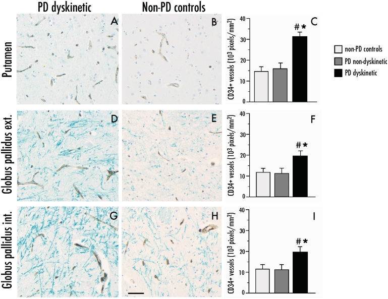
Parkinson’s Disease (PD) dyskinetic patients have an increased number of CD34-immunostained blood vessel profiles in the basal ganglia. Representative photomicrographs of CD34 expression in dyskinetic and non-dyskinetic patients in the putamen (A and B), external (ext.) and internal (int) globus pallidus in (D and E) and (G and H), respectively. Bar graphs to the right show the number of CD34-immunopositive pixels per sampling frame in the putamen (C), external and internal globus pallidus (F and I, respectively) from dyskinetic patients with Parkinson’s disease (PD dyskinetic, n = 11), non-dyskinetic patients with Parkinson’s disease (PD non-dyskinetic, n = 9) and neurologically healthy controls (non-PD controls, n = 6). One-factor ANOVA followed by post hoc Student–Newman–Keul’s, P < 0.05, *versus non-Parkinson’s disease controls, #versus non-dyskinetic Parkinson’s disease. Scale bar = 50 μm.

