
Figure 5.
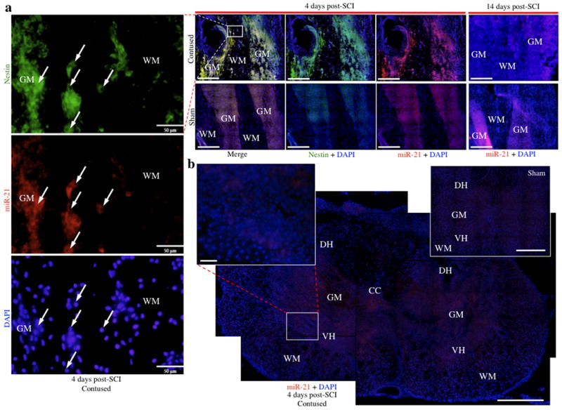
In situ hybridization analyses for miR21 expression at 4 and 14 days following SCI. a, Substantially increased expression of miR21 was observed within the grey matter at 4 days in contused animals and co-localized with nestin, indicated by yellow fluorescence (red = miR21; green = nestin). High magnification photomicrograph excerpts from the lesion site at 4 days show that nestin and miR21 co-localize to cells with small nuclei, and nestin appears globular as opposed to the fibrous morphology typically associated with astrocytes. Furthermore, miR21 is only observed within grey matter at the lesion site, with low peri-lesion expression. In contrast, decreased miR21 expression relative to sham animals was observed within grey matter tracts at the injury site 14 days post-SCI. b, MiR21 localizes throughout the grey matter tissue of coronal sections, including both the dorsal and ventral horns, with minimal expression in white matter. The coronal image of the lesion site is an overlaid composite of 4 coronal images of the same contused section, each containing 25 micrographs at 20× magnification stitched together per fluorescence channel, for an overall total of 200 micrographs stitched together to form one image (25 images per channel × 2 channels × 4 stitched coronal micrographs = 200 images). Abbreviations, central canal (CC), dorsal horn (DH), grey matter (GM), ventral horn (VH), and white matter (WH). Scale bars, 500 μm (a, b) and 50 μm (high magnification excerpts a, b).