Abstract
Francisella tularensis is a Gram-negative intracellular coccobacillus and the causative agent of the zoonotic disease tularemia. When compared with other bacterial pathogens, the extremely low infectious dose (<10 CFU), rapid disease progression, and high morbidity and mortality rates suggest that the virulent strains of Francisella encode for novel virulence factors. Surface-exposed molecules, namely outer membrane proteins (OMPs), have been shown to promote bacterial host cell binding, entry, intracellular survival, virulence and immune evasion. The relevance for studying OMPs is further underscored by the fact that they can serve as protective vaccines against a number of bacterial diseases. Whereas OMPs can be extracted from gram-negative bacteria through bulk membrane extraction techniques, including sonication of cells followed by centrifugation and/or detergent extraction, these preparations are often contaminated with periplasmic and/or cytoplasmic (inner) membrane (IM) contaminants. For years, the "gold standard" method for the biochemical and biophysical separation of gram-negative IM and outer membranes (OM) has been to subject bacteria to spheroplasting and osmotic lysis, followed by sucrose density gradient centrifugation. Once layered on a sucrose gradient, OMs can be separated from IMs based on the differences in buoyant densities, believed to be predicated largely on the presence of lipopolysaccharide (LPS) in the OM. Here, we describe a rigorous and optimized method to extract, enrich, and isolate F. tularensis outer membranes and their associated OMPs.
Keywords: Microbiology, Issue 40, Francisella, tularemia, outer membrane protein, sucrose density gradient centrifugation, membrane isolation, osmotic lysis, spheroplast
Protocol
Day 1: F. tularensis plate inoculation
Plate frozen stock cultures of F. tularensis onto Mueller-Hinton agar supplemented with 2.5% (vol/vol) donor calf serum, 2% (vol/vol) IsoVitaleX, 0.1% (wt/vol) glucose, and 0.025% (wt/vol) iron pyrophosphate. Grow F. tularensis at 37°C with 5% CO2 for approximately 24 h.
Day 2: F. tularensis liquid media inoculation
Prepare and autoclave 1 liter of cation-adjusted Mueller-Hinton medium (containing 1.23 mM calcium chloride dihydrate and 1.03 mM magnesium chloride hexahydrate). When cooled to 37°C, further supplement medium with 0.1% (wt/vol) glucose, 0.025% (wt/vol) iron pyrophosphate, and 2% (vol/vol) IsoVitaleX. Filter sterilize (0.22 μ) medium into two 500-ml aliquots.
Remove 12 ml of supplemented Mueller-Hinton medium and transfer into a sterile 50-ml Falcon tube.
Using a sterile 10-μl inoculation loop, scrape a large loopful of F. tularensis growth from agar plates (from Day 1) and transfer bacteria into 12 ml of Mueller-Hinton medium (see Step 2). Pipette solution multiple times to break-up clumps and prepare homogenous bacterial suspension. Do not vortex.
Using a sterile pipette, inoculate each 500-ml vessel with 5 ml of bacterial suspension from Step 3. Grow broth cultures at 37°C for 14 to 18 h with gentle shaking (190-200 rpm on a New Brunswick Innova 2300 series shaker).
Day 3: Spheroplasting, osmotic lysis, and sucrose density gradient centrifugation
Obtain F. tularensis cultures from the shaking incubator and remove a 1-ml aliquot from each 500-ml culture to check the respective optical densities. We have found optimum membrane extraction and isolation results when using F. tularensis cells in early logarithmic phase of growth, which correlates with an OD600 between 0.2 to 0.4 (~107 to 108 CFU/ml). Cultures with an OD600 less than 0.2 will not yield sufficient quantity of total membrane material for subsequent sucrose gradients. Conversely, cultures with an OD600 greater than 0.4 (nearing stationary phase) have been found to yield mixed-membrane fusions.
Centrifuge cultures at 7,500 x g for 30 min at 15°C to collect the cells. We recommend centrifugation of cultures in four 250-ml centrifuge bottles, because it facilitates subsequent pellet suspension.
Carefully remove the media supernatant from each centrifuge bottle and firmly tap the bottles on absorbent material to remove excess growth medium.
Within 10 min following completion of centrifugation, suspend each bacterial pellet in 8.75 ml of 0.75 M sucrose (in 5 mM Tris, pH 7.5). All four bacterial pellets should be suspended and transferred (35 ml total of bacterial suspension) to a sterile 250-ml flask (with a small stirbar) within 10 min.
While gently mixing the cell suspension on a stirplate, slowly add 70 ml (2 volumes) of 10 mM disodium EDTA (in 5 mM Tris, pH 7.8) over the course of 10 min. Add the EDTA solution with the tip of pipette below the cell suspension level to avoid elevated local concentrations of EDTA. The stirbar should be rotating at a rate sufficient to thoroughly mix the cell suspension but not fast enough to cause frothing or bubble formation. After the EDTA solution has been added, incubate the solution for 30 min at room temperature.
After the 30 min incubation, slowly add 11 ml (1/10th volume) of a 2 mg/ml lysozyme solution to a final concentration of 200 μg/ml. Continue to mix the cell suspension during the lysozyme solution addition. As noted above, add the lysozyme solution with the pipette tip below the cell suspension fluid level to avoid elevated local concentrations of lysozyme. Stock lysozyme solutions (2 mg/ml) can be prepared in single-use aliquots and stored at -20°C for three to four months. Incubate the resulting solution for 30 min at room temperature.
During the 30 min incubation, prepare a sterile 1L flask with a small stirbar and 530 ml of room temperature Cellgro Molecular Grade dH2O (4.5 volumes).
Osmotically lyse the cell suspension (from Step 6) by slow dilution (flow rate of approximately 11 ml/min) into 530 ml of Molecular Grade dH2O (from Step 7) over the course of 10 to 15 min with gentle mixing. Because of the larger solution volume in this step, increase the stirbar speed to ensure proper mixing. The stirbar should be rotating at a rate sufficient to thoroughly disperse the cell suspension once added to the dH2O but not fast enough to lead to frothing or bubble formation. Add the cell suspension with the tip of pipette resting on the bottom of the flask, adjacent to the stirbar, to facilitate uniform dilution of the cell suspension. We have found that 25-ml pipettes work best during this step. Carefully monitor the dilution process and interrupt the flow rate as necessary to disperse local accumulations of cells, cell clumps, and cell wisps. Incubate the resulting solution for 30 min at room temperature. Note that this step appears to be one of the most challenging and critical for overall success of the experiment.
After the 30 min incubation, transfer 40 ml aliquots of the osmotic lysis solution into 50-ml conical tubes and centrifuge at 7500 x g for 30 min at 10°C to remove intact cells and debris.
Following centrifugation, carefully remove 27 to 30 ml of supernatant from each conical tube and pool supernatants in a sterile 1L flask
Transfer 25 ml of pooled supernatant, each, into 16 ultracentrifuge tubes and centrifuge at 200,000 x g (44,400 rpm in F50L-8x39 FiberLite ultracentrifuge rotor) for 2 h at 4°C. Note that each rotor holds 8 tubes, so this step requires two rotors and two ultracentrifuges. Alternatively, the second set of 8 tubes should be held at 4°C until centrifuged.
Within 10 min of the completion of the ultracentrifuge run, prepare modified membrane resuspension buffer. Transfer 8 ml of membrane suspension buffer (25% [wt/wt] sucrose, 5 mM Tris, 30 mM MgCl2) to a sterile tube and add 1 tablet of EDTA-free protease inhibitor cocktail and 375 U of Benzonase. Gently mix solution to dissolve protease inhibitor tablet.
Following ultracentrifugation, carefully pour off supernatants, invert ultracentrifuge tubes on absorbent material, and firmly tap tubes to remove excess supernatant. Mark the location of each membrane pellet with a marker to aid in visualization. Gently resuspend eight membrane pellets with 600 μl each of modified membrane resuspension buffer. Transfer each membrane resuspension to the next set of eight tubes, gently resuspend these eight pellets, and pool the resulting membrane resuspensions in a sterile tube. The pelleted membranes are difficult to resuspend, so continue passing the modified membrane resuspension buffer over the pellet until it peels away from the wall of the tube. It is often necessary to use the micropipette tip to scrape the membrane pellet off the tube wall and aid in the resuspension process. When pipetting, avoid frothing or bubble formation. Note that this step appears to be one of the most challenging and critical for overall success of the experiment.
When the membrane pellets have been resuspended and removed from all 16 tubes, wash every four tubes with 600 μl of modified membrane resuspension buffer. The wash should be focused around the marked area (membrane pellet) of each tube. Transfer the wash into the membrane resuspension tube. Repeat the wash step for the remaining three sets of four tubes.
Incubate the membrane resuspension with gentle rocking for 30 min at room temperature to degrade DNA and aid in reduction of flocculent material.
Remove a small aliquot of the membrane suspension and perform protein quantification to determine total membrane yield. We typically prepare undiluted, 1:1 diluted (in 2.5% SDS), and 1:3 diluted (in 2.5% SDS) membrane samples, heat the samples at 90°C for 10 min, and use the BioRad DC Protein Assay to quantify the membrane resuspension protein concentration. Total protein yield is generally between 1.0 mg/ml and 1.6 mg/ml. The use of heat and SDS are required to release OMPs from the extracted membranes so that a more accurate protein quantification value can be obtained.
Prepare linear sucrose gradients by layering 1.8 ml each of sucrose solutions (wt/wt, prepared in 5 mM EDTA, pH 7.5) into 14 x 95 mm Ultra-Clear ultracentrifuge tubes in the following order: 55%, 50%, 45%, 40%, 35%, 30%. Layering of sucrose gradients is easiest to perform when using a bulb pipettor, not an automated pipettor.
Based on the membrane resuspension protein quantification (Step 16), layer 1.5 mg of membrane resuspension on top of each gradient. Each 14 x 95 mm ultracentrifuge tube has a maximum capacity of 12.5 ml and, given that 10.8 ml is accounted for by the sucrose solutions from Step 17, you should not load more than 1.7 ml of membrane resuspension per gradient. Individually weigh and carefully adjust the final weight of each sucrose gradient tube (by adding or removing membrane resuspension) so that all tubes weigh the same (± 0.01 g).
Transfer sucrose gradients into a SW-40Ti swinging bucket rotor and centrifuge at 256,000 x g (38,000 rpm) for a minimum of 17 h at 4°C.
Day 4: Collection of sucrose gradient fractions
After 17 h of centrifugation, stop the centrifuge run, and carefully remove sucrose gradients from the SW-40Ti rotor.
Carefully clean the bottom of the sucrose gradient tube with 70% ethanol, puncture the bottom of the tube with a 21g needle, and collect sequential 500 μl fractions from the gradient into sterile microfuge tubes. Repeat for subsequent gradients. The gradient should be allowed to drip by gravity flow, so as not to disturb the sucrose gradient. However, if the dripping ceases from the bottom of the gradient tube, a small amount of pressure may be applied to the top of the tube using one's finger.
Determine the refractive index of each sucrose gradient fraction using a refractometer and correlate the refractive index with a specific density in g/ml 1. Assess protein localization by preparing representative sucrose gradient fractions (density increments of 0.01; from 1.11 g/ml to 1.26 g/ml) for SDS-PAGE separation, transfer to nitrocellulose, and immunoblotting.
Representative Results
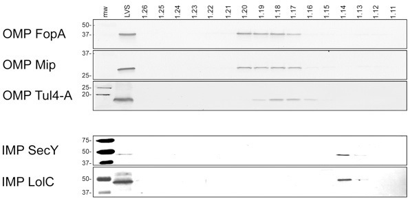
When performed correctly, this procedure results in the complete separation of IM and OM vesicles from both Type A (e.g. SchuS4) and Type B (e.g. LVS) strains of F. tularensis. As shown in representative immunoblots in Figure 1, F. tularensis OMPs localize between densities of 1.17 and 1.20 g/ml. By comparison, IMPs localize between densities of 1.13 and 1.14 g/ml.
 Figure 1. Immunoblotting of F. tularensis sucrose density gradients. Sequential fractions were collected from gradients and densities (g/ml) were calculated based upon refractive indices. Proteins were separated by SDS-PAGE, transferred to nitrocellulose, and immunoblotted to detect OMP and IMP fractionation. mw, prestained molecular weight standards with sizes (in kDa) noted on the left side of each blot. LVS, whole cell lysates of F. tularensis LVS. The corresponding sucrose gradient fraction densities are noted above their respective lanes. OMPs and IMPs, as noted in the left margin, were detected with polyclonal, monospecific antisera. Similar results were obtained for F. tularensis SchuS4.
Discussion
This protocol describes a variation of the spheroplasting, osmotic lysis, and sucrose density gradient centrifugation "gold standard" for the physical separation and enrichment of Gram-negative bacterial OMs from other cellular components 2, 3, 4. Whereas we previously described the application of this method for isolating OMs from both Type A and Type B strains of F. tularensis 5, this presentation offers a more detailed and optimized procedure for OM isolation. Indeed, this is an important advance for identifying potentially surface-exposed and presumptive virulence factors from this deadly pathogen. The significance of this procedure was demonstrated in studies by our lab showing that immunization of mice with purified OMPs afforded substantial protection against virulent Type A F. tularensis pulmonary challenge 6. As noted above, growth phase of the bacterial cells, careful monitoring during osmotic lysis, and gentle resuspension of membrane pellets are critical steps that appear to correlate with success/failure of this membrane separation procedure. While this optimized method is rigorous and subject to some experimental variability, the resulting OMs appear to be of remarkably enhanced purity as compared to those garnered from the use of detergents, lithium chloride, or sodium carbonate 7, 8, 9, 10, 11, 12.
Disclosures
No conflicts of interest declared.
Acknowledgments
The project described was supported by Grant Numbers P01 AI055637 and U54 AI057156 from NIAID/NIH. Its contents are solely the responsibility of the authors and do not necessarily represent the official views of the RCE Programs Office, NIAID, or NIH.
References
- Price CA. Academic Press; 1982. Centrifugation in density gradients; pp. 335–335. [Google Scholar]
- Osborn MJ, Gander JE, Parisi E, Carson J. Mechanism of assembly of the outer membrane of Salmonella typhimurium. Isolation and characterization of cytoplasmic and outer membrane. J Biol Chem. 1972;247(12):3962–3962. [PubMed] [Google Scholar]
- Nikaido H. Isolation of outer membranes. Methods Enzymol. 1994;235:225–225. doi: 10.1016/0076-6879(94)35143-0. [DOI] [PubMed] [Google Scholar]
- Mizuno T, Kageyama M. Separation and characterization of the outer membrane of Pseudomonas aeruginosa. J Biochem. 1978;84(1):179–179. doi: 10.1093/oxfordjournals.jbchem.a132106. [DOI] [PubMed] [Google Scholar]
- Huntley JF, Conley PG, Hagman KE, Norgard MV. Characterization of Francisella tularensis outer membrane proteins. J Bacteriol. 2007;189:561–561. doi: 10.1128/JB.01505-06. [DOI] [PMC free article] [PubMed] [Google Scholar]
- Huntley JF. Native outer membrane proteins protect mice against pulmonary challenge with virulent type A Francisella tularensis. Infect Immun. 2008;76(8):3664–3664. doi: 10.1128/IAI.00374-08. [DOI] [PMC free article] [PubMed] [Google Scholar]
- Bevanger L, Maeland JA, Naess AI. Agglutinins and antibodies to Francisella tularensis outer membrane antigens in the early diagnosis of disease during an outbreak of tularemia. J Clin Microbiol. 1988;26(3):433–433. doi: 10.1128/jcm.26.3.433-437.1988. [DOI] [PMC free article] [PubMed] [Google Scholar]
- Hubalek M. Towards proteome database of Francisella tularensis. J Chromatogr B Analyt Technol Biomed Life Sci. 2003;787(1):149–149. doi: 10.1016/s1570-0232(02)00730-4. [DOI] [PubMed] [Google Scholar]
- Pavkova I. Francisella tularensis live vaccine strain: proteomic analysis of membrane proteins enriched fraction. Proteomics. 2005;5(9):2460–2460. doi: 10.1002/pmic.200401213. [DOI] [PubMed] [Google Scholar]
- Sandstrom G, Tarnvik A, Wolf-Watz H. Immunospecific T-lymphocyte stimulation by membrane proteins from Francisella tularensis. J Clin Microbiol. 1987;25(4):641–641. doi: 10.1128/jcm.25.4.641-644.1987. [DOI] [PMC free article] [PubMed] [Google Scholar]
- Sjostedt A, Tarnvik A, Sandstrom G. The T-cell-stimulating 17-kilodalton protein of Francisella tularensis LVS is a lipoprotein. Infect Immun. 1991;59(9):3163–3163. doi: 10.1128/iai.59.9.3163-3168.1991. [DOI] [PMC free article] [PubMed] [Google Scholar]
- Twine SM. Francisella tularensis proteome: low levels of ASB-14 facilitate the visualization of membrane proteins in total protein extracts. J Proteome Res. 2005;4(5):1848–1848. doi: 10.1021/pr050102u. [DOI] [PubMed] [Google Scholar]


