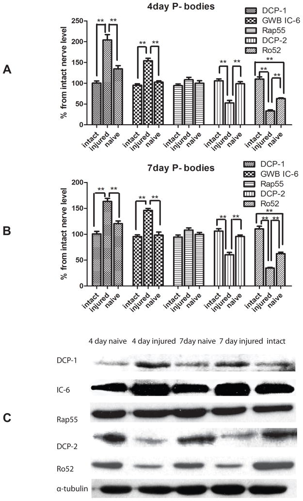
Figure 2.
Western Blot analyses of P-body components in the sciatic nerve after injury.
Western Blot analysis of P-body components in the sciatic nerve utilized the primary antibody against Dcp1, Dcp2, Rap55, Ro52, and GWB IC6. Pooled samples (6 nerves per each sample) were extracted from intact sciatic nerves, the contralateral naive nerve and the injured sciatic nerve at 4 days or 7 days after a conditioning nerve lesion. Protein levels were quantified by band densitomery and normalized to alpha-tubulin at 4 days (A) and 7 days (B) after conditional lesion. Immunostaining for Dcp1, the catalytic factor of Dcp2, showed an increase in expression in response to injury, whereas the expression of Dcp2, the decapping enzyme, decreased after injury at both time points. Immunostaining for GWB IC-6 showed much higher GWB IC-6 expression at 4 days and 7 days after injury, while Rap-55 showed no change in expression in response to injury. Ro52 expression level was significantly decreased following injury (N=3, * indicates P<0.05, ** indicates P<0.01). Representative immunoblots for Dcp1, Dcp2, Rap55, Ro52, and GWB IC6 are shown in C.

