Figure 4.
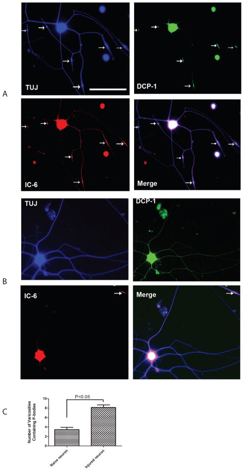
Number of VR containing P-bodies along the axon markedly increases after conditioning lesion.
Low density dissociated DRG cultures were labeled with antibodies to neuronal marker TUJ1 (Blue), Dcp1 (Green) and GWB IC-6 (Red). Merges of the images revealed the localization of the P-bodies in soma and axon. The appearance of P-bodies was limited to the VR along the axon. The fluorescent images in the upper panel show neuronal cultures after sciatic conditioning lesion (A). The images in the lower panel show neurons from naïve side (B). After lesion, the average number of VR containing P-bodies (white arrows) per neuron has significantly increased (C). Scale bar on microphotographs is 100μm.

