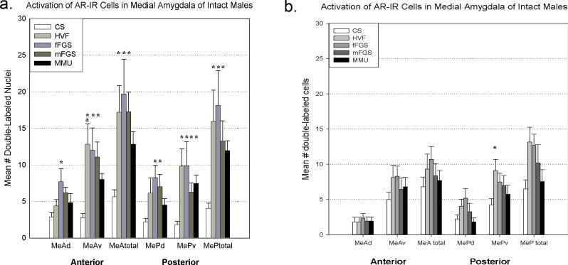
Figure 3.
Double-labeling of Fos and androgen receptors (AR) in the medial amygdala after exposure to various chemosensory stimuli gonad-intact (a) and testosterone-replaced (b) male hamsters. In gonad-intact males, conspecific stimuli yielded significantly more double-labeled cells than control (CS, clean swab) across the medial amygdala. The heterospecific stimulus (MMU, male mouse urine) produced significantly more double-labeled cells in the ventral portion of the posterior medial amygdala (MePv). Asterisk = significantly greater than control ( p<.05), a= significantly greater than MMU (p<.05). HVF= hamster vaginal fluid, fFGS= female flank gland secretion, mFGS= male flank gland secretion, MeA= anterior medial amygdala, MeP= posterior medial amygdala, d= dorsal subnucleus, v=ventral subnucleus.

