Abstract
Nitratifractor salsuginis Nakagawa et al. 2005 is the type species of the genus Nitratifractor, a member of the family Nautiliaceae. The species is of interest because of its high capacity for nitrate reduction via conversion to N2 through respiration, which is a key compound in plant nutrition. The strain is also of interest because it represents the first mesophilic and facultatively anaerobic member of the Epsilonproteobacteria reported to grow on molecular hydrogen. This is the first completed genome sequence of a member of the genus Nitratifractor and the second sequence from the family Nautiliaceae. The 2,101,285 bp long genome with its 2,121 protein-coding and 54 RNA genes is a part of the Genomic Encyclopedia of Bacteria and Archaea project.
Keywords: anaerobic, microaerobic, non-motile, Gram-negative, mesophilic, strictly chemolithoautotroph, Nautiliaceae, GEBA
Introduction
Strain E9I37-1T (= DSM 16511 = JCM 12458) is the type strain of Nitratifractor salsuginis, which in turn is the type and currently only species of the genus Nitratifractor [1]. The genus name is derived from the Neo-Latin word nitras meaning nitrate and the Latin word fractor meaning breaker, yielding the Neo-Latin word Nitratifractor meaning nitrate-breaker [1]. N. salsuginis strain E9I37-1T was isolated from a deep-sea hydrothermal vent chimney at the Iheya North hydrothermal field in the Mid-Okinawa Trough in Japan [1,2]. No further isolates of N. salsuginis have been obtained so far. Here we present a summary classification and a set of features for N. salsuginis E9I37-1T, together with the description of the complete genomic sequencing and annotation.
Classification and features
A representative genomic 16S rRNA sequence of strain E9I37-1T was compared using NCBI BLAST under default settings (e.g., considering only the high-scoring segment pairs (HSPs) from the best 250 hits) with the most recent release of the Greengenes database [3] and the relative frequencies, weighted by BLAST scores, of taxa and keywords (reduced to their stem [4]) were determined. The four most frequent genera were Nitratiruptor (48.5%), Nitratifractor (20.7%), Hydrogenimonas (15.7%) and Alvinella (15.1%) (eleven hits in total). Regarding the single hit to sequences from members of the species, the average identity within HSPs was 100.0%, whereas the average coverage by HSPs was 95.6%. Among all other species, the one yielding the highest score was Hydrogenimonas thermophila, which corresponded to an identity of 88.5% and an HSP coverage of 67.2%. (Note that the Greengenes database uses the INSDC (= EMBL/NCBI/DDBJ) annotation, which is not an authoritative source for nomenclature or classification.) The highest-scoring environmental sequence was AF420348 ('hydrothermal sediment clone AF420348') [5], which showed an identity of 96.7% and an HSP coverage of 97.8%. The five most frequent keywords within the labels of environmental samples which yielded hits were 'cave' (7.2%), 'biofilm' (5.7%), 'sulfid' (5.3%), 'spring' (4.8%) and 'structur' (3.1%) (239 hits in total). The five most frequent keywords within the labels of environmental samples which yielded hits of a higher score than the highest scoring species were 'hydrotherm' (8.6%), 'vent' (7.5%), 'pacif' (4.0%), 'microbi' (3.7%) and 'mat' (3.0%) (37 hits in total). These keywords are in accordance with the origin of the strain N. salsuginis E9I37-1T from a deep-sea hydrothermal vent chimney at the summits of the sulfide mounds in the sediment-hosted back-arc hydrothermal system Iheya North [1,2].
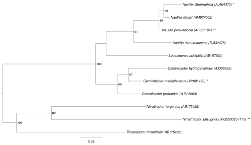
The 16S rRNA based tree in Figure 1 shows the phylogenetic neighborhood of N. salsuginis E9I37-1T. The sequences of the two identical 16S rRNA gene copies in the genome do not differ from the previously published 16S rRNA sequence (AB175500).
Figure 1.
Phylogenetic tree highlighting the position of N. salsuginis strain E9I37-1T relative to the other type strains within the family Nautiliaceae. The tree was inferred from 1,356 aligned characters [6,7] of the 16S rRNA gene sequence under the maximum likelihood criterion [8] and rooted in accordance with the current taxonomy. The branches are scaled in terms of the expected number of substitutions per site. Numbers to the right of bifurcations are support values from 200 bootstrap replicates [9] if larger than 60%. Lineages with type strain genome sequencing projects registered in GOLD [10] are labeled with an asterisk when unpublished, and with two asterisks when published [11]. The closest BLAST hit to N. salsuginis (see above) does not belong to Nautiliaceae, and this family does not appear as monophyletic in the last version of the 16S rRNA phylogeny from the All-Species-Living-Tree Project [12]. The species selection for Figure 1 was based on the current taxonomic classification (Table 1). However, an analysis including the type strains of Nautiliaceae and its neighboring families Campylobacteraceae, Helicobacteraceae and Hydrogenimonaceae (data not shown) did not provide evidence for the non-monophyly for any of these families.
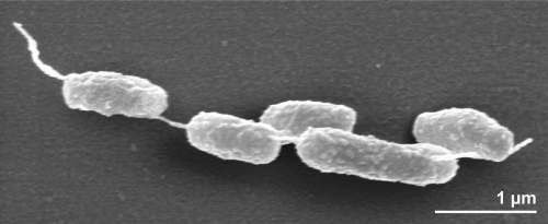
The cells of strain E9I37-1T are generally rod-shaped of 2.5 µm in length and 0.6 µm in width (Figure 2) and usually occur singly or in pairs (Figure 2) [1]. Strain E9I37-1T is a Gram-negative, non-motile and non spore-forming bacterium (Table 1). The organism is anaerobic to microaerophilic (0.09-0.55% O2 (v/v)) and chemolithoautotrophic, growing by respiratory nitrate reduction with H2 as the electron donor, forming N2 as a metabolic end product [1]. The main electron acceptors are NO3- or O2 [1]. Strain E9I37-1T uses S0 as a source of sulfur [1]. The doubling time of strain E9I37-1T was about 2.5 h [1]. The NaCl range for growth is between 1.5% and 3.5%, with an optimum at 3%; no growth was observed below 1.0% NaCl or above 4.0% NaCl [1]. The temperature range for growth is between 28ºC and 40ºC, with an optimum at 37ºC [1]. The pH range for growth is between 5.6 and 7.6, with an optimum at pH 7; no growth could be detected below pH 5.2 or above pH 8.1 [1]. Strain E9I37-1T was unable to use any organic compounds as energy or carbon sources [1]. The organism was sensitive to ampicillin, rifampicin, streptomycin, chloramphenicol (each at 50 µg ml-l) and kanamycin (200 µg ml-1), and insensitive to approximately 150 µg ml-1 kanamycin [1]. Enzymatic and genetic analyses demonstrated that strain E9I37-1T uses the reductive TCA (rTCA) cycle for carbon assimilation [21]. This was confirmed by the presence of all genes encoding the three key rTCA cycle enzymatic activities, namely ATP-dependent citrate lyase, pyruvate:ferredoxin oxidoreductase, and 2-oxoglutarate:ferredoxin oxidoreductase [21], but it was found to lack the gene for ribulose 1,5-bisphosphate carboxylase (RubisCO) activity, the key enzyme in the Calvin-Benson cycle [21].
Figure 2.
Scanning electron micrograph of N. salsuginis E9I37-1T
Table 1. Classification and general features of N. salsuginis E9I37-1T according to the MIGS recommendations [13].
| MIGS ID | Property | Term | Evidence code |
|---|---|---|---|
| Current classification | Domain Bacteria | TAS [14] | |
| Phylum Proteobacteria | TAS [15] | ||
| Class Epsilonproteobacteria | TAS [16,17] | ||
| Order Nautiliales | TAS [18] | ||
| Family Nautiliaceae | TAS [18] | ||
| Genus Nitratifractor | TAS [1] | ||
| Species Nitratifractor salsuginis | TAS [1] | ||
| Type strain E9I37-1 | TAS [1] | ||
| Gram stain | negative | TAS [1] | |
| Cell shape | rod shaped, occurring singly or in pairs | TAS [1] | |
| Motility | non-motile | TAS [1] | |
| Sporulation | none | TAS [1] | |
| Temperature range | 28-40ºC | TAS [1] | |
| Optimum temperature | 37°C | TAS [1] | |
| Salinity | 1.5-3.5% NaCl | TAS [1] | |
| MIGS-22 | Oxygen requirement | anaerobic and microaerobic | TAS [1] |
| Carbon source | probably CO2 | NAS | |
| Energy metabolism | strictly chemolithoautotrophic | TAS [1] | |
| MIGS-6 | Habitat | deep-sea hydrothermal vent chimneys | TAS [1] |
| MIGS-15 | Biotic relationship | not reported | NAS |
| MIGS-14 | Pathogenicity | not reported | NAS |
| Biosafety level | 1 | TAS [19] | |
| Isolation | deep-sea hydrothermal vent water of ‘E9’ chimney (inside part) | TAS [1,2] | |
| MIGS-4 | Geographic location | Iheya North hydrothermal field in the Mid-Okinawa Trough in Japan | TAS [1,2] |
| MIGS-5 | Sample collection time | 2002 or before | TAS [1,2] |
| MIGS-4.1 | Latitude | 27.78 | TAS [1,2] |
| MIGS-4.2 | Longitude | 126.88 | TAS [1,2] |
| MIGS-4.3 | Depth | 984 m | TAS [1,2] |
| MIGS-4.4 | Altitude | not reported | NAS |
Evidence codes - IDA: Inferred from Direct Assay (first time in publication); TAS: Traceable Author Statement (i.e., a direct report exists in the literature); NAS: Non-traceable Author Statement (i.e., not directly observed for the living, isolated sample, but based on a generally accepted property for the species, or anecdotal evidence). These evidence codes are from of the Gene Ontology project [20]. If the evidence code is IDA, the property was directly observed by one of the authors or an expert mentioned in the acknowledgements.
Chemotaxonomy
The major cellular fatty acids of strain E9I37-1Tare C18:1 (42.3% of the total fatty acid), C16:1 (30.7%) and C16:0 (24.3%), C14:0 3-OH (1.1%), C14:0 (0.9%) and C18:0 (0.7%) [1]. It should be noted that no information is given on the position of double bonds in the unsaturated fatty acids. No attempt has been made to examine the type strain for the presence of respiratory lipoquinones or to determine the polar lipid composition.
Genome sequencing and annotation
Genome project history
This organism was selected for sequencing on the basis of its phylogenetic position [22], and is part of the Genomic Encyclopedia of Bacteria and Archaea project [23]. The genome project is deposited in the Genome On Line Database [10] and the complete genome sequence is deposited in GenBank. Sequencing, finishing and annotation were performed by the DOE Joint Genome Institute (JGI). A summary of the project information is shown in Table 2.
Table 2. Genome sequencing project information.
| MIGS ID | Property | Term |
|---|---|---|
| MIGS-31 | Finishing quality | Finished |
| MIGS-28 | Libraries used | Three genomic libraries: one 454 pyrosequence standard library, one 454 PE library (12 kb insert size), one Illumina library |
| MIGS-29 | Sequencing platforms | Illumina GAii, 454 GS FLX Titanium |
| MIGS-31.2 | Sequencing coverage | 75.2 × Illumina; 31.5 × pyrosequence |
| MIGS-30 | Assemblers | Newbler version 2.4, Velvet, phrap |
| MIGS-32 | Gene calling method | Prodigal 1.4, GenePRIMP |
| INSDC ID | CP002452 | |
| Genbank Date of Release | January 24, 2011 | |
| GOLD ID | Gc01594 | |
| NCBI project ID | 46883 | |
| Database: IMG-GEBA | 2503538035 | |
| MIGS-13 | Source material identifier | DSM 16511 |
| Project relevance | Tree of Life, GEBA |
Growth conditions and DNA isolation
N. salsuginis E9I37-1T, DSM 16511, was grown anaerobically in DSMZ medium 1024 (Nitratiruptor and Nitratifractor medium) [24] at 37°C. DNA was isolated from 0.5-1 g of cell paste using Jetflex Genomic DNA Purification Kit (GENOMED 600100) following the standard protocol as recommended by the manufacturer. Cell lysis was enhanced by adding 20 µl proteinase K for two hours at 58°C. DNA is available through the DNA Bank Network [25].
Genome sequencing and assembly
The genome was sequenced using a combination of Illumina and 454 sequencing platforms. All general aspects of library construction and sequencing can be found at the JGI website [26]. Pyrosequencing reads were assembled using the Newbler assembler (Roche). The initial Newbler assembly consisting of 42 contigs in five scaffolds was converted into a phrap [27] assembly by making fake reads from the consensus, to collect the read pairs in the 454 paired end library. Illumina GAii sequencing data (158.03 Mb) was assembled with Velvet [28] and the consensus sequences were shredded into 1.5 kb overlapped fake reads and assembled together with the 454 data. The 454 draft assembly was based on 60.3 Mb 454 draft data and all of the 454 paired end data. Newbler parameters are -consed -a 50 -l 350 -g -m -ml 20. The Phred/Phrap/Consed software package [27] was used for sequence assembly and quality assessment in the subsequent finishing process. After the shotgun stage, reads were assembled with parallel phrap (High Performance Software, LLC). Possible mis-assemblies were corrected with gapResolution [26], Dupfinisher [29], or sequencing clones bridging PCR fragments with subcloning. Gaps between contigs were closed by editing in Consed, by PCR and by Bubble PCR primer walks (J.-F. Chang, unpublished). A total of 135 additional reactions were necessary to close gaps and to raise the quality of the finished sequence. Illumina reads were also used to correct potential base errors and increase consensus quality using a software Polisher developed at JGI [30]. The error rate of the completed genome sequence is less than 1 in 100,000. Together, the combination of the Illumina and 454 sequencing platforms provided 106.7 × coverage of the genome. The final assembly contained 274,574 pyrosequence and 2,079,398 Illumina reads.
Genome annotation
Genes were identified using Prodigal [31] as part of the Oak Ridge National Laboratory genome annotation pipeline, followed by a round of manual curation using the JGI GenePRIMP pipeline [32]. The predicted CDSs were translated and used to search the National Center for Biotechnology Information (NCBI) non-redundant database, UniProt, TIGR-Fam, Pfam, PRIAM, KEGG, COG, and InterPro databases. Additional gene prediction analysis and functional annotation was performed within the Integrated Microbial Genomes - Expert Review (IMG-ER) platform [33].
Genome properties
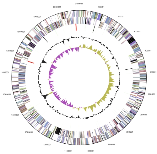
The genome consists of a 2,101,285 bp long chromosome with a G+C content of 53.9% (Table 3 and Figure 3). Of the 2,175 genes predicted, 2,121 were protein-coding genes, and 54 RNAs; 33 pseudogenes were also identified. The majority of the protein-coding genes (66.9%) were assigned with a putative function while the remaining ones were annotated as hypothetical proteins. The distribution of genes into COGs functional categories is presented in Table 4.
Table 3. Genome Statistics.
| Attribute | Value | % of Total |
|---|---|---|
| Genome size (bp) | 2,101,285 | 100.00% |
| DNA coding region (bp) | 1,916,093 | 91.19% |
| DNA G+C content (bp) | 1,132,843 | 53.91% |
| Number of replicons | 1 | |
| Extrachromosomal elements | 0 | |
| Total genes | 2,175 | 100.00% |
| RNA genes | 54 | 2.48% |
| rRNA operons | 2 | |
| Protein-coding genes | 2,121 | 97.52% |
| Pseudo genes | 33 | 1.52% |
| Genes with function prediction | 1,456 | 66.94% |
| Genes in paralog clusters | 144 | 6.62% |
| Genes assigned to COGs | 1,525 | 70.11% |
| Genes assigned Pfam domains | 1,616 | 74.30% |
| Genes with signal peptides | 411 | 18.90% |
| Genes with transmembrane helices | 501 | 23.03% |
| CRISPR repeats | 2 |
Figure 3.
Graphical circular map of the chromosome; From outside to the center: Genes on forward strand (color by COG categories), Genes on reverse strand (color by COG categories), RNA genes (tRNAs green, rRNAs red, other RNAs black), GC content, GC skew.
Table 4. Number of genes associated with the general COG functional categories.
| Code | value | %age | Description |
|---|---|---|---|
| J | 149 | 9.0 | Translation, ribosomal structure and biogenesis |
| A | 0 | 0.0 | RNA processing and modification |
| K | 64 | 3.9 | Transcription |
| L | 114 | 6.9 | Replication, recombination and repair |
| B | 0 | 0.0 | Chromatin structure and dynamics |
| D | 20 | 1.2 | Cell cycle control, cell division, chromosome partitioning |
| Y | 0 | 0.0 | Nuclear structure |
| V | 25 | 1.5 | Defense mechanisms |
| T | 69 | 4.2 | Signal transduction mechanisms |
| M | 133 | 8.1 | Cell wall/membrane/envelope biogenesis |
| N | 15 | 0.9 | Cell motility |
| Z | 0 | 0.0 | Cytoskeleton |
| W | 0 | 0.0 | Extracellular structures |
| U | 43 | 2.6 | Intracellular trafficking, secretion, and vesicular transport |
| O | 89 | 5.4 | Posttranslational modification, protein turnover, chaperones |
| C | 131 | 8.0 | Energy production and conversion |
| G | 58 | 3.5 | Carbohydrate transport and metabolism |
| E | 136 | 8.3 | Amino acid transport and metabolism |
| F | 50 | 3.0 | Nucleotide transport and metabolism |
| H | 97 | 5.9 | Coenzyme transport and metabolism |
| I | 37 | 2.3 | Lipid transport and metabolism |
| P | 81 | 4.9 | Inorganic ion transport and metabolism |
| Q | 18 | 1.1 | Secondary metabolites biosynthesis, transport and catabolism |
| R | 183 | 11.1 | General function prediction only |
| S | 136 | 8.3 | Function unknown |
| - | 650 | 29.9 | Not in COGs |
Acknowledgements
We would like to gratefully acknowledge the help of Andrea Schütze (DSMZ) for growing N. salsuginis cultures. This work was performed under the auspices of the US Department of Energy Office of Science, Biological and Environmental Research Program, and by the University of California, Lawrence Berkeley National Laboratory under contract No. DE-AC02-05CH11231, Lawrence Livermore National Laboratory under Contract No. DE-AC52-07NA27344, and Los Alamos National Laboratory under contract No. DE-AC02-06NA25396, UT-Battelle and Oak Ridge National Laboratory under contract DE-AC05-00OR22725, as well as German Research Foundation (DFG) INST 599/1-2.
References
- 1.Nakagawa S, Takai K, Inagaki F, Horikoshi K, Sako Y. Nitratiruptor tergarcus gen. nov., sp. nov. and Nitratifractor salsuginis gen. nov., sp. nov., nitrate-reducing chemolithoautotrophs of the epsilon-Proteobacteria isolated from a deep-sea hydrothermal system in the Mid-Okinawa Trough. Int J Syst Evol Microbiol 2005; 55:925-933 10.1099/ijs.0.63480-0 [DOI] [PubMed] [Google Scholar]
- 2.Takai K, Inagaki F, Nakagawa S, Hirayama H, Nunoura T, Sako Y, Nealson KH, Horikoshi K. Isolation and phylogenetic diversity of members of previously uncultivated ε-Proteobacteria in deep-sea hydrothermal fields. FEMS Microbiol Lett 2003; 218:167-174 [DOI] [PubMed] [Google Scholar]
- 3.DeSantis TZ, Hugenholtz P, Larsen N, Rojas M, Brodie E, Keller K, Huber T, Dalevi D, Hu P, Andersen G. Greengenes, a chimera-checked 16S rRNA gene database and workbench compatible with ARB. Appl Environ Microbiol 2006; 72:5069-5072 10.1128/AEM.03006-05 [DOI] [PMC free article] [PubMed] [Google Scholar]
- 4.Porter MF. An algorithm for suffix stripping. Program: electronic library and information systems. 1980; 14:130-137.
- 5.Teske A, Hinrichs KU, Edgcomb VP, Gomez AD, Kysela DT, Sogin ML, Jannasch HW. Microbial diversity of hydrothermal sediments in the Guaymas Basin: evidence for anaerobic methanotrophic communities. Appl Environ Microbiol 2002; 68:1994-2007 10.1128/AEM.68.4.1994-2007.2002 [DOI] [PMC free article] [PubMed] [Google Scholar]
- 6.Castresana J. Selection of conserved blocks from multiple alignments for their use in phylogenetic analysis. Mol Biol Evol 2000; 17:540-552 [DOI] [PubMed] [Google Scholar]
- 7.Lee C, Grasso C, Sharlow MF. Multiple sequence alignment using partial order graphs. Bioinformatics 2002; 18:452-464 10.1093/bioinformatics/18.3.452 [DOI] [PubMed] [Google Scholar]
- 8.Stamatakis A, Hoover P, Rougemont J. A rapid bootstrap algorithm for the RAxML Web servers. Syst Biol 2008; 57:758-771 10.1080/10635150802429642 [DOI] [PubMed] [Google Scholar]
- 9.Pattengale ND, Alipour M, Bininda-Emonds ORP, Moret BME, Stamatakis A. How many bootstrap replicates are necessary? Lect Notes Comput Sci 2009; 5541:184-200 10.1007/978-3-642-02008-7_13 [DOI] [PubMed] [Google Scholar]
- 10.Liolios K, Chen IM, Mavromatis K, Tavernarakis N, Hugenholtz P, Markowitz VM, Kyrpides NC. The Genomes On Line Database (GOLD) in 2009: status of genomic and metagenomic projects and their associated metadata. Nucleic Acids Res 2009; 38:D346-D354 10.1093/nar/gkp848 [DOI] [PMC free article] [PubMed] [Google Scholar]
- 11.Campbell BJ, Smith JL, Hanson TE, Klotz MG, Stein LY, Lee CK, Wu D, Robinson JM, Khouri HM, Eisen JA, et al. Adaptations to submarie hydrothermal environments exemplified by the genome of Natilia profundicola. PLoS Genet 2009; 5:e1000362 10.1371/journal.pgen.1000362 [DOI] [PMC free article] [PubMed] [Google Scholar]
- 12.Yarza P, Ludwig W, Euzéby J, Amman R, Schleifer KH, Glöckner FO, Rosselló-Mora R. Updates of the All-Species-Living-Tree Project based on 16S and 23S rRNA sequence analyses. Syst Appl Microbiol 2010; 33:291-299 10.1016/j.syapm.2010.08.001 [DOI] [PubMed] [Google Scholar]
- 13.Field D, Garrity G, Gray T, Morrison N, Selengut J, Sterk P, Tatusova T, Thomson N, Allen MJ, Angiuoli SV, et al. The minimum information about a genome sequence (MIGS) specification. Nat Biotechnol 2008; 26:541-547 10.1038/nbt1360 [DOI] [PMC free article] [PubMed] [Google Scholar]
- 14.Woese CR, Kandler O, Wheelis ML. Towards a natural system of organisms: proposal for the domains Archaea, Bacteria, and Eucarya. Proc Natl Acad Sci USA 1990; 87:4576-4579 10.1073/pnas.87.12.4576 [DOI] [PMC free article] [PubMed] [Google Scholar]
- 15.Garrity GM, Bell JA, Lilburn T. Phylum XIV. Proteobacteria phyl. nov. In: Garrity GM, Brenner DJ, Krieg NR, Staley JT (eds), Bergey's Manual of Systematic Bacteriology, Second Edition, Volume 2, Part B, Springer, New York, 2005, p. 1. [Google Scholar]
- 16.Validation List No 107. List of new names and new combinations previously effectively, but not validly, published. Int J Syst Evol Microbiol 2006; 56:1-6 10.1099/ijs.0.64188-0 [DOI] [PubMed] [Google Scholar]
- 17.Garrity GM, Bell JA, Lilburn T. Class V. Epsilonproteobacteria class. nov. In: Bergey’s Manual of Systematic Bacteriology, 2nd edn, vol. 2 (The Proteobacteria), part C (The Alpha-, Beta-, Delta-, and Epsilonproteobacteria), p. 1145. Edited by D. J. Brenner, N. R. Krieg, J. T. Staley & G. M. Garrity. New York: Springer. 2005. [Google Scholar]
- 18.Miroshnichenko ML, L'Haridon S, Schumann P, Spring S, Bonch-Osmolovskaya EA, Jeanthon C, Stackebrandt E. Caminibacter profundus sp. nov., a novel thermophile of Nautiliales ord. nov. within the class 'Epsilonproteobacteria', isolated from a deep-sea hydrothermal vent. Int J Syst Evol Microbiol 2004; 54:41-45 10.1099/ijs.0.02753-0 [DOI] [PubMed] [Google Scholar]
- 19.BAuA Classification of bacteria and archaea in risk groups. TRBA 2010; 466:150 [Google Scholar]
- 20.Ashburner M, Ball CA, Blake JA, Botstein D, Butler H, Cherry JM, Davis AP, Dolinski K, Dwight SS, Eppig JT, et al. Gene Ontology: tool for the unification of biology. Nat Genet 2000; 25:25-29 10.1038/75556 [DOI] [PMC free article] [PubMed] [Google Scholar]
- 21.Takai K, Campbell BJ, Cary SC, Suzuki M, Oida H, Nunoura T, Hirayama H, Nakagawa S, Suzuki Y, Inagaki F, et al. Enzymatic and genetic characterization of carbon and energy metabolisms by deep-sea hydrothermal chemolithoautotrophic isolates of Epsilonproteobacteria. Appl Environ Microbiol 2005; 71:7310-7320 10.1128/AEM.71.11.7310-7320.2005 [DOI] [PMC free article] [PubMed] [Google Scholar]
- 22.Klenk HP, Göker M. En route to a genome-based classification of Archaea and Bacteria? Syst Appl Microbiol 2010; 33:175-182 10.1016/j.syapm.2010.03.003 [DOI] [PubMed] [Google Scholar]
- 23.Wu D, Hugenholtz P, Mavromatis K, Pukall R, Dalin E, Ivanova NN, Kunin V, Goodwin L, Wu M, Tindall BJ, et al. A phylogeny-driven genomic encyclopaedia of Bacteria and Archaea. Nature 2009; 462:1056-1060 10.1038/nature08656 [DOI] [PMC free article] [PubMed] [Google Scholar]
- 24.List of growth media used at DSMZ: http://www.dsmz.de/microorganisms/media_list.php
- 25.Gemeinholzer B, Dröge G, Zetzsche H, Haszprunar G, Klenk HP, Güntsch A, Berendsohn WG, Wägele JW. The DNA Bank Network: the start from a German initiative. Biopreservation and Biobanking 2011; 9:51-55 10.1089/bio.2010.0029 [DOI] [PubMed] [Google Scholar]
- 26.JGI website. http://www.jgi.doe.gov/
- 27.The Phred/Phrap/Consed software package. http://www.phrap.com
- 28.Zerbino DR, Birney E. Velvet: algorithms for de novo short read assembly using de Bruijn graphs. Genome Res 2008; 18:821-829 10.1101/gr.074492.107 [DOI] [PMC free article] [PubMed] [Google Scholar]
- 29.Han C, Chain P. Finishing repeat regions automatically with Dupfinisher. In: Proceeding of the 2006 international conference on bioinformatics & computational biology. Arabnia HR, Valafar H (eds), CSREA Press. June 26-29, 2006: 141-146. [Google Scholar]
- 30.Lapidus A, LaButti K, Foster B, Lowry S, Trong S, Goltsman E. POLISHER: An effective tool for using ultra short reads in microbial genome assembly and finishing. AGBT, Marco Island, FL, 2008 [Google Scholar]
- 31.Hyatt D, Chen GL, LoCascio PF, Land ML, Larimer FW, Hauser LJ. Prodigal: prokaryotic gene recognition and translation initiation site identification. BMC Bioinformatics 2010; 11:119 10.1186/1471-2105-11-119 [DOI] [PMC free article] [PubMed] [Google Scholar]
- 32.Pati A, Ivanova NN, Mikhailova N, Ovchinnikova G, Hooper SD, Lykidis A, Kyrpides NC. GenePRIMP: a gene prediction improvement pipeline for prokaryotic genomes. Nat Methods 2010; 7:455-457 10.1038/nmeth.1457 [DOI] [PubMed] [Google Scholar]
- 33.Markowitz VM, Ivanova NN, Chen IMA, Chu K, Kyrpides NC. IMG ER: a system for microbial genome annotation expert review and curation. Bioinformatics 2009; 25:2271-2278 10.1093/bioinformatics/btp393 [DOI] [PubMed] [Google Scholar]


