Abstract
Background
The plexiform schwannoma, a peripheral nerve sheath tumor, is a very rare entity. But dermatologists should be familiar with since they may be the first who make diagnosis possible by taking a deep biopsy.
Main observation
A 24-year-old male presented with multiple asymptomatic subcutaneous nodules of the palmar side of his right hand. Histologic investigations revealed a plexiform schwannoma with numerous Antoni-A areas. There was no evidence of neurofibromatosis type 1 or 2.
Conclusions
Plexiform schwannoma of the hand is a rare nerve sheath tumor. In individual (symptomatic) cases hand surgery is an option that needs a critical indication. In every case histologic investigations are mandatory to confirm the diagnosis and not to overlook the malignant variant of this disease.
Keywords: nerve sheath tumors, plexiform schwannoma of the hand, neurofibromatosis
Introduction
In everyday practise benign nerve sheath tumors are rarely suggested clinically. Among 208 soft tissue tumors of hand and lower arm benign peripheral nerve tumors were seen in 11.5% with a correct clinical diagnosis in a single case only.[1] In a retrospective survey covering 397 benign and malignant peripheral nerve sheath tumors there have been 110 benign schwannomas.[2] Schwannomas (syn. neurilemmomas) in general are among the most common peripheral nerve sheath tumors together with neurofibromas.[3]
Imaging diagnostics like 3-dimensional ultrasound and MRI do not allow a sufficient discrimination of schwannomas form other nerve sheath tumors.[4] The only way to confirm the diagnosis is by histopathology.
We present an unusual case of a large unilateral plexiform schwannoma of the hand in a young male patient.
Case Report
A 24-year-old male patient experienced asymptomatic subcutaneous nodules of his right hand since 2000. There was a slow progress in size and number.
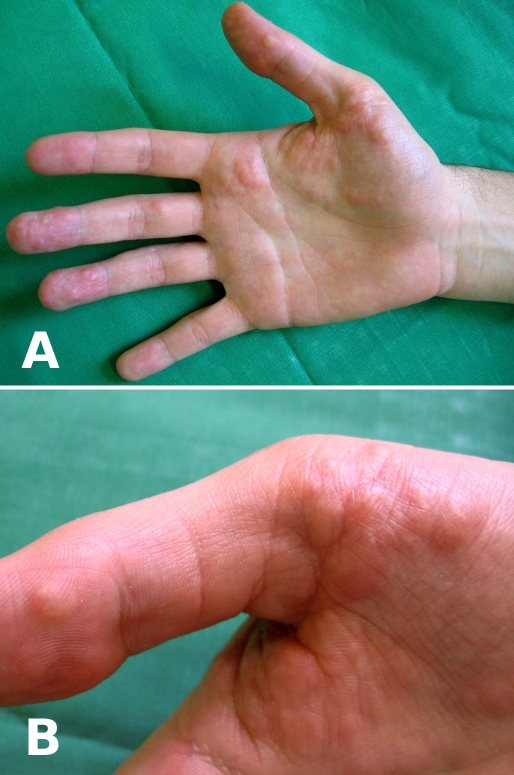
On examination we found soft, yellowish subcutaneous nodules of variable size of the volar site of the fingers and the palm of his right hand [Fig. 1A and B].
Figure 1.
Plexiform schwannoma - clinical presentation. (A) Overview. (B) Detail with subcutaneous nodular lesions.
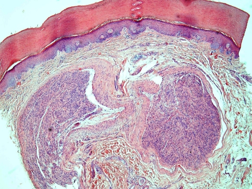
Histology of two biopsies showed spindle-shaped cell proliferations in the dermis and upper subcutaneous tissue with a capsule formation. Cells were characterized by neurogenic differentiation like Antoni-A-areas [Fig. 2].
Figure 2.
Histology of plexiform schwannoma with dermal encapsulated spindle-shaped tumor areas (Haematoxylin-Eosin x 4).
Clinical examination and consiliae did not reveal any sign of neurofibromatosis type 1 or type 2.
After confirmation of the diagnosis of a plexiform schwannoma of the right hand the patient was referred to the hand surgeon. He advised not to intervene by surgery because of the risk of paresis and the completely asyptomatic course without functional handicap in this patient.
Follow-up has been recommended by us.
Discussion
Schwannomas are benign tumors of the nerve sheath occuring either peripheral, visceral, intraspinal or intracranial. Peripheral schwannomas represent as asymptomatic and painless papules or nodules. Their colour can be yellowish to light brown. Cysts and haemorrhages may occur. Schwannomas of the hands are rare. Patient's mean age at diagnosis is 38.4 years.[5]
Histology is characterized by a biphasic pattern with tightly packed spindle-shaped cells (Antoni A area) and with loose, less cellular patterns with myxoid areas with thickened "hyalinized" vessel walls (Antoni B) and lipid-containing cells.[3] Mitoses are usually absent but pleomorphism of nuclei is common. Immunohistochemistry is helpful in differential diagnosis [Table 1]. The tumor cells express S-100 protein.[1,2] Complicating in differential diagnosis is the observation of collision tumors like schwannoma and perineurinoma.[6]
Table 1. Immunohistology of schwannoma and differential diagnoses.
| Tumor | S-100 | EMA | alpha-actin | desmin |
|---|---|---|---|---|
| Schwannoma | + | - | - | - |
| Perineuroma | - | + | - | - |
| Leiomyoma | - | - | + | + |
| Lipoma | - | - | - | - |
| Malignant nerve sheath tumor* | (+) | - | - | - |
| *) may show weak expression of neurendocrine markers such as neuron-specific enolase, neurofilament protein, myelin basic protein. | ||||
Plexiform schwannoma develops predominantly in young adults. Most often it represents as a pearl-like foci with an Antoni-A-pattern. There is a common association with neurofibromatosis type-2 (NF-2), but not NF-1.[7] Typical localisations are the nerve plexus, skin and subcutaneous tissue of the upper and lower extremities. Spinal and cranial nerves are usually spared by this tumor. The plexiform schwannoma without NF-2 - as described herein - is a rarity.[8–10] Major differential diagnoses include neurofibroma, perineurinoma and the malignant nerve sheath tumor.[1,2]
The tactics in treatment follow primarily the wait and see-rule. Surgery is optional in particular for symptomatic patients. Both long remissions and multiple rapid relapses have been observed after surgery.[5,11] Surgery bears the potential risk of nerve damage, therefore the indication has to be discussed together with both the patient and an experienced hand surgeon.
References
- Lincoski CJ, Harter GD, Bush DC. Benign nerve tumors of the hand and the forearm. Am J Orthop. 2007;36:E32–E36. [PubMed] [Google Scholar]
- Kim DH, Murovic JA, Tiel RL, Moes G, Kline DG. A series of 397 peripheral neural sheath tumors: 30-year experience at Louisiana State University Health Sciences Center. J Neurosurg. 2005;102:246–255. doi: 10.3171/jns.2005.102.2.0246. [DOI] [PubMed] [Google Scholar]
- Louis DS, Hankin FM. Benign nerve tumors of the upper extremity. Bull N Y Acad Med. 1985;61:611–620. [PMC free article] [PubMed] [Google Scholar]
- Amoretti N, Grimaud A, Hovorka E, Chevallier P, Roux C, Bruneton JN. Peripheral nerogenic tumors: is the use of different types of imaging diagnostically useful? Clin Imaging. 2006;30:201–205. doi: 10.1016/j.clinimag.2006.01.023. [DOI] [PubMed] [Google Scholar]
- Ozdemir O, Ozsoy MH, Kurt C, Coskunol E, Calli I. Schwannomas of the hand and wrist: long-term results and review of the literature. J Orthop Surg (Hong Kong) 2005;13:267–272. doi: 10.1177/230949900501300309. [DOI] [PubMed] [Google Scholar]
- Michal M, Kazakov DV, Belousova I, Bisceglia M, Zamecnik M, Mukensnabl P. A benign neoplasm with histopathological features of both schwannoma and retiform perineurinoma (bening schwannoma-perineurinoma): a report of six cases of a distinctive soft tissue tumor with a predilection for the fingers. Virchows Arch. 2004;445:347–353. doi: 10.1007/s00428-004-1102-5. [DOI] [PubMed] [Google Scholar]
- Lim HS, Jung J, Chung KY. Neurofibromatosis type 2 with multiple plexiform schwannomas. Int J Dermatol. 2004;43:336–340. doi: 10.1111/j.1365-4632.2004.01864.x. [DOI] [PubMed] [Google Scholar]
- Punia RS, Dhingra N, Mohan H. Cutaneous plexiform schwannoma of the finger not associated with neurofibromatosis. Am J Clin Dermatol. 2008;9:129–131. doi: 10.2165/00128071-200809020-00007. [DOI] [PubMed] [Google Scholar]
- Tadvi JS, Anand A, Humphrey A. Ancient schwannoma of the middle finger. J Hand Surg Eur Vol. 2007;32:722. doi: 10.1016/J.JHSE.2007.05.005. [DOI] [PubMed] [Google Scholar]
- Rockwell GM, Thoma A, Salama S. Schwannoma of the hand and wrist. Plast Reconstr Surg. 2003;111:1227–1232. doi: 10.1097/01.PRS.0000046039.28526.1A. [DOI] [PubMed] [Google Scholar]
- Talwalkar SC, Cutler L, Stilwell JH. Multiple plexiform schwannoma of the hand and forearm: a long-term follow-up. J Hand Surg Br. 2005;30:358–360. doi: 10.1016/j.jhsb.2005.04.008. [DOI] [PubMed] [Google Scholar]


