Abstract
Background
Parry-Romberg syndrome (PRS) or idiopathic hemifacial atrophy is a rare neurocutaneous syndrome. It is characterized by slowly progressive atrophy, located on one side of the face, primarily involving the skin, fat and connective tissue. PRS seems to overlap with "en coupe de sabre" morphea.
Main observations
We present a case of hemifacial atrophy in a 14-year-old boy treated with topical calcipotriol-betamethasone ointment. The diagnosis of PRS was established mainly based on the clinical findings and histological picture. The time to diagnosis was almost 9 years, similar to the mean time reported in the literature.
Conclusions
Understanding the pathogenesis and stopping disease progression is important as it can cause severe disfigurement and has neurological and psychiatric complications. Not much is known about the efficacy of agents used in the treatment of this syndrome making treatment decision very difficult. Possible complications, pathophysiology and therapeutic options are being discussed.
Keywords: betamethasone, en-coupe de sabre, propionate, calcipotriol, hemifacial atrophy, morphea, Parry-Romberg, scleroderma
Introduction
Parry-Romberg syndrome (PRS) is a rare disease entity characterized by atrophy of the skin, fat, connective tissue and muscles of one side of the face and thus is also called idiopathic progressive hemifacial atrophy.[1,2,3] It is an acquired condition of unknown aetiology presenting usually in childhood or adolescence, with gradual progression over several years.[2] Although it demonstrates self limited atrophy,[1] once the disease is established it does not exhibit spontaneous improvement.[2] PRS appears to overlap with "en coup de sabre" is a type of linear scleroderma (morphea) affecting the head.[2] The relationship between the two entities is not entirely clear,[3] but based on recent literature and the classification of Mayo Clinic,[3] we will consider PRS a clinical subtype of linear scleroderma. Extracutaneous manifestations of the disease have been described including neurological, articular, ocular, autoimmune and dental abnormalities.[2,4]
Case Report
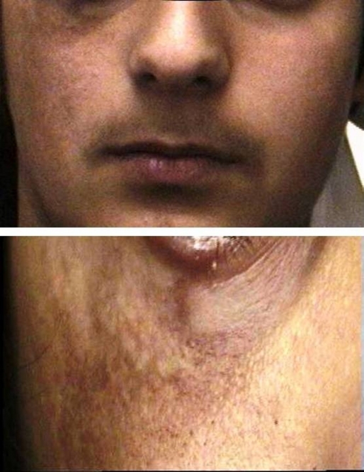
We present a case report of a 14-year-old boy with right hemifacial atrophy involving the zygomatic area, extending to the right side of the jaw, sparing the mouth, the temporal and parietal area. No family history of similar lesion was reported, and the other side of the face appeared intact. The onset has been estimated at the age of 5, demonstrating gradual progression over the following 8 years, leading to considerable wasting of the right side of the face by the time of initial presentation. The lesion was reported to have been stable in appearance for the last year by the patient and his parents. The psychological distress of the patient and the parental concern had led them to our dermatology clinic to seek potential treatment.
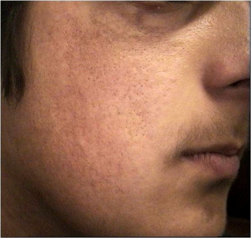
On presentation apart from the hemifacial wasting, there was hyperpigmentation of the affected skin, which also appeared to be drawn and tight, without any signs of cutaneous sclerosis. No vitiligo was noted and the hyperpigmentation was worse over the area were atrophy was more prominent. Associated alopecia was also observed with patchy loss of eyelashes of the lower eyelid, without enophthalmos, lid retraction, or visual disturbances [Fig. 1]. An ophthalmologic consultation confirmed the absence of ocular abnormalities. Apart from the loss of some eyelids there was no other associated alopecia or greying of hair. He did not have any facial (VII) nerve weakness or facial pain. Personal history of headaches, arthralgias, head trauma, seizures or limb weakness was also excluded. Laboratory examination, including autoimmune profile, did not reveal any abnormalities. No other asymmetry was noted on clinical examination or on the abdominal ultrasound that was performed. The patient did not exhibit any neurological symptoms, and a brain MRI confirmed that there was no neurological involvement. Skin biopsy obtained from the atrophic area of the right cheek, showed atrophy of the skin and subcutaneous tissue as well as localized fibrosis, without any other histologic changes consistent with morphea. We established the diagnosis of Parry-Romberg syndrome based on the clinical picture. Since the lesion had been stable, as reported by the patient, for the last year and due to the absence of extracutaneous manifestations, we decided to follow a conservative treatment approach. Topical treatment with calcipotriolbetamethasone diproprionate ointment was applied twice daily for 2 months followed by once daily application for a total period of six months, while the patient was followed up on a monthly basis. Hyperpigmentation subsided remarkably and the skin appeared less tight and much softer [Fig. 2]. No side effects were observed, but it was decided to continue treatment with only calcipotriol, once daily for another 3 months and re-evaluate the case. No further improvement was noted at the next follow up visit, or even after 6 months of calcipotriol daily treatment. There was no further wasting of the affected side though and thus it was decided to stop any treatment and follow up the patient twice yearly. He was also referred to a plastic surgeon for reconstructive surgery but denied any further interventions.
Figure 1.
Picture of the affected area on presentation.
Figure 2.
Picture of the affected area after treatment with Calcipotriol- Betamethasone ointment.
Discussion
The diagnosis of Parry-Romberg syndrome is based on the clinical manifestations of the disease.[2] The disease overlaps significantly with "en coup de sabre" morphea,[2,3,4] and recent articles based on case series of these rare conditions support the theory that in fact they are subtypes of the same disease.[3,4] A great proportion of the Parry Romberg population has some cutaneous facial sclerosis or en coup de sabre morphea, and correspondingly about one third of the patients that have "en coup de sabre" morphea also have PRS.[3] The median age of signs and symptoms onset is estimated to be around 10 years.[3] Time to diagnosis in both these conditions remains high, sometimes exceeding the 10 years, as in our case report where the diagnosis was established after 10.5 years from disease onset. Thus dermatologists need to include PRS in their differential diagnosis and always exclude coexisting morphea. Additionally although bilateral disease was initially believed to be extremely rare, more recently it has been noted that it is much more common that previously indicated.[3]
The aetiology of Parry-Romberg syndrome remains unknown, although various theories have been proposed. These include the trigeminal theory that considers the wasting process a result of trauma to the superior cervical ganglia,[1] and the autoimmune hypothesis due to the inflammatory changes seen at the disease onset and the high frequency of autoimmune antibodies observed.[2]
Recently extracutaneous involvement has been described in literature raising awareness that this is not only a cutaneous disease.[2,3,4] The clinical features could include apart from the hemifacial atrophy, hemiatrophy of the contralateral or ipsilateral arm, trunk or leg.[2] Atrophy of the tongue, dental and ocular abnormalities are not rare with enophthalmos, uveitis, episcleritis, ocular myopathy and other complications.[2] Most commonly articular manifestations are observed with arthritis[4] and neurological abnormalities presenting mainly with headaches, migraine, facial pain, and seizures.[2,3,4] The brain MRI can also demonstrate ipsilateral abnormalities mainly in the underlying grey or white matter and calcifications.[2,3]
Being more than a skin disease Parry Romberg syndrome requires early diagnosis and careful treatment approach. There are no published trials of treatment.[2] Various systemic treatments have been tried for PRS or "en coup de sabre" morphea including oral steroids, D-penicillamine, antimalarials, methotrexate, cyclophosphamide, cyclosporine, and azathioprine.[3,4] These aggressive immunosuppressive treatments are chosen based mainly on the disease activity as well as the extracutaneous complications. There is no standard treatment algorithm for PRS. For localized scleroderma, based on its clinical subtype and manifestations, there has recently been proposed a treatment algorithm, which mainly includes topical steroids, topical Vit D3 analogues, PUVA, UVA1, and methotrexate.[5,6,7,8] Due to the considerable overlap of "en coup de sabre" scleroderma and PRS, and the recent classification of PRS as a subtype of linear scleroderma we believe that the same treatment approach could be followed in these patients. Both topical steroids and Vit D3 analogues that are proposed to be used to uncomplicated localised scleroderma could be beneficial, as they both prevent fibroblast proliferation and have anti-inflammatory action.[9] In case of early diagnosis and while the disease is active causing gradual atrophy, possible extracutaneous manifestations should be excluded and consider initiation of early treatment in order to try preventing the disease evolution. The response to treatment for PRS is very difficult to evaluate though, and no trials are yet available to compare a drug's safety and efficacy in this orphan disease. Over the last years, plastic surgery has done big steps on confronting the severe disfigurement caused with very good cosmetic results, using fat, silicon and bone implants, flap/pedicle grafts,[2] and recently cell fat mixed with platelet gel.[10]
Conclusion
Although there has been considerable improvement in the diagnosis of Parry Romberg syndrome and its extracutaneous manifestations, there are no therapeutic guidelines to be followed. A valid treatment outcome measure needs to be developed and randomized clinical trials to be performed in order to establish the best treatment approach for this rare but severely disfiguring disease.
References
- Gulati S, Jain V, Garg G. Parry Romberg syndrome. Indian J Pediatr. 2006;73:448–449. doi: 10.1007/BF02758576. [DOI] [PubMed] [Google Scholar]
- Stone J. Parry-Romberg syndrome. Practical Neurology. 2006;6:185–188. [Google Scholar]
- Tollefson MM, Witman PM. En coup de sabre morphea and Parry-Romberg syndrome: a retrospective review of 54 patients. J Am Acad Dermatol. 2007;56:257–263. doi: 10.1016/j.jaad.2006.10.959. [DOI] [PubMed] [Google Scholar]
- Zulian F, Vallongo C, Woo P, Russo R, Ruperto N, Harper J, Espada G, Corona F, Mukamel M, Vesely R, Musiej-Nowakowska E, Chaitow J, Ros J, Apaz MT, Gerloni V, Mazur-Zielinska H, Nielsen S, Ullman S, Horneff G, Wouters C, Martini G, Cimaz R, Laxer R, Athreya BH. Localized scleroderma in childhood is not just a skin disease. Arthritis Rheum. 2005;52:2873–2881. doi: 10.1002/art.21264. [DOI] [PubMed] [Google Scholar]
- Kreuter A, Altmeyer P, Gambichler T. Treatment of localized scleroderma depends on the clinical subtype. Br J Dermatol. 2007;156:1363–1365. doi: 10.1111/j.1365-2133.2007.07866.x. [DOI] [PubMed] [Google Scholar]
- Kreuter A, Hyun J, Skrygan M, Sommer A, Bastian A, Altmeyer P, Gambichler T. Ultraviolet A1-induced downregulation of human beta-defensins and interleukin-6 and interleukin-8 correlates with clinical improvement in localized scleroderma. Br J Dermatol. 2006;155:600–607. doi: 10.1111/j.1365-2133.2006.07391.x. [DOI] [PubMed] [Google Scholar]
- Weibel L, Sampaio MC, Visentin MT, Howell KJ, Woo P, Harper JI. Evaluation of methotrexate and corticosteroids for the treatment of localized scleroderma (morphoea) in children. Br J Dermatol. 2006;155:1013–1020. doi: 10.1111/j.1365-2133.2006.07497.x. [DOI] [PubMed] [Google Scholar]
- Ozdemir M, Engin B, Toy H, Mevlitoglu I. Treatment of plaque-type localized scleroderma with retinoic acid and ultraviolet A plus the photosensitizer psoralen: a case series. J Am Acad Dermatol. 2008;22:519–521. doi: 10.1111/j.1468-3083.2007.02390.x. [DOI] [PubMed] [Google Scholar]
- Dytoc MT, Kossintseva I, Ting PT. First case series on the use of calcipotriol-betamethasone dipropionate for morphoea. Br J Dermatol. 2007;157:615–618. doi: 10.1111/j.1365-2133.2007.07971.x. [DOI] [PubMed] [Google Scholar]
- Cervelli V, Gentile P. Use of cell fat mixed with platelet gel in progressive hemifacial atrophy. Aesthetic Plast Surg. 2009;33:22–27. doi: 10.1007/s00266-008-9223-x. [DOI] [PubMed] [Google Scholar]


