Abstract
Background
New targeted therapies have been developed for inflammatory and neoplastic diseases.
Main observation
We report on a 73-year-old woman who developed multiple eruptive periungual and subungual pyogenic granulomas. Because of severe rheumatoid arthritis the patient was treated with monoclonal anti-CD20 antibodies. Eruptive granuloma pyogenicum developed after the second antibody application and remained more than 8 weeks after targeted therapy was over. New lesions, however, did not appear.
Conclusion
Eruptive granuloma pyogenicum of the nail apparatus is a possible new rare adverse effect of targeted therapies. To the best of our knowledge this is the first case in association with anti-CD20 antibody treatment.
Keywords: adverse events, eruptive granuloma pyogenicum, nails, rituximab, rheumatoid arthritis
Introduction
Pyogenic granuloma (PG), also called lobular capillary hemangioma and teleangiectatic granuloma, is a benign vascular proliferation of skin and mucous membranes. The histopathologic pattern of these lesions is one of capillary lobules separated by fibromyxoid stroma and inflammatory infiltrate, with lymphocytes being the most representative cells.
Bacterial and viral infections, hormonal stimuli, microscopic arteriovenous anastomoses and angiogenic growth factors are all hypothetical etiological factors. Traumas, long term irritation and tissue manipulation have been proposed as determining and maintenance factors.[1–3] Drug hypersensitivity reaction is another pathology associated with multiple PG.[4]
Multiple periungual and subungual PG are rare. We report an on a female patient with long-standing rheumatoid arthritis and immunosuppressive therapy.
Case Report
A 73-year-old woman with definite rheumatoid arthritis >10 years was referred to our Department. During last months she developed multiple easily bleeding lesions on hands and feet. She received immunosuppressive therapy for many years. Recently she was treated with monoclonal anti-CD20 antibody (Rituximab) for two months. Basic therapy was realized by weekly methotrexate 10 mg and prednisolone 5 mg/d. During monoclonal antibody therapy the somewhat painful lesions developed and remained after anti-CD20 antibody therapy was finished. However, thereafter no new lesions developed.
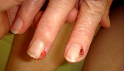
On examination we found periungual reddish papules, easily bleeding, with a diameter of up to 6 mm and some subungual similar lesions of the distal nail bed [Fig. 1]. The latter were seen on toes only. A periungual lesion was excised for histopathology. The histopathological examination showed angiomatous tissue composed with congested capillaries and venules which were embedded in an oedematous stroma containing a mild chronic inflammatory infiltrate.
Figure 1.
Eruptive pyogenic granulomas on fingers.
The diagnosis of multiple eruptive periungual and subungual pyogenic granulomas was confirmed.
Discussion
Nail pyogenic granuloma (PG) is common, often seen as an urgent case, given the recent onset as a bleeding nodule. Nail PGs are due to different causes that act through different pathogenetic mechanisms and may be treated in several ways. Nail PG is usually due to drugs, local trauma and peripheral nerve injury. Histopathology shows similar features in every type of PG, irrespective of cause. When PG is single, especially if it involves the nail bed, histological examination is necessary to rule out malignant melanoma. Treatment must be chosen according to the underlying cause.[5]
Multiple periungual and subungual PG, however, are very rare. In a pediatric patient multiple periungual PG have been reported after long term stay in intensive care unit. The authors suspected drugs and minor traumas as underlying cause.[6] An adult patient developed multiple perinugual PG on the feet after 5-fluorouracil treatment of rectal cancer, another during reverse transcriptase inhibitor therapy for human immunodeficiency virus infection [Table 1].[7,8]
Table 1. Case reports of multiple peringual pyogenic granuloma in adult patients (CR - complete remission).
| Reference | Patients | Aetiologicfactor | Outcome |
|---|---|---|---|
| Piraccini et al. (2010)5 | male, 50 years fingers |
skinsarcoidosis | CR after curettage and topical corticosteroids and antibiotics |
| male, 25years toes |
spondyloarthritis | CR after curettage and topical corticosteroids and antibiotics | |
| female, 24 years fingers and toes |
psoriasis | CR after curettage and topical corticosteroids and antibiotics | |
| Curr et al. (2006)7 | male, 65 years toes |
Systemic 5-fluoro-uracil for rectal carcinoma |
CR after curettage, diathermy, topical corticosteroids and antibiosis |
| Calista et al. (2000)15 | HIV-positive adults but age and gender not specified toes |
indinavir? | |
| Bouscart et al. (1998)16 | 42 patients, HIV-positive (38 men, 4 women), age not specified great toes |
indinavir | CR after months in case of drug withdrawal |
| Current case | female, 73 years | rituximab |
In the present case long-standing immunosuppression with impairment of peringugual and subungual skin integrity and minor traumas might be responsible for the development of multiple PG on fingers and toes. The patient was treated recently transiently with rituximab, an anti-CD20 chimeric murine/human monoclonal antibody. Among the vascular side-effects associated with rituximab administration, vasculitis has been rarely reported.[9] Our case seems to be the first with multiple eruptive PG associated with rituximab.
Treatment of PG can be done with surgery, cryotherapy, cautery, sclerotherapy, or laser. Cryotherapy often needs more than one session.[10,11] Topical phenol has been used successfully, but in several countries in Europe phenol is no longer listed as a medical drug because of possible toxic adverse effects.[12] C02, pulse-dye or Nd-YAG laser are very effective to clear such lesions but CO2 has a higher risk of scarring.[10,13,14] Pulsed-dye laser treatment is planned for the present patient.
References
- Tay YK, Weston WL, Morelli JG. Treatment of pyogenic granuloma in children with flashlamp-pumped pulsed dye laser. Pediatrics. 1997;99:368–370. doi: 10.1542/peds.99.3.368. [DOI] [PubMed] [Google Scholar]
- Naimer SA, Cohen A, Vardy D. Pyogenic granuloma of the penile shaft following circumcision. Pediatr Dermatol. 2002;19:39–41. doi: 10.1046/j.1525-1470.2002.00005.x. [DOI] [PubMed] [Google Scholar]
- Jordan DR, Brownstein S, Lee-Wing M, Ashenhurst M. Pyogenic granuloma following oculoplastic procedures: an imbalance in angiogenesis regulation? Can J Ophthalmol. 2001;36:260–268. doi: 10.1016/s0008-4182(01)80019-3. [DOI] [PubMed] [Google Scholar]
- Palmero ML, Pope E. Eruptive pyogenic granulomas developing after drug hypersensitivity reaction. J Am Acad Dermatol. 2009;60:855–857. doi: 10.1016/j.jaad.2008.11.021. [DOI] [PubMed] [Google Scholar]
- Piraccini BM, Bellavista S, Misciali C, Tosti A, de Berker D, Richert B. Periungual and subungual pyogenic granuloma. Br J Dermatol. 2010;163:941–953. doi: 10.1111/j.1365-2133.2010.09906.x. [DOI] [PubMed] [Google Scholar]
- Guhl G, Torrelo A, Hernández A, Zambrano A. Beau's lines and multiple periungueal pyogenic granulomas after long stay in an intensive care unit. Pediatr Dermatol. 2008;25:278–279. doi: 10.1111/j.1525-1470.2008.00657.x. [DOI] [PubMed] [Google Scholar]
- Curr N, Saunders H, Murugasu A, Cooray P, Schwarz M, Gin D. Multiple periungual pyogenic granulomas following systemic 5-fluorouracil. Australas J Dermatol. 2006;47:130–133. doi: 10.1111/j.1440-0960.2006.00248.x. [DOI] [PubMed] [Google Scholar]
- Williams LH, Fleckman P. Painless periungual pyogenic granulomata associated with reverse transcriptase inhibitor therapy in a patient with human immunodeficiency virus infection. Br J Dermatol. 2007;156:163–164. doi: 10.1111/j.1365-2133.2006.07512.x. [DOI] [PubMed] [Google Scholar]
- Kim MJ, Kim HO, Kim HY, Park YM. Rituximab-induced vasculitis: A case report and review of the medical published work. J Dermatol. 2009;36:284–287. doi: 10.1111/j.1346-8138.2009.00639.x. [DOI] [PubMed] [Google Scholar]
- Giblin AV, Clover AJ, Athanassopoulos A, Budny PG. Pyogenic granuloma - the quest for optimum treatment: audit of treatment of 408 cases. J Plast Reconstr Aesthet Surg. 2007;60:1030–1035. doi: 10.1016/j.bjps.2006.10.018. [DOI] [PubMed] [Google Scholar]
- Gilmore A, Kelsberg G, Safranek S. Clinical inquiries. What's the best treatment for pyogenic granuloma? J Fam Pract. 2010;59:40–42. [PubMed] [Google Scholar]
- Losa Iglesias ME, Becerro de Bengoa Vallejo R. Topical phenol as a conservative treatment for periungual pyogenic granuloma. Dermatol Surg. 2010;36:675–678. doi: 10.1111/j.1524-4725.2010.01528.x. [DOI] [PubMed] [Google Scholar]
- Khandpur S, Sharma VK. Successful treatment of multiple gingival pyogenic granulomas with pulsed-dye laser. Indian J Dermatol Venereol Leprol. 2008;74:275–277. doi: 10.4103/0378-6323.41389. [DOI] [PubMed] [Google Scholar]
- Galeckas KJ, Uebelhoer NS. Successful treatment of pyogenic granuloma using a 1,064-nm laser followed by glycerin sclerotherapy. Dermatol Surg. 2009;35:530–534. doi: 10.1111/j.1524-4725.2009.01081.x. [DOI] [PubMed] [Google Scholar]
- Calista D, Boschini A. Cutaneous side effects induced by indinavir. Eur J Dermatol. 2000;10:292–296. [PubMed] [Google Scholar]
- Bouscarat F, Bouchard C, Bouhour D. Paronychia and pyogenic granuloma of the great toes in patients treated with indinavir. N Engl J Med. 1998;338:1776–1777. doi: 10.1056/NEJM199806113382417. [DOI] [PubMed] [Google Scholar]


