Abstract
The antigenic diversity of HIV-1 has long been an obstacle to vaccine design, and this variability is especially pronounced in the V3 loop of the virus' surface envelope glycoprotein. We previously proposed that the crown of the V3 loop, although dynamic and sequence variable, is constrained throughout the population of HIV-1 viruses to an immunologically relevant β-hairpin tertiary structure. Importantly, there are thousands of different V3 loop crown sequences in circulating HIV-1 viruses, making 3D structural characterization of trends across the diversity of viruses difficult or impossible by crystallography or NMR. Our previous successful studies with folding of the V3 crown1, 2 used the ab initio algorithm 3 accessible in the ICM-Pro molecular modeling software package (Molsoft LLC, La Jolla, CA) and suggested that the crown of the V3 loop, specifically from positions 10 to 22, benefits sufficiently from the flexibility and length of its flanking stems to behave to a large degree as if it were an unconstrained peptide freely folding in solution. As such, rapid ab initio folding of just this portion of the V3 loop of any individual strain of the 60,000+ circulating HIV-1 strains can be informative. Here, we folded the V3 loop of the R2 strain to gain insight into the structural basis of its unique properties. R2 bears a rare V3 loop sequence thought to be responsible for the exquisite sensitivity of this strain to neutralization by patient sera and monoclonal antibodies4, 5. The strain mediates CD4-independent infection and appears to elicit broadly neutralizing antibodies. We demonstrate how evaluation of the results of the folding can be informative for associating observed structures in the folding with the immunological activities observed for R2.
Protocol
1. Methodology
The first step of the protocol is to select the V3 crown sequence you wish to fold in silico. For the R2 strain, the sequence for this fragment is KSIPMGPGRAFYT.
The 3D atomic structure of the peptide corresponding to this sequence should be built in the computer's virtual space. The ICM command for this is:buildpep "KSIPMGPGRAFYT" or File:New:Peptide can be selected under the pull down menu
Several parameters of the procedure are then set, including the number of search steps (length of the search), the simulation temperature, search strategy parameters including variable restraints to bias the search towards reasonable areas, energy terms, selection of different energy calculation methods, and parameters indicating how the search will be recorded such as whether to record a movie and how many intermediate scoring conformations should be recorded. All these parameters have been optimized by previous publications (see www.molsoft.com).
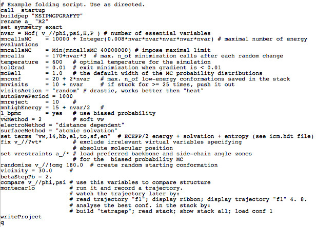
The folding is then initiated with the command:montecarlo or File:New:Peptide can be selected under the pull down menu Molecular Mechanics:Minimize:GlobalIn the former case, the parameters from step 3 should be set one by one in the command line. In the latter case, checkboxes for the most commonly chosen parameters are provided in a pane before the command is executed. For convenience, the same folding ICM script utilized for this experiment and previously published experiments is shown here:
 This script can be saved in a text file and run from the computer's operating system command line (usually LINUX) rapidly using the command:icm _foldingscript
2. Secrets to Success
The V3 loop is a constant 35 amino acids in length in almost every known strain, so the amino acid positions are numbered from 1 to 35 starting with the originating disulfide bonded cystine at 1 and terminating with the corresponding disulfide bonded cystine at 35. Since it is part of a loop, the boundaries of the crown of V3 should be carefully chosen. If too large a fragment is chosen, it is unlikely to behave as a freely flexible segment as if it were a free peptide, so the folding simulation will not correctly assess the structure. If too small a fragment is chosen, the informative tertiary structure may not form in the simulation. Our prior study showed that the fragment from position 10 to position 22 correlated with antibody bound crystallographic conformations, so this is the fragment of any V3 loop that should be chosen for folding.
Although using the graphical user interface and pull-down menus is user-friendly, the prior successful work on peptide folding in general using ICM and V3 loop crown folding in particular used the script above, simply modifying the sequence in the buildpep line and running the above script from the command line is recommended.
3. Representative Results
The results for the R2 folding are representative of the results for any V3 loop. To evaluate the results, the project file (it will be named "newProject1.icb" from the above script) should be opened and "Molecular Mechanics, Stack, View" chosen. A table of the stack conformations will appear. The stack conformations can be visualized graphically by clicking on the Plot/Histogram icon. "Molecular Mechanics, Stack, Play " will make a movie of the stack to visually appreciate the conformational preferences uncovered by the folding. For the R2 sequence, the conformation is beta-hairpin-like as expected for V3 loops2--especially in the fragment at positions 12 to 14 where a clear β-strand preference is seen throughout the stack, and very few alpha-helical conformations are seen. Furthermore, an energy gap of almost 3 units is seen between the lowest energy conformation and the second lowest energy conformation. From an energetic point of view, this means that the structure only flickers out of the lowest energy conformation less than one percent of the time: the folding results therefore suggest that the R2 V3 crown is a rigid, rather than flexible, structure. There may be additional important structural features in the ensemble of conformations, but they are difficult to systematically appreciate.
| JRFL | SF162_V3JRFL | SF162_V3R2 | |
| 447-52D GMT50 | 15 | 0.00061 | 0.00078 |
Table 1: Relative neutralization of JRFL and R2 V3 loops in masked and unmasked settings. The data was previously reported in Cardozo T., et al. ARHR (2008) and Pinter, A., et. al J. Virol (2004). Briefly, neutralizing activity was determined with a single-cycle infectivity assay using psVs generated with the env-defective luciferase-expressing pNL4-3.Luc.R-E- plasmid pseudotyped with the JRFL Env or the SF162 V3 variants described above: SF162_V3JRFL contains the JRFL V3 loop sequence in place of the SF162 V3 loop sequence. SF162_V3R2 contains the R2 V3 loop sequence in place of the SF162 V3 loop sequence. The psVs were incubated with serial dilutions of the 447-52D mAb for 1.5 h at 37°C, and then added to CD4+CCR5+ U87 target cells plated in 96-well plates in the presence of polybrene (10 mg=mL). After 24 hrs, cells were refed with RPMI medium containing 10% FBS and 10 mg=mL polybrene, followed by an additional 24-48 h of incubation. Luciferase activity was determined 48-72 h postinfection with a microtiter plate luminometer (HARTA, Inc.) using assay reagents from Promega, Inc. Geometric mean titers for 50% neutralization (GMT50) by 447-52D were determined by interpolation from neutralization curves and are averages of at least three independent assays.
Discussion
Ab initio folding reveals tertiary structural properties, the effects of which may not be evident from studying individual amino acids sequentially. Several previously obscure structural properties of the R2 V3 loop crown are revealed by the folding simulation. These observed structural preferences may correlate with observed functional properties of the R2 strain, namely sensitivity to neutralization and hyperinfectivity5.
First, a clear β-strand tendency in the N-terminal half of the R2 V3 crown at positions 12-14 is observed. This is the location of the R2 strain's unusual isoleucine-proline-methionine (IPM) sequence. From crystallographic structures of the 447-52D, 2219, Be48, 2557, 2558 and 537-D anti-V3 loop antibodies bound to V3 loop peptides, positions 12 to 14 of the V3 loop are bound by these antibodies, and, when bound, the segment adopts a local β-strand conformation. The folding simulation thus suggests that the R2 V3 loop crown prefers a conformation at positions 12 to 14 that is complementary to known anti-V3 loop neutralizing antibodies. This preference may explain the observed sensitivity to neutralization of this strain.
Secondly, the simulation shows a gap of 3 energy units between the lowest energy conformation and the second lowest energy conformation, suggesting a rigid structure. HIV-1's gp120 is an extremely dynamic molecule, requiring a large conformational shift from its unbound, immunologically protected (unliganded) form to its human receptor bound and immunologically exposed (liganded) form. Flexibility of the V3 loop may be required to effect this cooperative transition, and the rigid R2 V3 loop structure observed here may resist shifting out of the liganded form, much as a rigid block might be more difficult to tuck away into a small space compared to a softer flexible block. This resistance to adopting the unliganded form may expose V3 loop, the CD4 binding site and CD4 induced neutralization epitopes, all of which are characteristically exposed in the liganded form of gp120. Thus, the observed rigidity may also be conceptually connected to the observed neutralization sensitivity of the R2 strain.
Since the co-receptor interacting form of the V3 loop is also likely to be a β-hairpin like the forms targeted by neutralizing antibodies, the results suggest that the R2 V3 crown is locked into the co-receptor and neutralizing antibody complementary form, which in turn may promote the whole gp120 into its liganded form, making the strain highly infective. Thus, the observed conformational preference and rigidity both also can be conceptually associated with R2's hyperinfectivity.
This arrangement of a rigid, but exposed co-receptor and antibody complementary conformation of the V3 loop is distinct from that of a flexible, but buried or masked V3 loop that is typical of HIV strains. Folding of the JRFL strain (which exhibits the consensus subtype B HIV-1 V3 loop sequence) also shows a β-strand preference, but many closely distributed lowest energy conformations suggestive of V3 crown flexibility (data not shown). The prediction would be that this V3 loop sequence would be highly sensitive to neutralization by an appropriate antibody like 447-52D if it were exposed instead of buried. As expected, the JRFL and R2 sequences are equally sensitive when presented in an exposed setting within a chimeric SF162 pseudovirus in which the V3 loop is replaced by the JRFL or R2 V3 loops (Table 1), but the same JRFL V3 loop is completely insensitive to neutralization when presented in its native JRFL context.
These results suggest that the unusual IPM sequence of the R2 strain confers a rigid, antibody-binding-site-compatible, co-receptor-binding-site compatible shape in the key crown region of the V3 loop. The rigidity is hypothesized to obstruct the conformational flexibility of gp120 as it transitions between liganded and unliganded forms, locking it largely into its liganded form where the antibody-optimal, co-receptor-optimal shape in the V3 crown is exposed and available. The result is sensitivity to neutralization and hyperinfectivity of the R2 strain. In this way, the ab initio folding experiment described in this protocol is informative for HIV vaccine research: the folding results observed for R2 here may be important for HIV-1 vaccine immunogen design as a clue for how to lock gp120-based immunogens into preferred, neutralization sensitive conformations.
Disclosures
No conflicts of interest declared.
Acknowledgments
The work was supported by grants from the Bill and Melinda Gates Foundation (#38631) and the NIH, including DP2 OD004631 and R01 AI084119.
References
- Almond D, Kimura T, Kong X, Zolla-Pazner S&, Cardozo T. Dynamic characterization of the V3 loop crown. Antiviral Therapy. 2007;12:13–31. [Google Scholar]
- Almond D, Kimura T, Kong X, Swetnam J, Zolla-Pazner S, Cardozo T. Structural conservation predominates over sequence variability in the crown of HIV type 1's V3 loop. AIDS Res Hum Retroviruses. 2010;26 doi: 10.1089/aid.2009.0254. [DOI] [PMC free article] [PubMed] [Google Scholar]
- Abagyan R, Totrov M. Biased probability Monte Carlo conformational searches and electrostatic calculations for peptides and proteins. J Mol Biol. 1994;235:983–1002. doi: 10.1006/jmbi.1994.1052. [DOI] [PubMed] [Google Scholar]
- Quinnan GV, Zhang PF, Fu DW, Dong M, Alter HJ. Expression and characterization of HIV type 1 envelope protein associated with a broadly reactive neutralizing antibody response. AIDS Res Hum Retroviruses. 1999;15:561–5670. doi: 10.1089/088922299311088. [DOI] [PubMed] [Google Scholar]
- Zhang PF, Bouma P, Park EJ. A variable region 3 (V3) mutation determines a global neutralization phenotype and CD4-independent infectivity of a human immunodeficiency virus type 1 envelope associated with a broadly cross-reactive, primary virus-neutralizing antibody response. J Virol. 2002;76:644–655. doi: 10.1128/JVI.76.2.644-655.2002. [DOI] [PMC free article] [PubMed] [Google Scholar]


