Abstract
Purpose
Childhood cancer survivors experience an increased incidence of subsequent neoplasms (SNs). Those surviving the first SN (SN1) remain at risk to develop multiple SNs. Because SNs are a common cause of late morbidity and mortality, characterization of rates of multiple SNs is needed.
Patients and Methods
In a total of 14,358 5-year survivors of childhood cancer diagnosed between 1970 and 1986, analyses were carried out among 1,382 survivors with an SN1. Cumulative incidence of second subsequent neoplasm (SN2), either malignant or benign, was calculated.
Results
A total of 1,382 survivors (9.6%) developed SN1, of whom 386 (27.9%) developed SN2. Of those with SN2, 153 (39.6%) developed more than two SNs. Cumulative incidence of SN2 was 46.9% (95% CI, 41.6% to 52.2%) at 20 years after SN1. The cumulative incidence of SN2 among radiation-exposed survivors was 41.3% (95% CI, 37.2% to 45.4%) at 15 years compared with 25.7% (95% CI, 16.5% to 34.9%) for those not treated with radiation. Radiation-exposed survivors who developed an SN1 of nonmelanoma skin cancer (NMSC) had a cumulative incidence of subsequent malignant neoplasm (SMN; ie, malignancies excluding NMSC) of 20.3% (95% CI, 13.0% to 27.6%) at 15 years compared with only 10.7% (95% CI, 7.2% to 14.2%) for those who were exposed to radiation and whose SN1 was an invasive SMN (excluding NMSC).
Conclusion
Multiple SNs are common among aging survivors of childhood cancer. SN1 of NMSC identifies a population at high risk for invasive SMN. Survivors not exposed to radiation who develop multiple SNs represent a population of interest for studying genetic susceptibility to neoplasia.
INTRODUCTION
The relative 5-year survival rate after a diagnosis of childhood cancer, which was less than 30% in 1960, is now 79%.1 As of 2005, there were more than 328,000 survivors of childhood cancer in the United States.2 Perhaps the most serious late complication for these survivors is the development of subsequent neoplasms (SNs), many of which are subsequent malignant neoplasms (SMNs).3–10 SMNs are the most common cause of treatment-related death in long-term survivors (standardized mortality ratio, 15.2; 95% CI, 13.9 to 16.6).11 Combined with the increase of the cumulative incidence of SMNs in the Childhood Cancer Survivor Study (CCSS) population over the last decade (3.2% at 20 years from diagnosis to 9.3% at 30 years),6 it is clear that the development of SMNs is a central issue for aging survivors. However, it appears that the increasing cumulative incidence of a first SN does not fully describe the risk for this population. Survivors who experience their first SN may be at risk for the development of multiple SNs. The aging population of the CCSS allows a comprehensive assessment of this question.
PATIENTS AND METHODS
Identification and Contact of the Cohort
The CCSS is a retrospective, cohort study that provides longitudinal follow-up of 5-year survivors of childhood cancer who were treated at 26 institutions in the United States and Canada. Eligibility for participation in the CCSS included diagnosis of cancer before age 21 years; initial treatment between January 1, 1970, and December 31, 1986; and survival for at least 5 years from diagnosis. Participants were recruited from survivors treated for initial diagnoses of leukemia, CNS malignancy, Hodgkin's lymphoma, non-Hodgkin's lymphoma, Wilms tumor, neuroblastoma, soft tissue sarcoma, or bone tumors. The cohort methodology and study design have been previously described in detail.12,13 The CCSS was approved by institutional review boards at the 26 participating centers, and participants provided informed consent.
Collection of baseline data was initiated in 1994. All participants completed a questionnaire that included information on demographics, personal and family medical history, medical late effects experienced, and diagnosis of new neoplasms. Additionally, occurrence of SNs was collected in three subsequent follow-up questionnaires. Therapeutic exposures were ascertained through abstraction of medical and radiation therapy records of each participant by using a standardized protocol.13 Study questionnaires are available at http://ccss.stjude.org.
Definition and Ascertainment of Subsequent Neoplasms
SNs include new neoplasms (malignant and benign; Appendix Table A1, online only) collected from the cohort, not including recurrence of the primary childhood malignancy. SNs were initially ascertained through self- or proxy-report questionnaires and/or death certificates. Occurrences were subsequently confirmed by pathology report or, when not available, were confirmed by death certificate or other medical records reviewed by study investigators. Only SNs occurring 5 or more years after the childhood cancer diagnosis were evaluated.5,6 For this analysis, the term SN1 was defined as the first SN to occur after the primary diagnosis; SN2 represents the second SN; SN3, the third SN; and SN4, the fourth SN. Similarly, SMNs are numbered and defined.
Statistical Analysis
SNs were divided into three mutually exclusive subsets: SMNs, which included only malignant diagnoses utilized by the U.S. Surveillance Epidemiology and End Results (SEER) program with an International Classification of Diseases (ICD) –O behavior code of 3, thus excluding nonmelanoma skin cancers and nonmalignant meningiomas; nonmelanoma skin cancers (NMSCs, which included ICD-O morphology codes 8070, 8071, 8081, 8090, 8094); and all other benign neoplasms, which included meningiomas that were defined by an ICD-O behavior code of 0, 1, or 2, regardless of ICD-O site and morphology codes.
Frequency distributions of the entire cohort, those with SNs (any, ≥ 2, or ≥ 3) and SMNs (any, ≥ 2, or ≥ 3) were characterized by demographic and treatment variables. Exact information regarding location of the SN was not always available. The cumulative incidence of SN2, utilizing time from SN1 to the earliest of SN2, death, or last follow-up, was compared by using Gray's approach,14 in which death was incorporated as a competing risk event, in addition to describing the patterns of SN occurrence by each potential categoric risk factor. Cumulative incidence of SMN2 was analyzed in a similar manner from time of SMN1. Conditional cumulative incidence analyses for subsequent breast neoplasms and NMSCs after development of a first breast or NMSC SN were evaluated, with conditioning at 0, 6, 12, and 24 months of SN-free time from initial neoplasm. Five patients were excluded from cumulative incidence analyses on the basis of a missing date of SN diagnosis.
In multivariable analyses, we employed the method by Fine and Gray15 for modeling the subdistribution hazard rate of SN2 as a function of risk factors, assuming its proportionality, analogous to Cox regression. This allowed us to directly evaluate the risk factors associated with the cumulative incidence of SN2 while accounting for deaths as competing events. Analyses were adjusted for age at primary diagnosis, sex, ethnicity group, family history of cancer (in first-degree relative), education level, annual household income, smoking status, age at SN1, and time from primary cancer to SN1. In addition, other treatment and demographic factors that were significant at the .15 level in univariate analyses (Gray's test) were also included. Analyses were completed by using S-Plus CRR function (Spotfire S-Plus version 8.1; TIBCO, Seattle, WA).
In addition to the Fine and Gray15 method for estimating subdistribution hazard ratios (HRs), we employed Cox regression to estimate cause-specific HRs. Cause-specific hazards models with age as the time scale were utilized, and the outcome measure was defined as interval from age at SN1 to the minimum age at SN2, date of death, or date of last follow-up. We chose to emphasize the results of Fine and Gray analysis, because our primary interest was on covariate effects on cumulative incidence (and associated subdistribution HRs), but we also showed the cause-specific hazard analysis results (Appendix Table A2, online only), noting that the two methods gave similar conclusions for risk factors of SN2 and their effects.
RESULTS
We identified 14,358 survivors (median age at last follow-up, 31.9 years; range, 5.6 to 56.3 years), who accrued a total of 327,297 person-years of follow-up time, with a median of 23.0 years from diagnosis (range, 5.0 to 37.6 years). Within this cohort, radiation exposure was common (occurring in 67.9% of those who consented to release their medical records). Additional characteristics of the cohort are listed in Table 1 and in Appendix Table A3 (online only). Within this cohort, 1,382 people (9.6%) developed SN1, with a total of 9,387 person-years of follow-up time. Among these 1,382 SN1 occurrences, 386 (27.9%) developed an SN2. Of those with SN2, 153 (39.6%) developed greater than two SNs. In addition, 735 (5.1%) of the 14,358 members of the cohort developed SMN1, 68 (9.3%) of whom developed SMN2.
Table 1.
Characteristics of the CCSS Cohort: Entire Cohort, Survivors With Subsequent Neoplasms, and Survivors With Subsequent Malignant Neoplasms
| Subsequent Neoplasm |
Subsequent Malignant Neoplasm |
|||||||||||||
|---|---|---|---|---|---|---|---|---|---|---|---|---|---|---|
| Entire Cohort |
Any |
≥ 2 |
≥ 3 |
Any |
≥ 2 |
≥ 3 |
||||||||
| Characteristic* | No. | % | No. | % | No. | % | No. | % | No. | % | No. | % | No. | % |
| Total | 14,358 | 100 | 1,382 | 100 | 386 | 100 | 153 | 100 | 735 | 100 | 68 | 100 | 3 | 100 |
| Age at diagnosis, years | ||||||||||||||
| Mean | 8.3 | 11.0 | 12.5 | 13.4 | 11.1 | 12.4 | 15.7 | |||||||
| SD | 5.8 | 6.1 | 5.8 | 5.6 | 6.1 | 5.6 | 2.0 | |||||||
| Median | 6.8 | 11.9 | 14.2 | 15.1 | 12.2 | 14.1 | 14.7 | |||||||
| Range | 0.0-21.0 | 0.0-21.0 | 0.5-20.9 | 0.9-20.9 | 0.0-21.0 | 0.5-20.6 | 14.4-18.0 | |||||||
| Age at initial diagnosis, years | ||||||||||||||
| < 10 | 8,956 | 62.4 | 588 | 42.5 | 121 | 31.3 | 39 | 25.5 | 300 | 40.8 | 20 | 29.4 | 0 | 0.0 |
| ≥ 10 | 5,402 | 37.6 | 794 | 57.5 | 265 | 68.7 | 114 | 74.5 | 435 | 59.2 | 48 | 70.6 | 3 | 100 |
| Sex | ||||||||||||||
| Male | 7,713 | 53.7 | 589 | 42.6 | 153 | 39.6 | 69 | 45.1 | 289 | 39.3 | 15 | 22.1 | 2 | 66.7 |
| Female | 6,645 | 46.3 | 793 | 57.4 | 233 | 60.4 | 84 | 54.9 | 446 | 60.7 | 53 | 77.9 | 1 | 33.3 |
| Ethnicity | ||||||||||||||
| White non-Hispanic | 11,943 | 83.2 | 1,229 | 88.9 | 352 | 91.2 | 139 | 90.8 | 644 | 87.6 | 61 | 89.7 | 2 | 66.7 |
| Black non-Hispanic | 668 | 4.7 | 24 | 1.7 | 2 | 0.5 | 1 | 0.7 | 22 | 3.0 | 2 | 2.9 | 0 | 0.0 |
| Hispanic | 738 | 5.1 | 41 | 3.0 | 10 | 2.6 | 4 | 2.6 | 27 | 3.7 | 3 | 4.4 | 1 | 33.3 |
| Other | 1,009 | 7.0 | 88 | 6.4 | 22 | 5.7 | 9 | 5.9 | 42 | 5.7 | 2 | 2.9 | 0 | 0.0 |
| Family history of cancer in a first-degree relative | ||||||||||||||
| Yes | 2,356 | 16.4 | 360 | 26.0 | 128 | 33.2 | 54 | 35.3 | 184 | 25.0 | 25 | 36.8 | 1 | 33.3 |
| No | 12,002 | 83.6 | 1,022 | 74.0 | 258 | 66.8 | 99 | 64.7 | 551 | 75.0 | 43 | 63.2 | 2 | 66.7 |
| Smoking status | ||||||||||||||
| Never | 10,420 | 76.0 | 1,019 | 75.2 | 281 | 74.5 | 113 | 75.3 | 537 | 75.1 | 46 | 70.8 | 1 | 33.3 |
| Former | 1,325 | 9.7 | 177 | 13.1 | 47 | 12.5 | 25 | 16.7 | 102 | 14.3 | 6 | 9.2 | 1 | 33.3 |
| Current | 1,961 | 14.3 | 159 | 11.7 | 49 | 13.0 | 12 | 8.0 | 76 | 10.6 | 13 | 20.0 | 1 | 33.3 |
| Childhood cancer diagnosis | ||||||||||||||
| Acute lymphoblastic leukemia | 4,329 | 30.2 | 341 | 24.7 | 79 | 20.5 | 31 | 20.3 | 128 | 17.4 | 4 | 5.9 | 0 | 0.0 |
| Acute myeloid leukemia | 356 | 2.5 | 29 | 2.1 | 9 | 2.3 | 3 | 2.0 | 17 | 2.3 | 3 | 4.4 | 0 | 0.0 |
| Other leukemia | 145 | 1.0 | 13 | 0.9 | 4 | 1.0 | 2 | 1.3 | 11 | 1.5 | 2 | 2.9 | 0 | 0.0 |
| Astrocytomas | 1,182 | 8.2 | 83 | 6.0 | 20 | 5.2 | 6 | 3.9 | 36 | 4.9 | 6 | 8.8 | 0 | 0.0 |
| Medulloblastoma, PNET | 381 | 2.7 | 47 | 3.4 | 17 | 4.4 | 7 | 4.6 | 19 | 2.6 | 0 | 0.0 | 0 | 0.0 |
| Other CNS tumors | 314 | 2.2 | 31 | 2.2 | 11 | 2.8 | 6 | 3.9 | 15 | 2.0 | 3 | 4.4 | 0 | 0.0 |
| Hodgkin's disease | 1,927 | 13.4 | 446 | 32.3 | 175 | 45.3 | 78 | 51.0 | 252 | 34.3 | 27 | 39.7 | 1 | 33.3 |
| Non-Hodgkin's lymphoma | 1,080 | 7.5 | 81 | 5.9 | 20 | 5.2 | 8 | 5.2 | 45 | 6.1 | 2 | 2.9 | 0 | 0.0 |
| Kidney tumors | 1,256 | 8.7 | 51 | 3.7 | 10 | 2.6 | 2 | 1.3 | 33 | 4.5 | 2 | 2.9 | 0 | 0.0 |
| Neuroblastoma | 954 | 6.6 | 44 | 3.2 | 3 | 0.8 | 0 | 0.0 | 32 | 4.4 | 1 | 1.5 | 0 | 0.0 |
| Soft tissue sarcoma | 1,246 | 8.7 | 112 | 8.1 | 16 | 4.1 | 5 | 3.3 | 75 | 10.2 | 6 | 8.8 | 0 | 0.0 |
| Ewing sarcoma | 403 | 2.8 | 48 | 3.5 | 7 | 1.8 | 2 | 1.3 | 36 | 4.9 | 2 | 2.9 | 0 | 0.0 |
| Osteosarcoma | 733 | 5.1 | 52 | 3.8 | 15 | 3.9 | 3 | 2.0 | 34 | 4.6 | 10 | 14.7 | 2 | 66.7 |
| Other bone tumors | 52 | 0.4 | 4 | 0.3 | 0 | 0.0 | 0 | 0.0 | 2 | 0.3 | 0 | 0.0 | 0 | 0.0 |
| Radiation exposure | ||||||||||||||
| Any | 8,546 | 67.9 | 1,120 | 88.3 | 336 | 92.3 | 139 | 95.9 | 574 | 84.9 | 45 | 71.4 | 1 | 33.3 |
| None | 4,013 | 31.9 | 147 | 11.6 | 28 | 7.7 | 6 | 4.1 | 101 | 14.9 | 18 | 28.6 | 2 | 66.7 |
| Unknown | 33 | 0.3 | 1 | 0.1 | 0 | 0.0 | 0 | 0.0 | 1 | 0.1 | 0 | 0.0 | 0 | 0.0 |
Abbreviations: CCSS, Childhood Cancer Survivor Study; PNET, primitive neuroectodermal tumor; SD, standard deviation.
Percentages for individual characteristics were calculated on total number of participants who provided information for those characteristics.
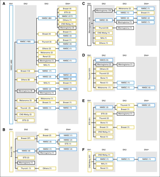
NMSC was the most common SN (1,104 total episodes; Appendix Table A4, online only) and occurred as SN1 in 485 survivors (Fig 1A), 61 (12.6%) of whom subsequently developed an invasive SMN. Other important patterns were identified. Of 176 participants who had a breast neoplasm as SN1, 37 developed a new breast neoplasm as SN2 (Fig 1B). Multiple subsequent meningiomas were common (Fig 1C). Thyroid cancer, soft-tissue sarcomas, and CNS malignancies were frequently observed as SN1 and preceded a variety of additional SNs (Figs 1D to 1F).
Fig 1.
Survivors with multiple neoplasms after first subsequent neoplasm (SN) when SN1 is (A) nonmelanoma skin cancer (NMSC), (B) breast cancer, (C) meningioma, (D) thyroid cancer, (E) soft tissue sarcoma (STS), and (F) CNS malignancies (Malig). Gold, blue, and black boxes represent subsequent malignant neoplasms (SMNs), NMSCs, and benign neoplasms, respectively. NHL, non-Hodgkin's lymphoma.
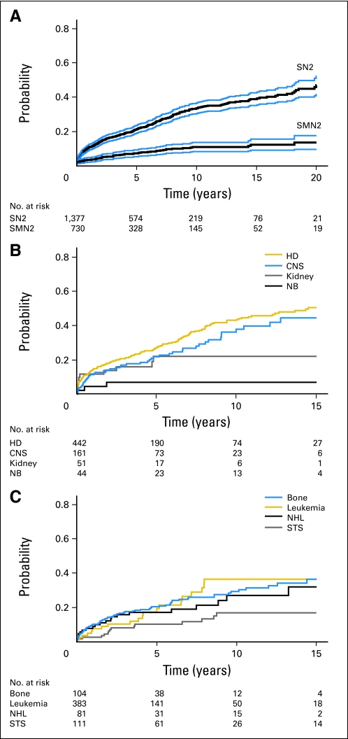
Among survivors with SN1, the cumulative incidence of SN2 was 33.4% (95% CI, 30.3% to 36.5%) at 10 years, was 38.8% (95% CI, 35.1% to 42.5%) at 15 years, and was 46.9% (95% CI, 41.6% to 52.2%) at 20 years (Fig 2A). When SN1 was an SMN, the cumulative incidence of developing SMN2 after SMN1 was 12.4% (95% CI, 9.1% to 15.6%) at 15 years (Fig 2A). The cumulative incidence of SN2 was highest among survivors of a primary Hodgkin's lymphoma (50.3% at 15 years; 95% CI, 44.1% to 56.6%) or CNS malignancy (44.5% at 15 years; 95% CI, 33.4% to 55.6%; Figs 2B and 2C). Among survivors exposed to radiation as therapy for their primary cancer, the cumulative incidence of SN2 was 41.3% (95% CI, 37.2% to 45.4%) at 15 years from SN1 compared with 25.7% (95% CI, 16.5% to 34.9%) for those not treated with radiation (Appendix Table A5, online only). Similarly, among patients with SMN1 who were not exposed to radiation, the cumulative incidence of SMN2 was 22.8% at 10 years.
Fig 2.
(A) Cumulative incidence of second subsequent neoplasm (SN2) after occurrence of first SN (top line) with 95% CI and of second subsequent malignant neoplasm (SMN2; bottom line) after occurrence of first SMN (SMN1) with 95% CI; (B, C) cumulative incidence of SN2 after SN1 by primary pediatric cancer diagnosis. HD, Hodgkin's disease; NB, neuroblastoma; NHL, non-Hodgkin's lymphoma; STS, soft tissue sarcoma.
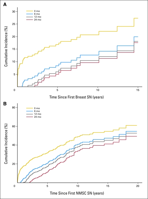
Within this cohort, there were 252 breast lesions (n = 189 invasive, n = 61 in situ, and n = 2 benign). The cumulative incidence of developing a second breast SN was 20.7% (95% CI, 14.7% to 26.7%) at 10 years from the time of development of the initial breast SN (Fig 3A). Because of a significant rate of synchronous breast lesions, cumulative incidences of a second breast neoplasm conditioned on time from first breast SN of 6, 12, and 24 months were 12.6% (95% CI, 7.3% to 18.0%), 10.4% (95% CI, 5.2% to 15.7%), and 9.6% (95% CI, 4.4% t0 14.8%), respectively, at 10 years from initial breast SN. The cumulative incidence of developing a second NMSC at 10 years from the first NMSC was 49.0% (95% CI, 43.5% to 54.5%). Because multiple lesions at presentation were common, when conditioned on time from first NMSC (Fig 3B) of 6, 12, and 24 months, the cumulative incidences of a second NMSC were 40.8% (95% CI, 34.7% to 46.9%), 38.2% (95% CI, 32.0% to 44.4%), and 33.8% (95% CI, 27.3% to 40.3%), respectively, at 10 years from first NMSC.
Fig 3.
Conditional cumulative incidence of (A) second subsequent breast neoplasm after occurrence of first subsequent breast neoplasm and of (B) second subsequent nonmelanoma skin cancer (NMSC) after first subsequent NMSC, conditioned on time of 0 (gold line), 6 (blue line), 12 (gray line), or 24 (black line) months (mo) from first subsequent breast neoplasm or NMSC. SN, subsequent neoplasm.
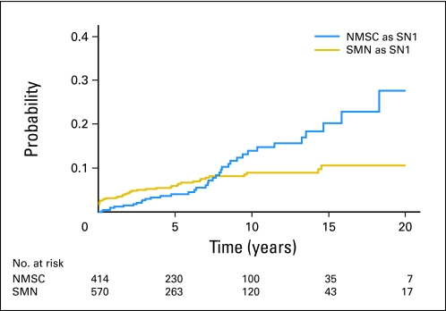
Among survivors who received radiation and developed an SN, 414 had NMSC as SN1, whereas 570 had an invasive SMN as SN1. The cumulative incidence of an additional invasive malignancy (ie, SMN) was 20.3% (95% CI, 13.0% to 27.6%) at 15 years after NMSC compared with 10.7% (95% CI, 7.2% to 14.2%) after SMN1 (Fig 4) and was potentially influenced by the survival estimates for those with an invasive SMN as SN1 (45.4% at 15 years; 95% CI, 37.0% to 53.8%) that were lower than for those with NMSC as SN1 (79.0% at 15 years; 95% CI, 68.4% to 89.6%).
Fig 4.
Cumulative incidence of a subsequent malignant neoplasm among radiotherapy-exposed patients after nonmelanoma skin cancer (NMSC) as first subsequent neoplasm (SN; blue line) and subsequent malignant neoplasm (SMN) as SN1 (gold line).
The limitation of incomplete information regarding treatment for SNs and SMNs is recognized. Table 2 provides multivariable models evaluating risk factors (ie, demographic, treatment exposure for primary cancer, health behaviors, family history) associated with the cumulative incidences of multiple SNs and multiple SMNs after SN1 and SMN1, respectively. Exposure to radiation for the primary cancer was associated with cumulative incidence of SN2 (subdistribution HR, 2.16; 95% CI, 1.32 to 3.55). However, radiation appeared protective from SMN2 (subdistribution HR, 0.48; 95% CI, 0.25 to 0.94; cause-specific HR, 0.41; 95% CI, 0.20 to 0.85). Note that the radiation-exposed survivors had a lower survival estimate at 10 years from SMN1 (56.1%; 95% CI, 49.8% to 62.4%) compared with those who had no radiation exposure (64.3%; 95% CI, 49.6% to 79.0%). Additionally, older age at SN1 (≥ 30 years) was associated with higher cumulative incidence of developing an SN2 compared with those younger than 30 years of age (HR, 1.9; 95% CI, 1.28 to 2.82). Women were more likely than men to develop SMN2 (HR, 2.53; 95% CI, 1.23 to 2.51). However, when breast neoplasia as SMN2 was removed from analysis, association with female sex did not reach statistical significance (HR, 1.57; 95% CI, 0.70 to 3.50). Having a first-degree relative with cancer and being a current smoker were associated with an increased cumulative incidence for multiple SMNs, but the associations were not statistically significant at the.05 level.
Table 2.
Multivariable-Adjusted Subdistribution Hazard Ratios for Development of SN2 From Time of SN1 and for SMN2 From Time of SMN1
| Characteristic | SN1 → SN2 |
SMN1 → SMN2 |
||||
|---|---|---|---|---|---|---|
| Hazard Ratio | 95% CI | P | Hazard Ratio | 95% CI | P | |
| Age at diagnosis, years | ||||||
| ≥ 10 | 1.31 | 0.91 to 1.88 | .15 | 1.72 | 0.49 to 6.02 | .40 |
| < 10 | 1.00 | 1.00 | ||||
| Sex | ||||||
| Female | 1.00 | 0.78 to 1.28 | .99 | 2.53 | 1.23 to 5.21 | .01 |
| Male | 1.00 | 1.00 | ||||
| Ethnicity | ||||||
| Other | 1.13 | 0.68 to 1.89 | .64 | 0.43 | 0.05 to 3.40 | .42 |
| Black non-Hispanic | 0.44 | 0.11 to 1.82 | .26 | 2.12 | 0.54 to 8.23 | .28 |
| Hispanic | 0.83 | 0.37 to 1.85 | .65 | 1.26 | 0.35 to 4.46 | .72 |
| White non-Hispanic | 1.00 | 1.00 | ||||
| First-degree relative with cancer | ||||||
| Yes | 1.05 | 0.81 to 1.36 | .73 | 1.68 | 0.91 to 3.11 | .10 |
| No | 1.00 | 1.00 | ||||
| Radiation exposure | ||||||
| Yes | 2.16 | 1.32 to 3.55 | .01 | 0.48 | 0.25 to 0.94 | > .03 |
| No | 1.00 | 1.00 | ||||
| Age at SN1, years | ||||||
| < 30 | 1.00 | — | — | |||
| ≥ 30 | 1.90 | 1.28 to 2.82 | .001 | — | — | |
| Time from primary to SN1 | — | — | ||||
| Time from primary to SN1 by 10 years | 1.07 | 0.82 to 1.40 | .61 | — | — | |
| Education | ||||||
| < High school | 1.00 | 0.78 to 1.28 | .99 | 1.09 | 0.37 to 3.18 | .87 |
| High school graduate | 1.02 | 0.64 to 1.63 | .94 | 0.73 | 0.29 to 1.83 | .50 |
| > High school | 1.00 | 1.00 | ||||
| Household income, $ per year | ||||||
| ≥ 20,000 | 1.42 | 0.98 to 2.07 | .06 | 1.98 | 0.77 to 5.13 | .16 |
| < 20,000 | 1.00 | 1.00 | ||||
| Smoking | ||||||
| Current | 1.01 | 0.68 to 1.48 | .97 | 1.98 | 0.92 to 4.24 | .08 |
| Former | 0.91 | 0.64 to 1.30 | .61 | 0.85 | 0.35 to 2.09 | .73 |
| Never | 1.00 | 1.00 | ||||
| Alkylator score | ||||||
| 1-2 | 0.87 | 0.65 to 1.17 | .36 | — | — | |
| 3-4 | 1.04 | 0.76 to 1.42 | .81 | — | — | |
| 0 | 1.00 | — | ||||
| Treatment era | ||||||
| 1970-1979 | 1.13 | 0.83 to 1.53 | .44 | |||
| 1980-1986 | 1.00 | |||||
| P53-associated SN1* | ||||||
| Yes | — | — | 1.19 | 0.67 to 2.09 | .55 | |
| No | — | 1.00 | ||||
| Primary sarcoma | ||||||
| Yes | — | — | 1.20 | 0.61 to 2.35 | .60 | |
| No | — | 1.00 | ||||
| Age at SMN1 | ||||||
| Age at SMN1 by 10 years | — | — | 0.94 | 0.37 to 2.38 | .89 | |
| Time from primary to SMN1 | ||||||
| Time from primary to SMN1 by 10 years | — | — | 1.03 | 0.34 to 3.15 | .96 | |
NOTE. Boldface indicates values are significant.
Abbreviations: SMN, subsequent malignant neoplasms; SN, subsequent neoplasms.
P53-associated tumors included those of the breast, brain, lung, colorectal, and gastric regions as well as all sarcomas, adrenocortical carcinomas, acute lymphoblastic leukemia, and non-Hodgkin's lymphomas.
DISCUSSION
It is well established that the cumulative incidence of SNs increases with increased time from diagnosis.7,11,16 We now describe the experience of long-term survivors of childhood cancer after SN1 and the risk for multiple occurrences of SNs, either benign or malignant. Greater than one quarter (28%) of the CCSS population with SN1 experienced an additional neoplasm, such that, within 20 years from diagnosis of SN1, the estimated cumulative incidence of SN2 was 47%. Although previous studies have identified occurrences of multiple SMNs, the number of patients reported in any one study has been too small (range, 2 to 32) to allow detailed description and analysis.6,8,17–20 Although not included in the current analysis, the experience of retinoblastoma survivors would certainly suggest that underlying genetic susceptibility (eg, RB1 gene alteration in the case of certain patients with retinoblastoma) plays a role in development of multiple SNs.16,21 Considering the young age (median, 32 years) of the CCSS cohort, these survivors have yet to reach ages when sharp increases in the incidence of cancer in the general population occur. Therefore, it appears that the multiple tissue injuries accrued as a result of cancer therapy, along with the impact of genetic susceptibility of some survivors to multiple cancers, set the stage in the second decade of survival (median follow-up, 18 years) for a significant increase in the number of survivors with multiple cancers.
Certain patterns of SNs raise concern. Of 176 survivors who experienced a breast neoplasm as SN1, 37 (21%) experienced one or more subsequent primary breast neoplasms. In all, 42 women experienced multiple breast neoplasms, with a cumulative incidence of 20.7% at 10 years from diagnosis of the initial lesion. In many of these occurrences, women had bilateral (synchronous) lesions at presentation not thought to be metastatic, or occurrence of a tumor in the contralateral breast shortly after the primary diagnosis. Nonetheless, among those who were tumor free at 1 year from the initial diagnosis, 10% will still develop a subsequent breast neoplasm 10 years from the initial diagnosis. Although increased risk for breast cancer after treatment for childhood cancer has been well documented,17 this pattern for multiple cancers raises new concerns. Similarly, of 142 participants who had a nonmalignant meningioma, 126 occurred as SN1, and 12 survivors (10%) experienced multiple meningiomas. Therefore, not only is the incidence of meningioma increasing with time from treatment exposure18 but also, with time, survivors may be at increasing risk for multiple meningiomas as well.
Among radiation-exposed individuals who developed NMSC as SN1 in this analysis, one in five developed an invasive neoplasm within 15 years, an incidence almost twice that of those also exposed to radiation but who had an invasive neoplasm (SMN) as SN1. Therefore, NMSC may represent a clinical marker for early identification of a population at high risk for a future malignant neoplasm. Occurrence of NMSC as a first SN may identify survivors with genetic susceptibility to radiation injury and/or deficient DNA repair. In the general population, there is evidence for an association between a NMSC diagnosis and increased risk for subsequent cancer.19,20 Future studies regarding genetic susceptibility should include evaluation of polymorphisms in genes associated with DNA repair.22 In addition, a surprising number of patients with SMN1 who were not exposed to radiation developed SMN2 (cumulative incidence, 22.8% at 10 years). This may identify a population of patients with an underlying cancer predisposition syndrome. We note that SMN1 as SN1 may also identify a genetically susceptible population, but, because of the higher rate of mortality after SMN1 (compared with NMSC as SN1), these patients don't have the same opportunity to develop SMN2. Additionally, though many survivors had multiple NMSCs at presentation, conditional cumulative incidence analysis identifies the high risk that remains in these survivors for future additional NMSCs.
Repeated investigations have identified that female sex, young age at primary cancer diagnosis, radiation exposure, family history of cancer, as well as a primary diagnosis of sarcoma or Hodgkin's lymphoma increase risk for SMN.5,6,23 In this analysis, radiation exposure and older age at SN1 (≥ 30 years) appear to be the most important risk factors for development of multiple SNs. Although the association with radiation is plausible given the established association between radiation and SMN1, we hypothesize that older age at SN1 identifies a population of survivors who are reaching ages at which, in the general population, cancer risks increase. The appearance of a protective effect of radiation for development of SMN2 after SMN1 is likely artifactual as a result of death as a competing risk for SMN2 development. Survivors with SMN1 who had received radiation were more likely to die and, thus, less likely to have SMN2. Furthermore, because both the subdistribution HR (0.48) and cause-specific HR (0.41) for developing SMN2 after SMN1 indicated protective effects of having had radiation for the original childhood cancer, we infer that risk of death and risk of SMN2 were positively correlated, perhaps highly, after SMN1. That is, deaths after SMN1 removed survivors who were at high risk for SMN2 from risk sets disproportionately more in the radiation-exposed subgroup. Had this not been the case, the cause-specific HR would not have been in the protective direction. Although females were at increased risk for SMN2 after SMN1, patients with first-degree relatives with cancer and patients with a smoking history may also be at increased risk. However, these findings were of borderline statistical significance. Additionally, having a sarcoma or a p53 (Li-Fraumeni) –associated SMN1 was not found to be associated with developing multiple malignancies.
Limitations of this study should be considered. The majority of SNs are initially ascertained by self report, which may result in underestimation of true incidence rates. Additionally, although CCSS has collected detailed chemotherapy exposures and radiation dosimetry and volume measures relating to the primary cancer, only limited information was available regarding treatment of SNs, restricting risk factor analyses to primary cancer exposures only. Clearly, exposures resulting from treatment of subsequent neoplasms could impact subsequent cancer risk. The limitation of not having the SN treatment information to include in the risk factor analysis does not detract from the findings and clinical implications of the high cumulative incidence of multiple subsequent neoplasms. Finally, systematic and precise determination of SN occurrence as in field versus out of the radiation field was not possible in many of the occurrences, because exact information regarding location of the SN was not always available either from the respondent or as noted on the pathology report.
In conclusion, with increased follow-up time, survivors of childhood cancer are at increasing risk for multiple subsequent neoplasms. The CCSS cohort, with its large population and comprehensive follow-up over two decades, provides a unique resource for evaluation of patterns of SNs as the cohort ages. Although some of the traditional risk factors for SMNs, such as radiation exposure and female sex, are evident among those with multiple neoplasms, future studies should utilize the CCSS biospecimen collection as a resource to identify causes of genetic susceptibility in these populations. Diagnosis of a NMSC may identify those survivors with a predisposition for subsequent invasive neoplasms. Thus, in addition to annual screening dermatologic examination, compliance with additional guidelines for screening for invasive malignancy (ie, mammography, colonoscopy) is important.
Appendix
Table A1.
Benign Neoplasm Subtypes Identified Within the CCSS Cohort
| Tumor Group | Specific Histopathologic Diagnosis | Count |
|---|---|---|
| Breast | Intraductal carcinoma, noninfiltrating NOS | 46 |
| Comedocarcinoma, noninfiltrating | 5 | |
| Noninfiltrating intraductal papillary adenocarcinoma | 3 | |
| Lobular carcinoma in situ | 3 | |
| Carcinoma in situ NOS | 2 | |
| Intraductal carcinoma and lobular carcinoma in situ | 2 | |
| Neoplasm, uncertain whether benign or malignant | 2 | |
| Myeloid leukemia | Myelodysplastic syndrome NOS | 3 |
| Chronic myeloproliferative disease | 1 | |
| Melanoma | Melanoma in situ | 7 |
| Nonmelanoma skin* | Squamous cell carcinoma in situ NOS | 12 |
| Bowen's disease | 2 | |
| Nonmalignant meningioma | Meningioma NOS | 138 |
| Meningotheliomatous meningioma | 8 | |
| Fibrous meningioma | 5 | |
| Transitional meningioma | 5 | |
| Meningiomatosis NOS | 3 | |
| Renal | Oxyphilic adenoma | 1 |
| Soft tissue sarcoma | Neurilemmoma NOS | 19 |
| Fibrous histiocytoma NOS | 1 | |
| Hemangioblastoma | 1 | |
| Other | Squamous cell carcinoma in situ NOS | 18 |
| Neoplasm, uncertain whether benign or malignant | 7 | |
| Neoplasm, benign | 4 | |
| Transitional cell carcinoma in situ | 3 | |
| Carcinoma in situ NOS | 2 | |
| Adenocarcinoma in situ | 2 | |
| Bowen's disease | 1 | |
| Sex cord-stromal tumor | 1 | |
| Granulosa cell tumor NOS | 1 | |
| Leydig cell tumor NOS | 1 | |
| Gonadoblastoma | 1 | |
| Teratoma NOS | 1 | |
| Subependymal giant cell astrocytoma | 1 | |
| Myxopapillary ependymoma | 1 | |
| Ganglioglioma | 1 | |
| Total | 314 |
NOTE. Subtypes are identified as International Classification of Diseases O-2 codes ending in 0, 1, or 2.
Abbreviations: CCSS, Childhood Cancer Survivor Study; NOS, not otherwise specified.
Does not include malignant nonmelanoma skin cancers (ie, International Classification of Diseases O behavior code 3; n = 1,090).
Table A2.
Multivariable-Adjusted Hazard Ratios for Development of SN2 From Time of SN1 and, for SMN2, From Time of SMN1 With Age As the Time Scale, Using Cause-Specific Hazards Model
| Characteristic | SN1 → SN2 |
SMN1 → SMN2 |
||||
|---|---|---|---|---|---|---|
| Hazard Ratio | 95% CI | P | Hazard Ratio | 95% CI | P | |
| Age at diagnosis, years | ||||||
| < 10 | 1.00 | 1.00 | ||||
| ≥ 10 | 1.60 | 1.07 to 2.40 | .0222 | 2.17 | 0.52 to 9.13 | .2906 |
| Sex | ||||||
| Female | 0.96 | 0.76 to 1.22 | .7666 | 2.63 | 1.07 to 6.46 | .035 |
| Male | 1.00 | 1.00 | ||||
| Ethnicity | ||||||
| Other | 1.10 | 0.66 to 1.83 | .7256 | NA | ||
| Black non-Hispanic | 0.58 | 0.14 to 2.38 | .4531 | 1.37 | 0.16 to 11.54 | .7693 |
| Hispanic | 1.00 | 0.49 to 2.04 | .9978 | 1.88 | 0.53 to 6.75 | .3314 |
| White non-Hispanic | 1.00 | 1.00 | ||||
| First-degree relative with cancer | ||||||
| Yes | 1.14 | 0.89 to 1.47 | .2923 | 1.75 | 0.91 to 3.36 | .0924 |
| No | 1.00 | 1.00 | ||||
| Radiation exposure | ||||||
| Yes | 2.01 | 1.27 to 3.17 | .0028 | 0.41 | 0.20 to 0.85 | .0159 |
| No | 1.00 | 1.00 | ||||
| Age at SN1, years | ||||||
| < 30 | 1.00 | — | — | |||
| ≥ 30 | 1.82 | 1.19 to 2.78 | .0059 | — | ||
| Time from Primary to SN1 | ||||||
| Time from Primary to SN1 by 10 years | 1.46 | 1.07 to 1.98 | .0154 | — | — | |
| Education | ||||||
| < High school | 1.41 | 0.87 to 2.28 | .166 | 1.24 | 0.34 to 4.58 | .7416 |
| High school graduate | 0.93 | 0.64 to 1.34 | .6843 | 1.11 | 0.41 to 2.99 | .8426 |
| > High school | 1.00 | 1.00 | ||||
| Household income, $ per year | ||||||
| ≥ 20,000 | 1.39 | 0.96 to 2.01 | .0828 | 1.47 | 0.54 to 3.98 | .4538 |
| < 20,000 | 1.00 | 1.00 | ||||
| Smoking | ||||||
| Current | 0.87 | 0.60 to 1.25 | .4479 | 1.72 | 0.72 to 4.11 | .224 |
| Former | 0.92 | 0.66 to 1.29 | .6332 | 0.92 | 0.34 to 2.51 | .8701 |
| Never | 1.00 | 1.00 | ||||
| P53-associated SN1 | ||||||
| Yes | — | — | 1.46 | 0.76 to 2.80 | .2525 | |
| No | — | 1.00 | ||||
| Primary sarcoma | ||||||
| Yes | — | — | 1.10 | 0.52 to 2.34 | .8067 | |
| No | — | 1.00 | ||||
| Age at SMN1 | ||||||
| Age at SMN1 by 10 years | — | — | 1.09 | 0.30 to 3.93 | .9002 | |
| Time from primary to SMN1 | ||||||
| Time from primary to SMN1 by 10 years | — | — | 1.06 | 0.33 to 3.40 | .9178 | |
Abbrevations: NA, not applicable; SMN, subsequent malignant neoplasm; SN, subsequent neoplasm.
Table A3.
Additional Characteristics of the Childhood Cancer Survivor Study Cohort: Entire Cohort, Survivors With Subsequent Neoplasm, and Survivors With Subsequent Malignant Neoplasms
| Subsequent Neoplasm |
Subsequent Malignant Neoplasm |
|||||||||||||
|---|---|---|---|---|---|---|---|---|---|---|---|---|---|---|
| Entire Cohort |
Any |
Multiple |
≥ 3 |
Any |
Multiple |
≥ 3 |
||||||||
| Characteristic* | No. | % | No. | % | No. | % | No. | % | No. | % | No. | % | No. | % |
| Total | 14,358 | 100 | 1,382 | 100 | 386 | 100 | 153 | 100 | 735 | 100 | 68 | 100 | 3 | 100 |
| Treatment era | ||||||||||||||
| 1970-1979 | 7,749 | 54.0 | 457 | 33.1 | 98 | 25.4 | 36 | 23.5 | 257 | 35.0 | 18 | 26.5 | 1 | 33.3 |
| 1980-1986 | 6,609 | 46.0 | 925 | 66.9 | 288 | 74.6 | 117 | 76.5 | 478 | 65.0 | 50 | 73.5 | 2 | 66.7 |
| Education | ||||||||||||||
| < High school | 4,795 | 35.3 | 216 | 16.6 | 40 | 10.9 | 14 | 9.6 | 123 | 17.8 | 11 | 16.7 | 0 | 0.0 |
| High school graduate | 2,315 | 17.0 | 194 | 14.9 | 50 | 13.6 | 19 | 13.0 | 113 | 16.4 | 12 | 18.2 | 1 | 33.3 |
| > High school | 6,485 | 47.7 | 889 | 68.4 | 277 | 75.5 | 113 | 77.4 | 455 | 65.8 | 43 | 65.2 | 2 | 66.7 |
| Annual household income, $ per year | ||||||||||||||
| < 20,000 | 2,668 | 21.0 | 217 | 17.6 | 40 | 11.2 | 16 | 11.0 | 117 | 18.1 | 6 | 9.8 | 0 | 0.0 |
| > 20,000 | 10,047 | 79.0 | 1,015 | 82.4 | 317 | 88.8 | 130 | 89.0 | 531 | 81.9 | 55 | 90.2 | 3 | 100 |
| Treatment of primary diagnose | ||||||||||||||
| RT only | 39 | 0.3 | 4 | 0.3 | 1 | 0.3 | 1 | 0.7 | 2 | 0.3 | 0 | 0.0 | 0 | 0.0 |
| Chemotherapy only | 831 | 6.6 | 25 | 2.0 | 5 | 1.4 | 2 | 1.4 | 14 | 2.1 | 3 | 4.8 | 0 | 0.0 |
| Surgery only | 913 | 7.3 | 37 | 2.9 | 7 | 1.9 | 1 | 0.7 | 22 | 3.3 | 4 | 6.3 | 0 | 0.0 |
| RT + chemotherapy | 1,471 | 11.7 | 158 | 12.5 | 46 | 12.6 | 16 | 11.0 | 55 | 8.1 | 3 | 4.8 | 1 | 33.3 |
| RT + surgery | 1,485 | 11.8 | 269 | 21.2 | 104 | 28.6 | 47 | 32.4 | 129 | 19.1 | 12 | 19.0 | 0 | 0.0 |
| RT + chemotherapy + surgery | 5,551 | 44.1 | 689 | 54.4 | 185 | 50.8 | 75 | 51.7 | 388 | 57.5 | 30 | 47.6 | 0 | 0.0 |
| Chemotherapy + surgery | 2,284 | 18.2 | 85 | 6.7 | 16 | 4.4 | 3 | 2.1 | 65 | 9.6 | 11 | 17.5 | 2 | 66.7 |
| Alkylating score | ||||||||||||||
| 0 | 5,805 | 50.1 | 568 | 49.8 | 174 | 53.2 | 66 | 50.0 | 266 | 44.3 | 26 | 46.4 | 0 | 0.0 |
| 1-2 | 4,463 | 38.5 | 399 | 35.0 | 93 | 28.4 | 37 | 28.0 | 224 | 37.3 | 18 | 32.1 | 2 | 100 |
| 3-4 | 1,310 | 11.3 | 174 | 15.2 | 60 | 18.3 | 29 | 22.0 | 111 | 18.5 | 12 | 21.4 | 0 | 0.0 |
| Epipodophyllotoxin | ||||||||||||||
| None | 11,363 | 91.2 | 1,182 | 94.0 | 346 | 95.8 | 142 | 97.9 | 632 | 94.5 | 60 | 95.2 | 3 | 100 |
| 1-1,000 | 356 | 2.9 | 29 | 2.3 | 6 | 1.7 | 2 | 1.4 | 12 | 1.8 | 0 | 0.0 | 0 | 0.0 |
| 1,001-4,000 | 352 | 2.8 | 24 | 1.9 | 5 | 1.4 | 0 | 0.0 | 14 | 2.1 | 2 | 3.2 | 0 | 0.0 |
| ≥ 4,000 | 382 | 3.1 | 23 | 1.8 | 4 | 1.1 | 1 | 0.7 | 11 | 1.6 | 1 | 1.6 | 0 | 0.0 |
| Time from primary to secondary cancer, years | ||||||||||||||
| < 15 | 412 | 29.9 | 412 | 29.9 | 110 | 28.7 | 48 | 31.4 | 293 | 40.0 | 29 | 42.6 | 0 | 0.0 |
| ≥ 15 | 967 | 70.1 | 967 | 70.1 | 273 | 71.3 | 105 | 68.6 | 440 | 60.0 | 39 | 57.4 | 3 | 100 |
Abbreviation: RT, radiation therapy.
Percentages for individual characteristics were calculated on total number of participants providing information for those characteristics.
Table A4.
Total Number of Subsequent Neoplasms and Subsequent Malignant Neoplasms by Type of Neoplasm
| Type of Neoplasm | Subsequent Neoplasm |
Subsequent Malignant Neoplasm |
||
|---|---|---|---|---|
| No. | % | No. | % | |
| Total | 2,210 | 100 | 806 | 100 |
| Bone sarcoma | 45 | 2.04 | 45 | 5.58 |
| Breast | 252 | 11.40 | 189 | 23.45 |
| Lymphoblastic leukemia | 10 | 0.45 | 10 | 1.24 |
| Myeloid leukemia | 31 | 1.40 | 27 | 3.35 |
| Non-Hodgkin's lymphoma | 21 | 0.95 | 21 | 2.61 |
| Hodgkin's lymphoma | 9 | 0.41 | 9 | 1.12 |
| Malignant CNS | 76 | 3.44 | 76 | 9.43 |
| Melanoma | 56 | 2.53 | 49 | 6.08 |
| Nonmelanoma skin | 1,104 | 49.95 | — | |
| Nonmalignant meningioma | 159 | 7.19 | — | |
| Renal | 21 | 0.95 | 20 | 2.48 |
| Soft tissue sarcoma | 94 | 4.25 | 73 | 9.06 |
| Thyroid | 131 | 5.93 | 131 | 16.25 |
| Other | 201 | 9.10 | 156 | 19.35 |
Table A5.
Specific Neoplasm Subtypes Identified Within the CCSS Cohort As SN1 or SN2-Positive for Survivors Who Did Not Receive Radiation Therapy
| Tumor Group | SN1 |
SN2-Positive |
||
|---|---|---|---|---|
| Specific Histopathologic Diagnosis | Count | Specific Histopathologic Diagnosis | Count | |
| Bone sarcoma | Osteosarcoma NOS | 2 | Osteosarcoma NOS | 2 |
| Breast | Infiltrating duct carcinoma | 11 | Infiltrating duct carcinoma | 6 |
| Infiltrating duct and lobular carcinoma | 4 | Infiltrating duct and lobular carcinoma | 1 | |
| Intraductal carcinoma, noninfiltrating NOS | 4 | |||
| Phyllodes tumor, malignant | 3 | |||
| Infiltrating ductular carcinoma | 2 | |||
| Mucinous adenocarcinomas | 1 | |||
| Lobular carcinoma NOS | 1 | |||
| Lymphoblastic leukemia | Acute lymphoblastic leukemia NOS | 2 | ||
| Adult T-cell leukemia/lymphoma | 2 | |||
| Myeloid leukemia | Acute promyelocytic leukemia | 2 | ||
| Acute myeloid leukemia | 1 | |||
| Chronic myeloproliferative disease | 1 | |||
| Myelodysplastic syndrome NOS | 1 | |||
| Non-Hodgkin's lymphoma | Malignant lymphoma, large-cell, diffuse NOS | 1 | ||
| Hodgkin's lymphoma | Hodgkin's disease, nodular sclerosis NOS | 2 | ||
| Hodgkin's disease, lymphocytic predominance, nodular | 1 | |||
| Malignant CNS | Astrocytoma, anaplastic | 3 | Primitive neuroectodermal tumor | 1 |
| Ependymoma NOS | 1 | |||
| Astrocytoma NOS | 1 | |||
| Fibrillary astrocytoma | 1 | |||
| Pilocytic astrocytoma | 1 | |||
| Melanoma | Oligodendroglioma NOS | 1 | ||
| Malignant melanoma NOS | 8 | Malignant melanoma NOS | 3 | |
| Melanoma in situ | 1 | |||
| Superficial spreading melanoma | 2 | |||
| Nonmelanoma skin | Basal cell carcinoma NOS | 30 | Basal cell carcinoma NOS | 11 |
| Squamous cell carcinoma NOS | 1 | Squamous cell carcinoma NOS | 1 | |
| Squamous cell carcinoma in situ NOS | 1 | |||
| Renal | Meningioma NOS | 2 | Renal cell carcinoma | 2 |
| Meningotheliomatous meningioma | 1 | |||
| Soft tissue sarcoma | Fibrous histiocytoma, malignant | 3 | Fibrosarcoma NOS | 1 |
| Renal cell carcinoma | 2 | Neurilemmoma NOS | 1 | |
| Leiomyosarcoma NOS | 2 | Leiomyosarcoma NOS | 1 | |
| Dermatofibrosarcoma NOS | 2 | |||
| Oxyphilic adenoma | 1 | |||
| Rhabdomyosarcoma NOS | 1 | |||
| Thyroid | Kaposi's sarcoma | 1 | ||
| Papillary carcinoma NOS | 5 | Papillary carcinoma NOS | 1 | |
| Papillary carcinoma, follicular variant | 4 | Papillary carcinoma, follicular variant | 2 | |
| Other | Squamous cell carcinoma in situ NOS | 3 | Basal cell carcinoma NOS | 1 |
| Squamous cell carcinoma NOS | 3 | Squamous cell carcinoma NOS | 1 | |
| Adenocarcinoma NOS | 3 | |||
| Serous cystadenoma, borderline malignancy | 2 | |||
| Mucinous adenocarcinoma | 2 | |||
| Choriocarcinoma NOS | 2 | |||
| Neoplasm, malignant | 1 | |||
| Carcinoma in situ NOS | 1 | |||
| Carcinoma NOS | 1 | |||
| Transitional cell carcinoma in situ | 1 | |||
| Transitional cell carcinoma NOS | 1 | |||
| Papillary transitional cell carcinoma | 1 | |||
| Adenocarcinoma in situ | 1 | |||
| Bronchiolo-alveolar adenocarcinoma | 1 | |||
| Endometrioid carcinoma | 1 | Endometrioid carcinoma | 1 | |
| Mucoepidermoid carcinoma | 1 | |||
| Serous surface papillary carcinoma | 1 | |||
| Infiltrating duct carcinoma | 1 | |||
| Germinoma | 1 | |||
| Teratoma, malignant NOS | 1 | |||
| Malignant lymphoma NOS | 1 | |||
| Leukemia NOS | 1 | |||
| Total | 147 | 36 | ||
Abbreviations: CCSS, Childhood Cancer Survivor Study; NOS, not otherwise specified; SN, subsequent neoplasm.
Footnotes
Supported by the National Cancer Institute grant No. U24-CA55727 (L.L.R.), by the Cancer Center Support (CORE) grant No. CA 21765 (to St Jude Children's Research Hospital), and by the American Lebanese-Syrian Associated Charities.
Presented in part at the 46th Annual Meeting of the American Society of Clinical Oncology, June 4-8, 2010, Chicago, IL.
Authors' disclosures of potential conflicts of interest and author contributions are found at the end of this article.
AUTHORS' DISCLOSURES OF POTENTIAL CONFLICTS OF INTEREST
The author(s) indicated no potential conflicts of interest.
AUTHOR CONTRIBUTIONS
Conception and design: Gregory T. Armstrong, Wendy Leisenring, Sue Hammond, Smita Bhatia, Joseph P. Neglia, Marilyn Stovall, Deokumar Srivastava, Leslie L. Robison
Collection and assembly of data: Gregory T. Armstrong, Wei Liu, Wendy Leisenring, Sue Hammond, Smita Bhatia, Joseph P. Neglia, Marilyn Stovall, Deokumar Srivastava, Leslie L. Robison
Data analysis and interpretation: Gregory T. Armstrong, Wei Liu, Wendy Leisenring, Yutaka Yasui, Sue Hammond, Smita Bhatia, Joseph P. Neglia, Marilyn Stovall, Deokumar Srivastava, Leslie L. Robison
Manuscript writing: All authors
Final approval of manuscript: All authors
REFERENCES
- 1.Horner MJ, Reis LAG, Krapcho M, et al. SEER Cancer Statistics Review, 1975-2006. Bethesda, MD: National Cancer Institute; 2009. http://seer.cancer.gov/csr/1975_2006/ [Google Scholar]
- 2.Mariotto AB, Rowland JH, Yabroff KR, et al. Long-term survivors of childhood cancers in the United States. Cancer Epidemiol Biomarkers Prev. 2009;18:1033–1040. doi: 10.1158/1055-9965.EPI-08-0988. [DOI] [PubMed] [Google Scholar]
- 3.Meadows AT, D'Angio GJ, Evans AE, et al. Oncogenesis and other late effects of cancer treatment in children. Radiology. 1975;114:175–180. doi: 10.1148/114.1.175. [DOI] [PubMed] [Google Scholar]
- 4.Mike V, Meadows AT, D'Angio GJ. Incidence of second malignant neoplasms in children: Results of an international study. Lancet. 1982;2:1326–1331. doi: 10.1016/s0140-6736(82)91524-0. [DOI] [PubMed] [Google Scholar]
- 5.Neglia JP, Friedman DL, Yasui Y, et al. Second malignant neoplasms in five-year survivors of childhood cancer: Childhood cancer survivor study. J Natl Cancer Inst. 2001;93:618–629. doi: 10.1093/jnci/93.8.618. [DOI] [PubMed] [Google Scholar]
- 6.Meadows AT, Friedman DL, Neglia JP, et al. Second neoplasms in survivors of childhood cancer: Findings from the Childhood Cancer Survivor Study cohort. J Clin Oncol. 2009;27:2356–2362. doi: 10.1200/JCO.2008.21.1920. [DOI] [PMC free article] [PubMed] [Google Scholar]
- 7.Meadows AT, Baum E, Fossati-Bellani F, et al. Second malignant neoplasms in children: An update from the Late Effects Study Group. J Clin Oncol. 1985;3:532–538. doi: 10.1200/JCO.1985.3.4.532. [DOI] [PubMed] [Google Scholar]
- 8.Bhatia S, Robison LL, Oberlin O, et al. Breast cancer and other second neoplasms after childhood Hodgkin's disease. N Engl J Med. 1996;334:745–751. doi: 10.1056/NEJM199603213341201. [DOI] [PubMed] [Google Scholar]
- 9.Reulen RC, Winter DL, Frobisher C, et al. Long-term cause-specific mortality among survivors of childhood cancer. JAMA. 2010;304:172–179. doi: 10.1001/jama.2010.923. [DOI] [PubMed] [Google Scholar]
- 10.Friedman DL, Whitton J, Leisenring W, et al. Subsequent neoplasms in 5-year survivors of childhood cancer: The Childhood Cancer Survivor Study. J Natl Cancer Inst. 2010;102:1083–1095. doi: 10.1093/jnci/djq238. [DOI] [PMC free article] [PubMed] [Google Scholar]
- 11.Armstrong GT, Liu Q, Yasui Y, et al. Late mortality among 5-year survivors of childhood cancer: A summary from the Childhood Cancer Survivor Study. J Clin Oncol. 2009;27:2328–2338. doi: 10.1200/JCO.2008.21.1425. [DOI] [PMC free article] [PubMed] [Google Scholar]
- 12.Robison LL, Mertens AC, Boice JD, et al. Study design and cohort characteristics of the Childhood Cancer Survivor Study: A multi-institutional collaborative project. Med Pediatr Oncol. 2002;38:229–239. doi: 10.1002/mpo.1316. [DOI] [PubMed] [Google Scholar]
- 13.Robison LL, Armstrong GT, Boice JD, et al. The Childhood Cancer Survivor Study: A National Cancer Institute-supported resource for outcome and intervention research. J Clin Oncol. 2009;27:2308–2318. doi: 10.1200/JCO.2009.22.3339. [DOI] [PMC free article] [PubMed] [Google Scholar]
- 14.Gray R. A class of K-sample tests for comparing the cumulative incidence of a competing risk. Ann Stat. 1988;16:1141–1154. [Google Scholar]
- 15.Fine JP, Gray RJ. A proportional hazards model for the subdistribution of a competing risk. J Amer Stat Assoc. 1999;94:496–509. [Google Scholar]
- 16.Abramson DH, Melson MR, Dunkel IJ, et al. Third (fourth and fifth) nonocular tumors in survivors of retinoblastoma. Ophthalmology. 2001;108:1868–1876. doi: 10.1016/s0161-6420(01)00713-8. [DOI] [PubMed] [Google Scholar]
- 17.Kenney LB, Yasui Y, Inskip PD, et al. Breast cancer after childhood cancer: A report from the Childhood Cancer Survivor Study. Ann Intern Med. 2004;141:590–597. doi: 10.7326/0003-4819-141-8-200410190-00006. [DOI] [PubMed] [Google Scholar]
- 18.Armstrong GT, Liu Q, Yasui Y, et al. Long-term outcomes among adult survivors of childhood central nervous system malignancies in the Childhood Cancer Survivor Study. J Natl Cancer Inst. 2009;101:946–958. doi: 10.1093/jnci/djp148. [DOI] [PMC free article] [PubMed] [Google Scholar]
- 19.Chen J, Ruczinski I, Jorgensen TJ, et al. Nonmelanoma skin cancer and risk for subsequent malignancy. J Natl Cancer Inst. 2008;100:1215–1222. doi: 10.1093/jnci/djn260. [DOI] [PMC free article] [PubMed] [Google Scholar]
- 20.Friedman GD, Tekawa IS. Association of basal cell skin cancers with other cancers (United States) Cancer Causes Control. 2000;11:891–897. doi: 10.1023/a:1026591016153. [DOI] [PubMed] [Google Scholar]
- 21.de Bree R, Moll AC, Imhof SM, et al. Subsequent tumors in retinoblastoma survivors: The role of the head and neck surgeon. Oral Oncol. 2008;44:982–985. doi: 10.1016/j.oraloncology.2007.12.005. [DOI] [PubMed] [Google Scholar]
- 22.Travis LB, Rabkin CS, Brown LM, et al. Cancer survivorship: Genetic susceptibility and second primary cancers—Research strategies and recommendations. J Natl Cancer Inst. 2006;98:15–25. doi: 10.1093/jnci/djj001. [DOI] [PubMed] [Google Scholar]
- 23.Jenkinson HC, Hawkins MM, Stiller CA, et al. Long-term population-based risks of second malignant neoplasms after childhood cancer in Britain. Br J Cancer. 2004;91:1905–1910. doi: 10.1038/sj.bjc.6602226. [DOI] [PMC free article] [PubMed] [Google Scholar]


