

Abstract
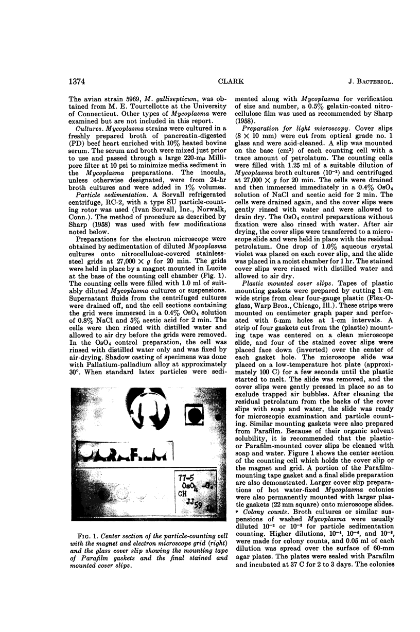
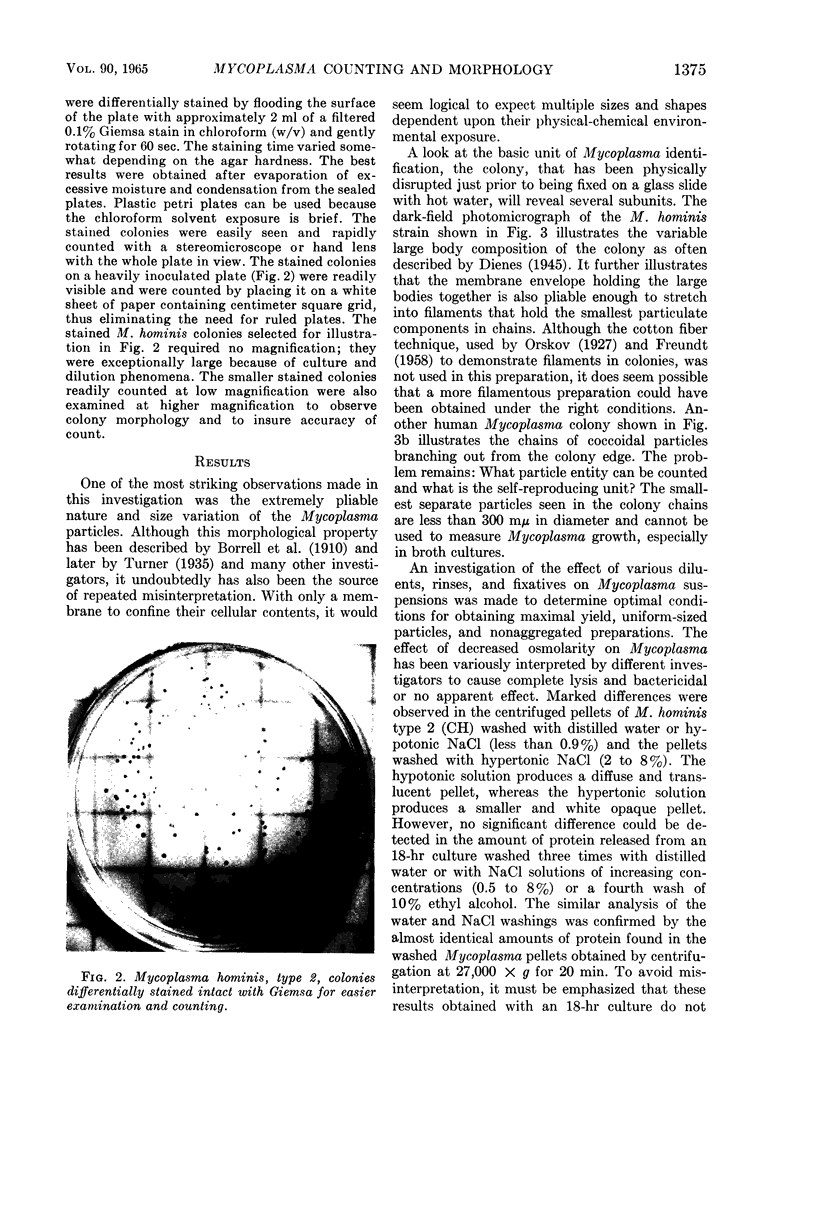
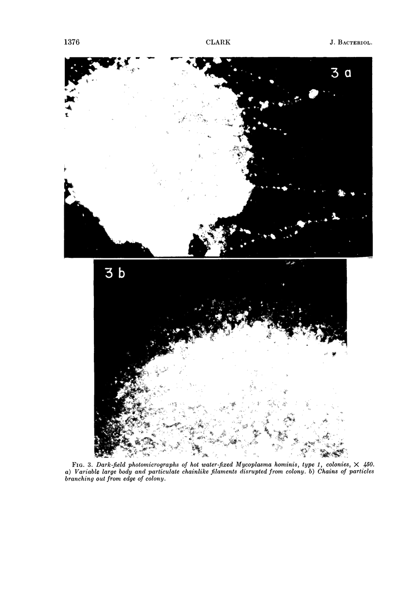
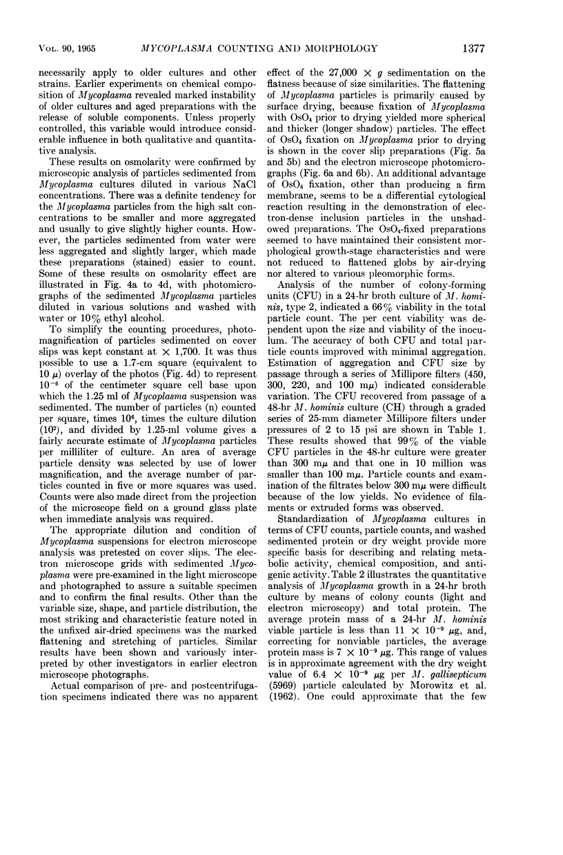
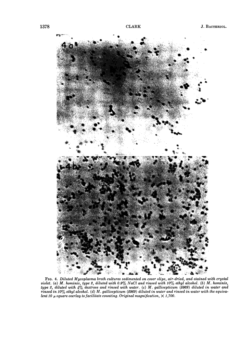
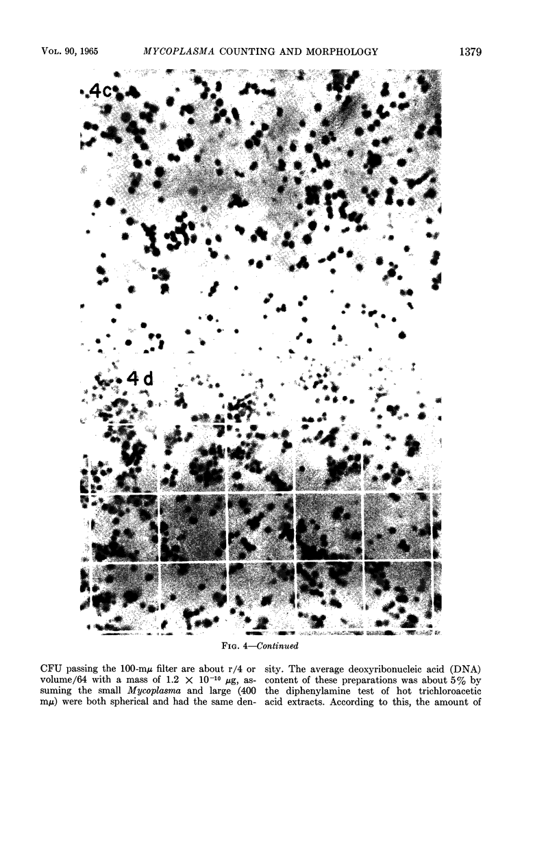
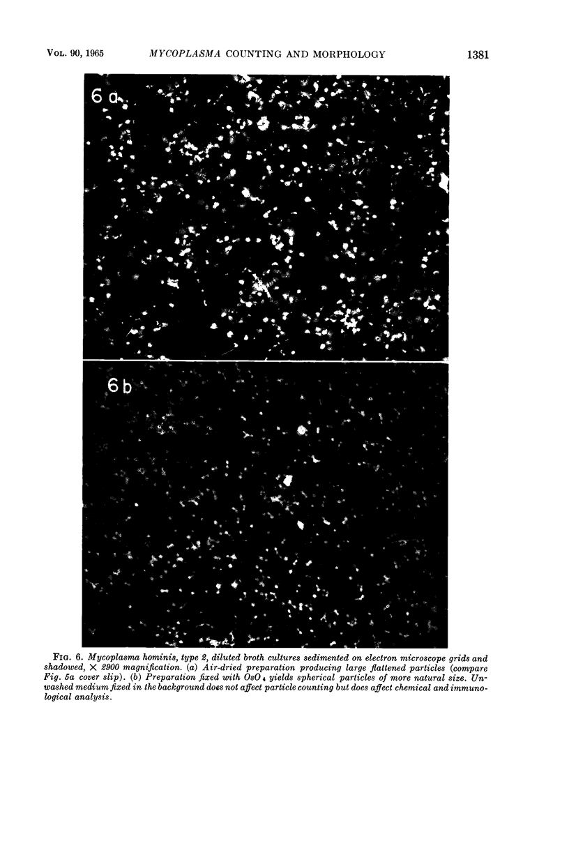
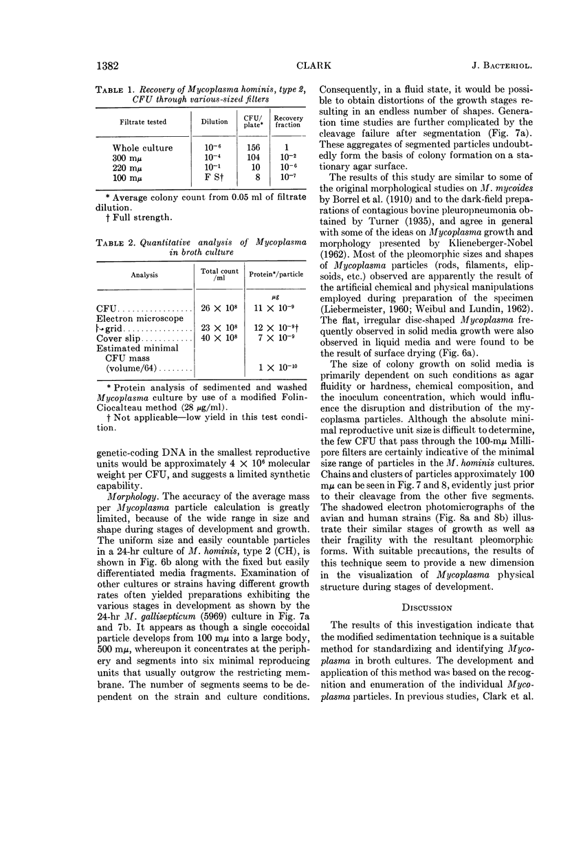
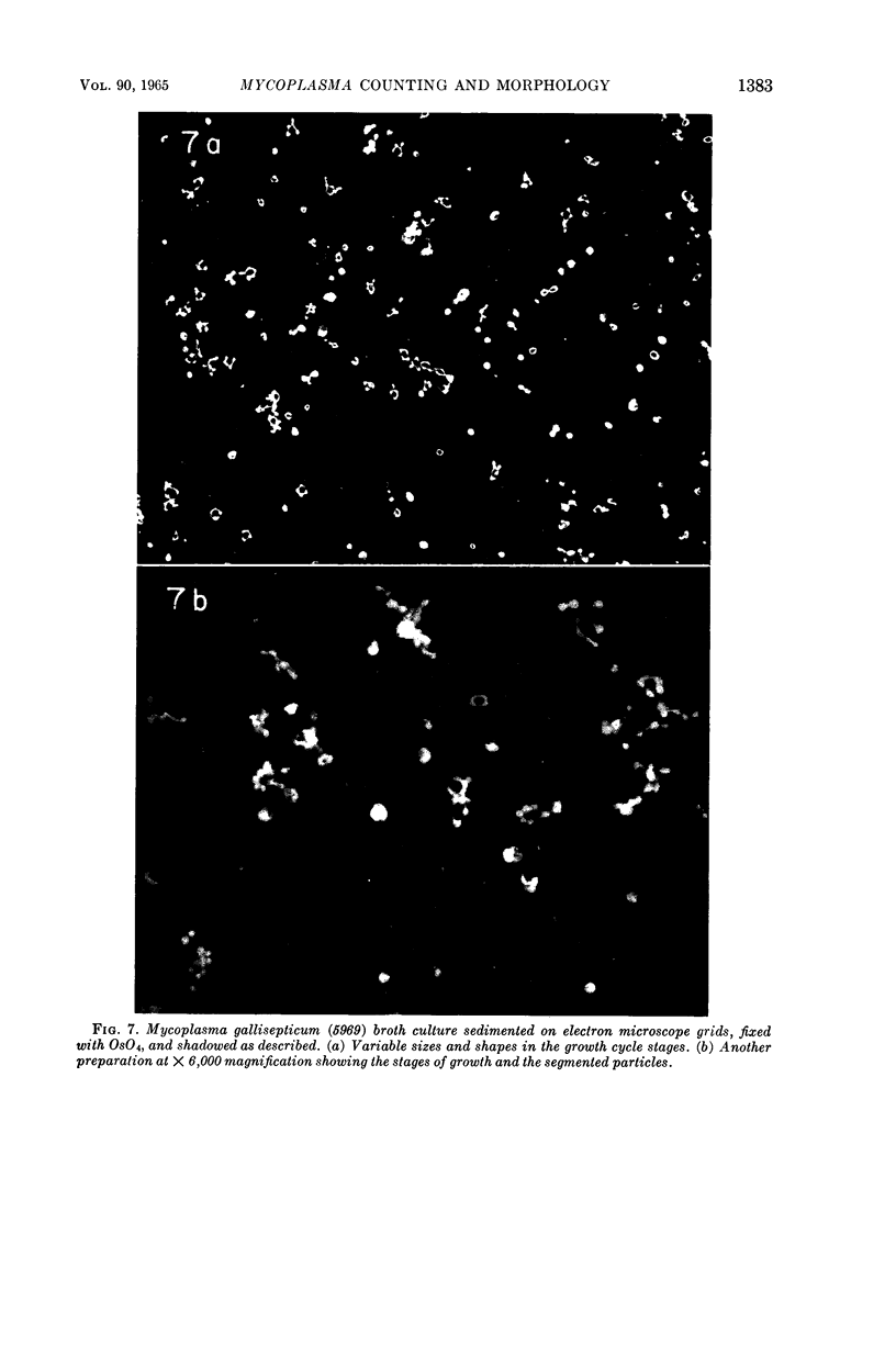
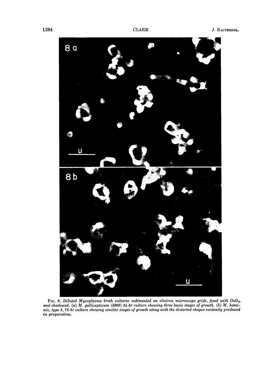
Clark, Harold W. (The George Washington University School of Medicine, Washington, D.C.). Sedimentation counting and morphology of Mycoplasma. J. Bacteriol. 90:1373–1386. 1965.—The sedimentation technique for counting viral particles was applied to the quantitation and morphological identification of Mycoplasma in broth cultures. Mycoplasma, apparently in their native form, firmly adhered to the surface, when sedimented on glass cover slips or onto electron microscope grids. The sedimented cover slip preparations stained with crystal violet could be readily counted in the light microscope. The cultures sedimented onto electron microscope grids were readily counted at low magnification and provided excellent preparations for morphological examination at higher magnifications. It was found that air-dried Mycoplasma particles were enlarged considerably because of excessive flattening. Fixation of sedimented Mycoplasma particles in diluted OsO4 prior to air drying yielded a more realistic morphology, with various sizes and shapes in the stages of the growth cycle exhibited. A new technique of differentially staining Mycoplasma colonies on agar plates was developed to facilitate the quantitation of viable colony-forming units for comparison with total counts. The use of plastic or Parafilm gaskets for dry mounting was developed to facilitate the handling and examination of the stained cover slip preparations. The results of this investigation indicated that the growth cycle of some Mycoplasma species includes a stage of hexadic fission with the cleavage of minimal reproductive units (less than 100 mμ) containing a limited deoxyribonucleic acid genetic coding molecule (approximately 4 × 10⁶).
Full text
PDF
Images in this article
Selected References
These references are in PubMed. This may not be the complete list of references from this article.
- CLARK H. W., FOWLER R. C., BROWN T. M. Preparation of pleuropneumonia-like organisms for microscopic study. J Bacteriol. 1961 Mar;81:500–502. doi: 10.1128/jb.81.3.500-502.1961. [DOI] [PMC free article] [PubMed] [Google Scholar]
- Dienes L. Morphology and Nature of the Pleuropneumonia Group of Organisms. J Bacteriol. 1945 Oct;50(4):441–458. [PMC free article] [PubMed] [Google Scholar]
- LIEBERMEISTER K. Morphology of the PPLO and L forms of Proteus. Ann N Y Acad Sci. 1960 Jan 15;79:326–343. doi: 10.1111/j.1749-6632.1960.tb42694.x. [DOI] [PubMed] [Google Scholar]
- MOROWITZ H. J., TOURTELLOTTE M. E., GUILD W. R., CASTRO E., WOESE C. The chemical composition and submicroscopic morphology of Mycoplasma gallisepticum, avian PPLO 5969. J Mol Biol. 1962 Feb;4:93–103. doi: 10.1016/s0022-2836(62)80041-2. [DOI] [PubMed] [Google Scholar]
- WEIBULL C., LUNDIN B. M. Size and shape of pleuropneumonia-like organisms grown in liquid media. J Bacteriol. 1962 Sep;84:513–519. doi: 10.1128/jb.84.3.513-519.1962. [DOI] [PMC free article] [PubMed] [Google Scholar]