Abstract
Ewing's sarcoma/primitive neuroectodermal tumor (ES/PNET) is an unusual malignancy with aggressive behavior. ES/PNET in the cerebellopontine angle (CPA) is extremely uncommon, and we report on a rare case here. A 31-year-old man presented with one month history of left facial palsy, hearing loss, swallowing difficulty, and hoarseness. Magnetic resonance images showed a large mass in the left CPA and a small one in the right cerebellar hemisphere. The patient underwent a surgery for the CPA mass lesion, and the pathology was compatible with ES/PNET. Radiation therapy and chemotherapy were administered. In contrast to the initial radiologic findings resembling vestibular schwannoma or meningioma, ES/PNET had several distinct clinical features. A patient with a CPA mass and presenting unusual clinical features should be suspected of having a rare malignancy.
Keywords: Ewing's sarcoma, Primitive neuroectodermal tumor, Cerebellopontine angle
INTRODUCTION
Ewing's sarcoma/peripheral primitive neuroectodermal tumor (ES/PNET) is a rare malignant tumor, the designation given to a family of small cell neoplasm. It usually affects those in childhood and adolescence and arises in bone, soft tissue, or very rarely, in the central nervous system1,11). ES/PNET may develop in the cerebellopontine angle (CPA), which is an extremely rare location, and to the best of our knowledge, there are only eight documented cases. We report this rare case together with a literature review.
CASE REPORT
A 31-year-old man presented with a one-month history of left facial palsy, hearing loss, swallowing difficulty, and hoarseness. A neurological examination revealed left facial palsy (House-Brackmann Grade IV), left facial hypesthesia, and left-side hearing loss. Cerebellar dysfunction and gait disturbance were not found. Audiometry revealed a threshold of 45 dB, and speech discrimination was 0%.
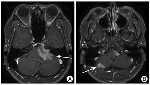
Magnetic resonance (MR) images showed a 4.3 cm sized, well-demarcated mass in the left CPA. The mass was extended to the left internal acoustic canal and compressed the brain stem. Another 1.5 cm sized mass was observed in the convexity of the right cerebellar hemisphere. The tumors were hyper-intense on a T2-weighted image without peritumoral edema and enhanced heterogeneously (Fig. 1). Hydrocephalus was not observed. Based on the radiological findings, the clinical diagnosis was vestibular schwannoma having atypical features with a cerebellar convexity meningioma.
Fig. 1.
A magnetic resonance (MR) image showing two heterogenously enhanced masses in the left cerebellopontine angle (CPA) and the convexity of the right cerebellar hemisphere. The large mass was 4.2 cm in size and well demarcated, compressing the brain stem to the opposite side (A). The small mass was 1.5 cm in size, and the mass effect was not combined (B).
The left CPA mass was surgically removed to relieve brain stem compression and to obtain pathological specimens via the retrosigmoid approach. The facial nerve was identified and seemed to be swollen (Fig. 2). The mass was very gray-colored, fragile, hypervascular, and quite different from benign tumors. There was no clear dissection plane of the arachnoid membrane and the mass encased a vessel.
Fig. 2.
The patient underwent a surgery for the left CPA mass via the retrosigmoid approach, and a subtotal resection was done. The mass was gray-colored, fragile, and hypervascular. The dissection plane of the arachnoid membrane was not well delineated (A). The facial nerve (arrow) was identified and seemed to be swollen (B). An encasement of a vessel (curved arrow) by the mass was observed (C).
On histopathological examination, the mass was composed of small round cells without nuclear pleomorphism. Cytoplasm was scanty with positive staining for periodic acid Schiff (PAS) and negative staining for diastase digested PAS. Necrosis and mitosis were frequently observed. An immunohistochemical study demonstrated a positive reaction for vimentin, neuron-specific enolase, and MIC2 antigen (CD99); however, no reaction was noticed with glial fibrillary acidic protein, desmin, synaptophysin, smooth muscle actin, leukocyte common antigen, and epithelial membrane antigen. The Ki-67 proliferative index was 25% (Fig. 3). These findings were compatible with ES/PNET4).
Fig. 3.
On hematoxylin and eosin staining, the mass was composed of small round cells with striking uniformity (A). On immunohistochemical staining, a positive reaction was demonstrated for vimentin (B), neuron-specific enolase (C), and CD99 (D). The mass was compatible with ES/PNET.
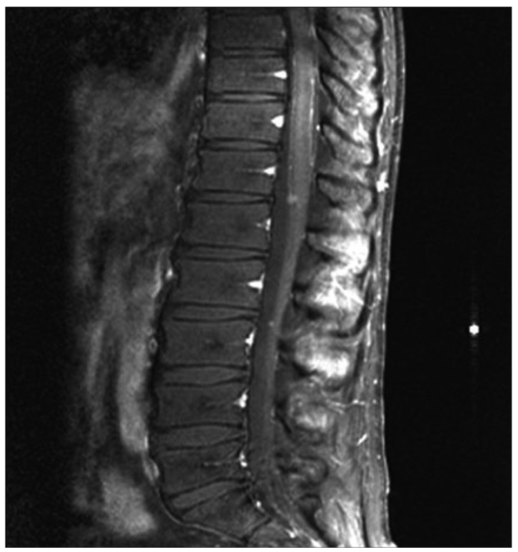
A whole spine MR imaging study after surgery revealed a metastatic nodule in the thoracolumbar junction (Fig. 4). An adjuvant craniospinal irradiation of 54 Gy total dose with 34 fractions was performed, followed by four cycles of chemotherapy. A regimen of procarbazine, lomustine, and vincristine was initially tried, and then the author (Kim JH) changed it to lomustine, cisplatin, and vincrisitine for neutropenic fever. Two cycles of chemotherapy were added with vincristine, adriamycin, cyclophosphamide, and ifosfamide and etoposide because the recurring mass was found on a postoperative, seven-month follow-up MRI. The extent of the mass was enlarged despite the adjuvant chemotherapy, so the patient underwent a second operation and gamma knife radiosurgery. The disease continued to progress, and the patient finally died of tumor bleeding 13 months after diagnosis.
Fig. 4.

A whole spine MRI revealed a well enhanced metastatic nodule at the thoracolumbar junction.
DISCUSSION
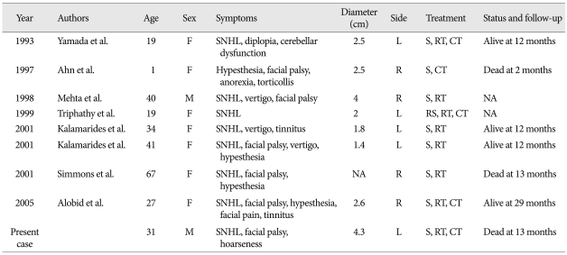
ES/PNET has been known as a malignant tumor with very aggressive behavior. ES/PNET is extremely rare in the CPA, and there are only eight well-documented cases since Yamada et al. reported the first case in 1993 (Table 1)2,3,9,12,14-16). As the radiological feature may be indistinct from that of other benign tumors, it is important to differentiate rare malignancy from others for establishing a management plan for further treatment11).
Table 1.
Summary of clinical features of ES/PNET in the cerebellopontine angle reported in the literature
SNHL : sensorineural hearing loss, NA : not available in the original article, S : surgery, RT : radiotherapy, CT : chemotherapy, RS : radiosurgery, R : right, L : left, F : female, M : male
The presented case showed multiple cranial nerve palsies that progressed for a relatively short period, less than one year. These clinical features are quite unusual for common CPA tumors, such as vestibular schwannoma or meningioma, for several reasons. Vestibular schwannoma and meningioma usually affect the patients in their seventh and sixth decades, respectively8,17). Clinical manifestations of vestibular schwannoma are usually associated with eighth nerve such as hearing difficulties (93%), tinnitus (83%), and dizziness (54%); however, involvement of other cranial nerves, such as facial hypesthesia-palsy (26%) and swallowing difficulty (5.6%), is relatively uncommon6). The CPA meningioma often presents with cerebellar dysfunction (67%), hearing difficult (58%), and facial hypesthesia-paresthesia (51%); symptoms associated with facial nerve (9%) and low cranial nerves (15%) are less likely8). In the literature regarding ES/PNET, six out of eight patients presented with facial palsy or low cranial nerve palsy among their initial symptoms, with or without eighth nerve dysfunction. The encasement of vessels rather than displacement is also uncommon in the most benign CPA mass lesions5). Therefore, when a patient with a CPA mass presents with unusual features distinct from those of vestibular schwannoma or meningioma, one should be concerned about the malignant disease and should consider an operation to confirm a histopathological diagnosis.
In this case, the tumor was progressed and eventually the patient died of tumor bleeding, although he had been treated with a multidisciplinary approach. It is known that the best outcome can be achieved when maximal surgical resection followed by adjuvant chemotherapy and radiotherapy are performed1,7,9,13). In previous literature, not all patients with ES/PNET in the CPA found dead on their last follow-up, however, there were no long-term survivors neither. Although multiagent chemotherapy with vincristine, cyclophosphamide, doxorubicin, and ifosfamide and etoposide is commonly used for localized ES/PNET, there is no standard regimen for recurrent and metastatic disease, or especially for intracranial ES/PNET10). Therefore, further investigation about the chemotherapeutic regimen may be mandatory.
CONCLUSION
ES/PNET is a rare tumor and usually affects bone or soft tissue. Rarely, ES/PNET arises in the CPA, which may make it indistinguishable from common benign tumors. Unusual clinical features in the presence of the CPA mass could be clues for a malignant tumor, and it is essential to confirm a histopathological diagnosis to establish a management plan.
References
- 1.Ahmad R, Mayol BR, Davis M, Rougraff BT. Extraskeletal Ewing's sarcoma. Cancer. 1999;85:725–731. [PubMed] [Google Scholar]
- 2.Ahn MS, Jackler RK. Exophytic brain tumors mimicking primary lesions of the cerebellopontine angle. Laryngoscope. 1997;107:466–471. doi: 10.1097/00005537-199704000-00008. [DOI] [PubMed] [Google Scholar]
- 3.Alobid I, Gastón F, Morello A, Guilemany JM, Mullol J. Peripheral primitive neuroectodermal tumor of the cerebellopontine angle. Acta Otolaryngol. 2005;125:426–429. doi: 10.1080/00016480410025234. [DOI] [PubMed] [Google Scholar]
- 4.Antunes NL, Lellouch-Tubiana A, Kalifa C, Delattre O, Pierre-Kahn A, Rosenblum MK. Intracranial Ewing sarcoma/'peripheral' primitive neuroectodermal tumor of dural origin with molecular genetic confirmation. J Neurooncol. 2001;51:51–56. doi: 10.1023/a:1006432919281. [DOI] [PubMed] [Google Scholar]
- 5.Bonneville F, Sarrazin JL, Marsot-Dupuch K, Iffenecker C, Cordoliani YS, Doyon D, et al. Unusual lesions of the cerebellopontine angle : a segmental approach. Radiographics. 2001;21:419–438. doi: 10.1148/radiographics.21.2.g01mr13419. [DOI] [PubMed] [Google Scholar]
- 6.Coscarón Blanco E, Muñoz Herrera A, Serradilla López JM, Maillo Sánchez A, Paniagua Escudero JC. [Clinical picture of 8th pair schwannoma. Is it expressive enough?] Acta Otorrinolaringol Esp. 2007;58:43–47. [PubMed] [Google Scholar]
- 7.Furuno Y, Nishimura S, Kamiyama H, Numagami Y, Saito A, Kaimori M, et al. Intracranial peripheral-type primitive neuroectodermal tumor. Neurol Med Chir (Tokyo) 2008;48:72–76. doi: 10.2176/nmc.48.72. [DOI] [PubMed] [Google Scholar]
- 8.Gerganov V, Bussarsky V, Romansky K, Popov R, Djendov S, Dimitrov I. Cerebellopontine angle meningiomas. Clinical features and surgical treatment. J Neurosurg Sci. 2003;47:129–135. discussion 135. [PubMed] [Google Scholar]
- 9.Kalamarides M, Dewolf E, Couvelard A, Shahidi A, Bouccara D, Cyna-Gorse F, et al. Extraaxial primitive neuroectodermal tumor mimicking a vestibular schwannoma: diagnostic and therapeutic difficulties. Report of two cases. J Neurosurg. 2001;94:612–616. doi: 10.3171/jns.2001.94.4.0612. [DOI] [PubMed] [Google Scholar]
- 10.Ludwig JA. Ewing sarcoma : historical perspectives, current state-of-the-art, and opportunities for targeted therapy in the future. Curr Opin Oncol. 2008;20:412–418. doi: 10.1097/CCO.0b013e328303ba1d. [DOI] [PubMed] [Google Scholar]
- 11.Mallucci CL, Ward V, Carney AS, O'Donoghue GM, Robertson I. Clinical features and outcomes in patients with non-acoustic cerebellopontine angle tumours. J Neurol Neurosurg Psychiatry. 1999;66:768–771. doi: 10.1136/jnnp.66.6.768. [DOI] [PMC free article] [PubMed] [Google Scholar]
- 12.Mehta JS, Sharr MM. An unusual cause of acute labyrinthine failure. J Laryngol Otol. 1998;112:193–195. doi: 10.1017/s0022215100140290. [DOI] [PubMed] [Google Scholar]
- 13.Scurr M, Judson I. How to treat the Ewing's family of sarcomas in adult patients. Oncologist. 2006;11:65–72. doi: 10.1634/theoncologist.11-1-65. [DOI] [PubMed] [Google Scholar]
- 14.Simmons MA, Luff DA, Banerjee SS, Ramsden RT. Peripheral primitive neuroectodermal tumour (pPNET) of the cerebellopontine angle presenting in adult life. J Laryngol Otol. 2001;115:848–852. doi: 10.1258/0022215011909161. [DOI] [PubMed] [Google Scholar]
- 15.Tripathy LN, Forster DM, Timperley WR. Adult primitive neuroectodermal tumour. A case report and review of the literature. Br J Neurosurg. 1999;13:90–92. doi: 10.1080/02688699944302. [DOI] [PubMed] [Google Scholar]
- 16.Yamada S, Aiba T, Hara M. Cerebellopontine angle medulloblastoma : case report and literature review. Br J Neurosurg. 1993;7:91–94. doi: 10.3109/02688699308995062. [DOI] [PubMed] [Google Scholar]
- 17.Yoshimoto Y. Systematic review of the natural history of vestibular schwannoma. J Neurosurg. 2005;103:59–63. doi: 10.3171/jns.2005.103.1.0059. [DOI] [PubMed] [Google Scholar]


