Abstract
Myxococcus xanthus Social (S) motility occurs at high cell densities and is powered by the extension and retraction of Type IV pili which bind ligands normally found in matrix exopolysaccharides (EPS). Previous studies showed that FrzS, a protein required for S-motility, is organized in polar clusters that show pole-to-pole translocation as cells reverse their direction of movement. Since the leading cell pole is the site of both the major FrzS cluster and type IV pilus extension/retraction, it was suggested that FrzS might regulate S-motility by activating pili at the leading cell pole. Here, we show that FrzS regulates EPS production, rather than type IV pilus function. We found that the frzS phenotype is distinct from that of Type IV pilus mutants such as pilA and pilT, but indistinguishable from EPS mutants, such as epsZ. Indeed, frzS mutants can be rescued by the addition of purified EPS, 1% methylcellulose, or co-culturing with wildtype cells. Our data also indicate that the cell density requirement in S-motility is likely a function of the ability of cells to construct functional multicellular clusters surrounding an EPS core.
Introduction
Bacteria modulate activity in response to cell density and surface cues, allowing groups of cells to accomplish more than individuals [1], [2]. Myxococcus xanthus is a Gram-negative soil bacterium that utilizes multicellularity during vegetative swarming, predation of prey microorganisms and, when nutrients are reduced, aggregation into fruiting bodies that contain spores [3], [4]. Motility is required for all of these functions. Unlike other surface motile bacteria such as Bacillus subtilis and Proteus mirabilis, M. xanthus does not produce flagella but glides on surfaces through a combination of Social (S) and Adventurous (A) motility [5], [6], [7]. The two systems can be synergistic, but confer distinct advantages depending on the culture conditions: S-motility promotes group movement on soft, nutrient rich surfaces like 0.3–0.5% agar, while A-motility functions best on firmer surfaces, like 1.5–2.0% agar [8], [9]. Conditions that promote S-motility result in smooth colony edges that lack isolated cells. In contrast, conditions that promote A-motility result in colony edges containing many individual gliding cells as well as groups.
S-motility occurs by extension and retraction of Type IV pili, but also requires extracellular polysaccharide matrix (EPS) [10], [11], [12]. EPS is rich in glucosamine and N-acetylglucosamine and has been proposed to serve as an anchor for pilus binding and retraction [13]. Isolated M. xanthus cells do not show S-motility on an agar surface; however, cells regain S-motility at high cell density [14]. This observation is the basis for the hypothesis that pili from one cell bind to EPS on the surface of a close neighboring cell, propelling cell movement [7], [13], [14]. However, distance may not be the only factor inhibiting S-motility at low cell densities, as the cell density requirement can be completely abolished by the addition of 1% methylcellulose [15]. These results indicate that the Type IV pili are synthesized and functional even at low cell densities.
A number of genetic loci have been identified that are required for S-motility, including a 37 gene cluster homologous to known eps biosynthesis genes [12], [16], [17]. Polysaccharide slime has also been implicated in A-motility, but a causal connection between polysaccharide production and A-motility has not been established. Based on recent data, A-motility has been proposed to be powered by proton motive force (PMF) and driven by distributed motor proteins that move along a helical track, creating differential drag forces that distort the cell surface and generate surface waves that push cells forward [18].
To achieve directed motility, M. xanthus cells need to periodically reverse. Cell reversals in M. xanthus involve the inversion of cell polarity so that the lagging cell pole becomes the new leading cell pole and the old leading cell pole becomes the new lagging cell pole [19], [20]. Since the pili required for S-motility are found only at the leading cell pole, precursor and regulatory proteins required for S-motility must either be transported from pole-to-pole when cells reverse or be present at both cell poles, but subject to periodic activation/inactivation by regulators [21]. For example, monomers of the major pilin subunit, PilA are localized throughout the membrane but are assembled only at the leading cell pole [22]. By contrast, FrzS, an S-motility protein controlled by the frizzy (Frz) chemosensory system, periodically translocates from pole to pole during cellular reversals [23], [24]. It is therefore of interest to determine the role of FrzS in S-motility and the reason for its periodic pole-to-pole translocation.
FrzS contains two principal domains: an N-terminal pseudo-receiver domain that lacks the critical aspartyl residue that is typically phosphorylated in two component signaling systems, and a C-terminal coiled-coil domain, predicted to be involved in the dynamic assembly or disassembly of protein complexes [25]. The coiled-coil domain of FrzS promotes oligomerization when overexpressed in E. coli. [24]. Although FrzS lacks the canonical sites for activation of the receiver domain by a cognate kinase, two receiver domain point mutations result in mislocalization (Y102A, H92F) of the protein and loss of function [26]. These domain structures suggested that FrzS might function as an intermediary, transducing signals from the Frz chemosensory system to activate pilus assembly at the leading cell pole [23], [24]. Unexpectedly, the results presented in this study show that FrzS, rather than being a regulator of pilus production, is actually a regulator of EPS production.
Results
frzS mutant cells show restored S-motility when mixed with wildtype cells
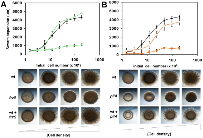
S-motility in wildtype M. xanthus strain DZ2 is best observed when cells are incubated at high cell density on nutrient rich, 0.5% agar CYE plates, as A-motility does not function under these conditions. On this medium, M. xanthus cells glide away from the initial inoculum in a cell density dependent manner, producing thin spreading swarms. In most cases, capturing an image by 24 h is sufficient to demonstrate normal S-motility or S-motility defects. To quantify S-motile swarming, we prepared two-fold serial dilutions of exponential phase cultures, and then spotted 3 µl aliquots of cells onto 0.5% agar CYE plates at cell densities ranging from 2×10⁶ to 2×10⁸ cells. We measured the size of the initial spots and the swarm diameters after 24 h at 32°C to determine swarm expansion as a function of initial cell density. The results are shown in Fig. 1. In this assay, wildtype cells showed cooperative swarming as cell densities were increased: (i) poor swarming was observed at low cell densities; (ii) as cell densities were increased, there was a sharp increase in the rate of swarm expansion; (iii) at the highest cell densities, a maximum swarm expansion rate was achieved (see Fig. 1). The maximum swarm expansion rate for wildtype cells was 4.3 mm/day, and the cell number at which half the maximum swarm expansion occurs was 2×10⁷ cells. We consider this number to be important for quantifying cooperative social swarming as any decrease in cooperation should shift the curve to the right increasing the number of cells required for rapid swarm expansion.
Figure 1. Quantitative social motility assay.
Serial dilutions of mid exponential-phase cultures ranging from 2×10⁶ to 2×10⁸ cells/assay were prepared either in mono-cultures or co-cultures, then aliquoted onto 0.5% agar CYE plates. Swarm expansion was imaged and measured after 24 h incubation at 32°C. (A) Cell density-dependent social motility was assayed in wildtype (black ♦), frzS (green ▴) and a wildtype+frzS co-culture (green ▵), with the error bars representing one standard deviation. Corresponding images below show wildtype swarms, defective frzS swarms with small abortive flares, and full recovery of motility in the co-culture. (B) Analysis of wildtype (black ♦), S-motility defective pilA (orange ▪) and partial recovery of a wildtype+pilA co-culture (orange □) with corresponding images from the even number data points shown below. Scale bar is 1 mm.
In contrast, S-motility mutants like pilA (DZ4469) showed little swarm expansion, even at high initial cell densities, and exhibited smooth colony edges (see Fig. 1B). Interestingly, co-culturing wildtype with pilA (1∶1) showed an intermediate swarm expansion rate of 3.0 mm/day, indicating that wildtype cells are not inhibited by the presence of non-motile pilA cells in the swarm (see Fig. 1B). The behavior of the wildtype-pilA co-culture indicates that pilA cells are not counted as part of the functional swarming group, i.e., an initial cell population of 2×10⁷ cells in which 50% are wildtype behaves the same as an initial cell population of 1×10⁷ cells that are 100% wildtype. In contrast, frzS swarms (DZ4335) differed from both wildtype cells and pilA cells. Instead of smooth colony edges like the pilA mutants, frzS swarms showed wrinkled colony edges, consisting of tiny flares emerging from the swarms (see Fig. 1A, lower panel). After 24 h, the net swarm expansion was extremely small, only about 1 mm. However, co-culturing of wildtype and frzS cells (1∶1) resulted in a swarm expansion rate similar to wildtype levels, 4.3 mm/day. The result that wildtype-frzS mixed swarms expand at the same rate as wildtype alone is consistent with and suggestive of the possibility that wildtype fully rescues the swarming defect of the frzS mutant. This complementation was not observed in other motility mutants, including pilT (DK10409), tgl (DZ4191), cglB (DZ4477), mglA (TM12), frzF (DZ4483), and difA (YZ601) strains (data not shown). All of these mixtures showed a phenotype similar to wildtype cells mixed 1∶1 with buffer, indicating that non-motile cells in this assay do not inhibit wildtype swarming, and that the phenotype of wildtype-frzS co-cultures can only be due to increased activity of one or both strains.
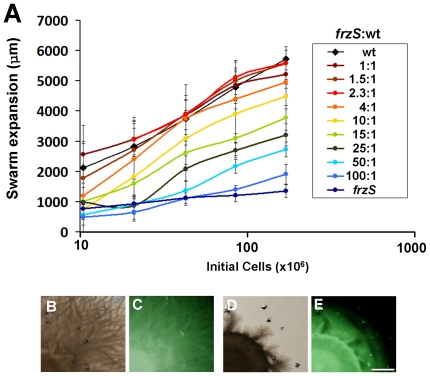
The extracellular complementation by wildtype is robust as up to a 4∶1 frzS to wildtype cell ratio resulted in maximal swarm expansion (see Fig. 2). Higher ratios of frzS to wildtype cells reduced swarm expansion incrementally, with a 9∶1 ratio yielding a phenotype similar to a 1∶1 pilA-wildtype mixture, and a 100∶1 ratio required to yield a phenotype similar to frzS alone. To further determine if frzS cells migrate with wildtype cells from the initial inoculum through to the colony edge in the 0.5% agar S-motility assay, we analyzed co-cultures of wildtype and frzS with DZ4538, a frzSY102A::gfp mutant that expresses a C-terminal Green Fluorescent Protein (GFP) tag [26]. We examined these co-cultures with fluorescence stereo microscopy and imaged the colonies after 24 h incubation (see Fig. 2B–E). The images show that indeed the frzSY102A::GFP strain was motile in the co-culture since fluorescence was visible from the colony center out to the periphery in both mixtures.
Figure 2. Complementation of the frzS Social motility defect.
(A) Swarm expansion data for frzS-wildtype co-cultures at the ratios listed. (B,C) Transmission and fluorescence stereomicroscopy of 1∶1 co-cultures of wildtype with frzSY102A::gfp and (D,E) ΔfrzS with frzSY102A::gfp showing migration of the GFP-labeled cells corresponding to the colony edge. Scale bar is 1 mm.
Isolated frzS cells are motile in 1% methylcellulose
Previous studies of FrzS led to the hypothesis that FrzS regulates S-motility by activating PilT ATPase retraction at the leading cell pole [20], [24]. However, the ability of frzS cells to show S-motility swarming when co-cultured with wildtype cells indicates that they may harbor a defect other than pili function. We used the 1% methylcellulose assay developed by Sun et al. to analyze the behavior of isolated frzS cells [15], [27]. Cells were harvested from exponential phase cultures, mixed with 1% methylcellulose and observed immediately with phase contrast microscopy. By tracking cells at 1 min intervals for 20 min, isolated frzS cells were observed to show motility behavior similar to wildtype (see Fig. 3). Thus, frzS cells produce functional Type IV pili that can propel cells forward. In contrast, isolated pilT cells were completely non-motile in 1% methylcellulose.
Figure 3. Motility assay in methylcellulose.
Mid-exponential phase cells were resuspended in 1% methylcellulose on glass slides and S-motility observed by time-lapse microscopy. Images show 7 cell tracks for (A) frzS mutant; (B) wildtype strain DZ2: and (C) pilT mutant over 9 min (Top). Images at 0, 3, 6, and 9 min are shown below. Cells were tracked in three independent experiments each with frzS, wildtype and pilT, but movement was only observed with frzS and wildtype DZ2. Scale bar is 5 µm.
frzS cells are defective in EPS production and cell-cell agglutination
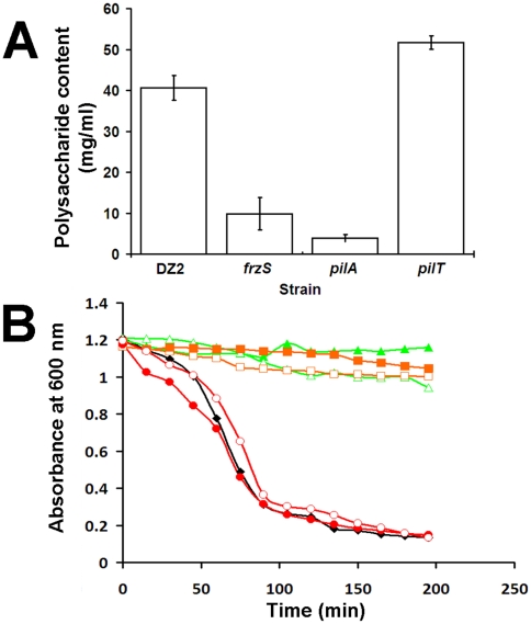
Since examination of frzS cells showed evidence of functional type IV pili, we hypothesized that frzS might be defective in EPS production or secretion. We therefore examined the frzS mutant for EPS production using Congo red dye, previously shown to bind to M. xanthus EPS [28]. Cells were harvested from exponentially growing cultures, mixed with Congo red dye and centrifuged. Unbound dye was measured at 490 nm, and polysaccharide content was determined relative to a cellulose standard curve. Figure 4A shows that frzS cells bind Congo red dye but only at 25% of wildtype levels. pilA mutants, which have a more severe defect in EPS production, bound EPS at 10% of wildtype levels. In contrast, a pilT mutant, defective at pili retraction, overproduces EPS at a level 127% of wildtype. These data indicate that frzS is defective in EPS production, but do not indicate what distinguishes frzS from the other S-motility mutants.
Figure 4. EPS analysis and agglutination assay.
(A) Congo red dye was added to mid-exponential phase cultures of the given strains, cells were centrifuged and the resulting supernatants analyzed in a spectrophotometer at 490 nm. Values were normalized to wildtype binding. An increase is indicative of more Congo red bound by the cells and increased EPS production, such as observed in pilT. pilA and frzS cells show reduced EPS production. Data is from three independent trials. (B) Cells were also analyzed with agglutination assays performed on wildtype (black ♦), frzS (green ▴), pilA (orange ▪), and pilT (red •), and a 1∶1 mixture of wildtype with the mutant (open symbols). In wildtype, absorbance at OD600 at the top of a cuvette decreases over time as cells agglutinate and settle to the bottom of the cuvette. pilA and frzS inhibit agglutination by wildtype, whereas pilT was observed to behave similar to wildtype. Data shown is representative of three trials.
EPS is also required for cell-cell agglutination, which is observed when M. xanthus cells in suspension bind to each other and settle to the bottom of a cuvette [29], [30]. In wildtype cells, this is observed as an exponential drop in absorbance at 600 nm (see Fig. 4B). However, frzS cells were defective in agglutination (see Fig. 4B) and remained suspended in liquid similar to EPS deficient mutants like pilA. These results differ from previously published agglutination data for a frzS insertion mutant (DZ4219) [8]. We therefore retested both mutant strains for agglutination, EPS production and social motility (see Fig. S1). In our hands, both frzS mutant strains were found to be defective in these assays. Interestingly, when either frzS or pilA were mixed in a 1∶1 ratio with wildtype cells, the co-culture showed significantly reduced agglutination relative to wildtype (see Fig. 4B). This surprising result indicates that EPS defective cells interfere with the ability of wildtype cells to form aggregates necessary for agglutination. This was not a cell density effect since wildtype cells by themselves did not show reduced agglutination when cell densities were reduced by half (data not shown). The pilT mutant, which overproduces EPS, agglutinates more rapidly than wildtype and stimulates wildtype agglutination (see Fig. 4B). These data indicate that frzS cells are not only defective in EPS-dependent agglutination, but they are also not much better than pilA cells at agglutination, even when mixed with wildtype.
Purified EPS can restore S-motility to frzS mutant cells
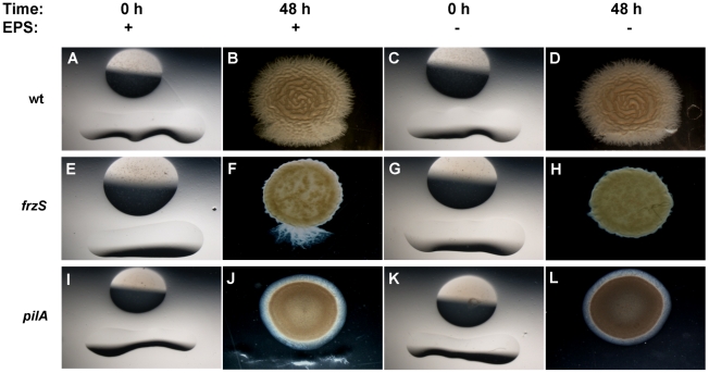
To determine whether exposure to EPS is sufficient for the restoration of S-motility to frzS swarms, we purified EPS from wildtype cells and pipetted this preparation as a linear streak adjacent to spots of wildtype, frzS, or pilA cells on 0.5% agar plates (Fig. 5). In this assay, wildtype cells increased their swarm expansion as they contacted the zone containing EPS. Significantly, the frzS cells, which were otherwise non-motile, also showed a burst of swarming but only in the EPS zone. The pilA mutant did not respond to the EPS nor did frzS cells exposed to a control buffer (Fig. 5). This indicates that purified EPS is sufficient to restore S-motility to frzS swarms, and that frzS is not required for pili retraction during soft agar swarming.
Figure 5. Complementation of S-motility with purified EPS.
Washed cells of wildtype (A–D), frzS (E–H), and pilA (I–L) were spotted onto 0.5% agar CYE plates adjacent to a thin line of either an aliquot of EPS purified from wildtype (left two columns) or a buffer control (right two columns). Images were captured at 0 and 48 h, showing stimulation of motility in wildtype and frzS cells, but not pilA cells. Stimulation of motility can be observed as downward outgrowth from the colony. Images show representative images from multiple trials.
Additionally, we purified EPS from wildtype, pilT, pilA and frzS cells and tested these EPS preparations for their ability to stimulate frzS, pilA, and difA mutant swarming (see Table 1). All of these strains produced some EPS and they all stimulated S-motility in the frzS mutant. However, none of the EPS preparations were able to stimulate motility in the pilA or difA mutants. This is consistent with previous observations that showed pilA and difA mutants have S-motility defects in both pili function and EPS production [31], [32].
Table 1. EPS complementation.
| Test strain | EPS Source | ||||
| DZ2 | pilT | frzS | pilA | Buffer | |
| frzS | ++ | ++ | ++ | + | − |
| pilA | − | − | − | − | − |
| difA | − | − | − | − | − |
EPS was purified from several strains and stimulation of motility assayed in frzS, pilA and difA.
Multicellular localization of EPS
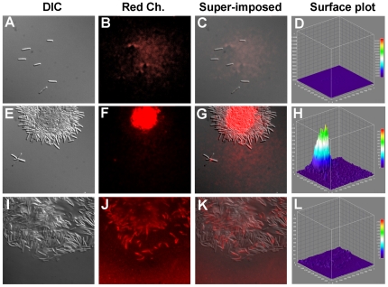
To determine whether EPS production has an impact on cellular organization, we examined wildtype M. xanthus cells harvested from exponential phase cultures and cells 4 h after incubation on a plastic surface in submerged culture with Differential Interference Contrast (DIC) and fluorescence microscopy (see Fig. 6A–L). Liquid grown cells were dispersed and stained poorly with wheat germ agglutinin conjugated to a Texas red fluorophore (WGA-TR). However, after surface incubation, wildtype cells were observed as both isolated cells and tightly packed cell clusters organized around a central bundle that stains brightly with WGA-TR (see Fig. 6E–H). Most cells were observed in large clusters of up to 103 cells with a high intensity feature that localized to the center of the cell clusters rather than to individual cells. Interestingly, very small clusters of cells (for example, see panel 5E) showed 4–5 cells radially organized around a small WGA-TR binding feature (see panel 5F), reminiscent of rosette structures observed in Caulobacter crescentus [33]. In contrast, the frzS cells showed fewer clusters after surface incubation, and these clusters were more disorganized than those of wildtype (see Fig. 6I–L). WGA-TR staining of frzS clusters showed reduced and decentralized staining, with the stain more likely to co-localize with single cells, rather than at distinct multicellular foci.
Figure 6. Fluorescence microscopy analysis of EPS production.
DIC and fluorescence analysis of (A–D) wildtype M. xanthus cells from a liquid culture. (E–H) Wildtype M. xanthus cells after 4 h incubation with a solid surface in submerged culture, showing the formation of small and large rosette structures with centralized WGA-TR staining. (I–L) frzS cells after 4 h incubation with a solid surface, showing disorganized clustering and dispersed low levels of WGA-TR staining. Each data set depicts from left to right: DIC, red channel fluorescence, superimposed overlay, and surface plot of signal intensity.
Construction and analysis of EPS biosynthetic mutants
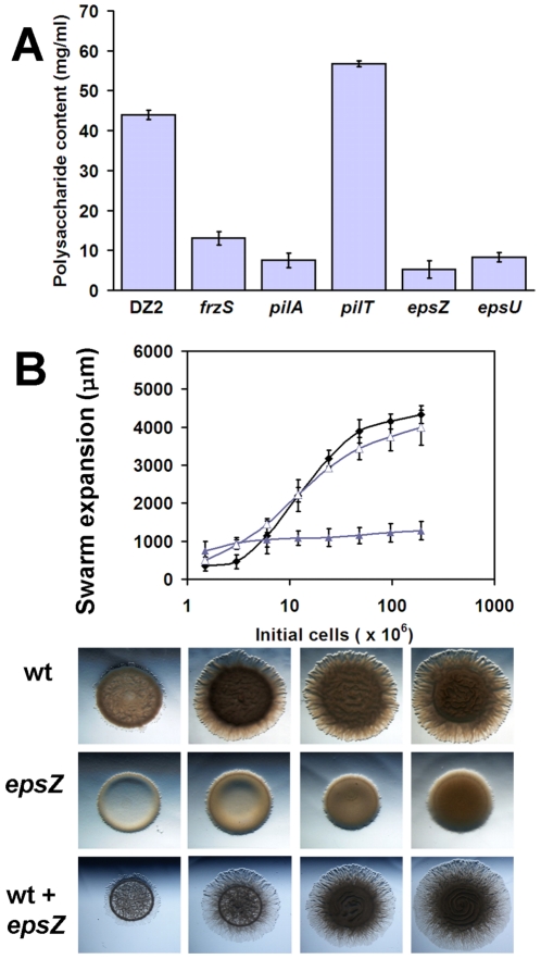
To compare the observed frzS phenotype to that of mutants in EPS production, we constructed mutations in several of the genes predicted to be required for EPS production. We targeted mxan_7415 (epsZ), and mxan_7422 (epsU), genes that are predicted to catalyze glycosyl transferase reactions critical for EPS biosynthesis and mxan_3227 and mxan_3229, genes predicted to provide the same function in the production of capsular polysaccharide (CPS) [12], [34]. Insertion mutants were constructed in theses strains as described previously [24]. Analysis of these strains indicates that epsU (DZ4830) and epsZ (DZ4831) are defective in EPS production (Fig. 7A), whereas no defect was detectable in the mxan_3227 and mxan_3229 loci (data not shown). Congo red dye-binding assays showed that epsZ and epsU mutants contained polysaccharide less than or equal to that found in pilA extracts.
Figure 7. EPS mutant analysis.
(A) Congo red analysis of EPS extracts from the given strains. (B) Quantitative social motility with wildtype (black ♦), S-motility defective epsZ (purple ▴), and a 1∶1 co-culture of the two strains (purple ▵) showing increased swarming relative to epsZ alone, with corresponding images shown below.
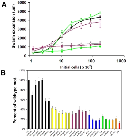
The epsZ mutant was defective in S-motility, but showed no defect in swarm expansion when incubated in a 1∶1 co-culture with wildtype strain DZ2 (Fig. 7B). Since frzS and epsZ mutants could both be complemented by wildtype cells, we hypothesized that if they were functioning within the same pathway, then they should not be able to complement each other. Alternatively, if frzS and epsZ effect different aspects of S-motility, then they should be able to complement each other and restore S-motility. We therefore analyzed S-motility in different combinations of mixed cultures to determine the effect of co-culturing cells (Fig. 8). For example, a 1∶1 frzS-epsZ mixture showed a slight elevation in S-motility relative to each strain in mono-culture, but motility levels were still low, at less than 30% of wildtype levels and therefore not statistically significant (see Fig. 8A). We expanded this analysis to include mixtures with wildtype DZ2, difA, epsZ, epsU, frzS, pilA and pilT (see Fig. 8B). All strains were analyzed in mono-culture for their maximum swarm expansion, ranging from 42% (difA) to 12% (pilT) of wildtype levels. Next, we examined all 21 possible co-culture combinations, to determine if it is possible to identify two S-motility defective mutants that, when combined, show fully functional S-motility. Our results are summarized as follows: (i) Mixtures of wildtype with frzS, epsZ and espU showed wildtype levels of swarming. (ii) All strains mixed with epsZ showed motility levels equivalent to the most proficient of the two strains. (iii) Strains with the lowest swarm expansions, pilA and pilT showed a slight increase in swarm expansion when mixed. (iv) Most other combinations showed swarm expansions that were intermediate to the individual mono-culture phenotypes. This analysis indicates that there are no clear complementation groups among these S-motility defective strains, but that in certain cases the more defective S-motility mutant can be partially complemented by the more motile strain. The EPS+ phenotype alone is not enough for complementation in this assay (e.g. frzS mixed with pilT), and the EPS producing strain must also be capable of moving across the surface. These results indicate that EPS− cells are not able to bind and glide away with EPS from donor EPS+ cells.
Figure 8. Pathway analysis.
(A) Quantitative S-motility assay examining wildtype (black ♦), frzS (green ▴), wildtype+frzS (green ▵), epsZ (purple ▪), wildtype+epsZ (purple □ ) and epsZ+frzS (gray ○). (B) Combinatorial quantitative social motility analysis of DZ2, difA, epsZ, epsU, frzS, pilA, and pilT strains. Mono-culture and co-culture combinations of these strains are shown, indicating that frzS, epsZ, and epsU are in the same functional pathway as they are both complemented by the presence of wildtype, but are not able to complement each other.
Discussion
The data presented here suggest a new model for the role of FrzS and cell density in M. xanthus S-motility. We began with the development of a quantitative S-motility assay that allowed us to analyze the contribution of different cell densities and S-motility genes on multicellular swarming. These studies showed that frzS cells are S-motile when mixed with wildtype cells, despite the inability of frzS to function in mono-culture. This behavior phenocopies the EPS mutants, epsZ and epsU. Additionally, isolated frzS cells are S-motile in 1% methylcellulose. In contrast, mutants defective in pili function do not show restored motility in co-cultures with wildtype cells. These results show that, rather than regulating extention/retraction of the Type IV pili, FrzS functions in the regulation of EPS biosynthesis and/or secretion. frzS cells are able to produce low levels of EPS, since we could purify EPS from frzS cultures and, when concentrated, use this EPS to rescue S-motility swarming in the same frzS mutant (Table 1). It is possible that FrzS regulates secretion of EPS, but since EPS secretion, polymerization, and translocation may be carried out by the same enzyme, all of these functions may be affected [35]. Since FrzS was previously shown to form polar clusters that move from pole-to pole when cells reverse [20], it is possible that FrzS directly interacts with EPS secretion channels along the cell membrane or at the cell poles, activating them in response to a signal received, presumably through the Frz pathway. Another possibility is that FrzS is involved in sensing cell clusters. Cell clustering is weak and disorganized in a frzS mutant, and it is not yet clear if this disorganized state causes the defect in EPS production, or is the result of it.
The data presented here also suggest major revisions in our understanding of S-motility in M. xanthus. Previously, it was shown that S-motility has a cell density requirement, which was interpreted to be a function of cell-cell distance [14]. Thus, isolated cells showed no S-motility but cells that were making cell-cell contact or in the immediate vicinity of another cell became S-motile. It was suggested that the presence of neighboring cells coated with EPS (within a 5 µm radius) provided an anchor for pilus binding and retraction and therefore a basis for cell movement. Our data support a different hypothesis. We suggest that secreted EPS may be the usual anchor for the Type IV pili, rather than the presence of neighboring cells per se. Our conclusions are based on the following: (i) Cells showed full S-motility in the absence of neighboring cells when immersed in 1% methylcellulose; and (ii) Cells at high cell density showed maximal S-motility swarming, while cells at a lower cell density, but still in intimate contact, showed almost no S-motility. For example, we examined S-motility at cell densities ranging from 0.25 to 25 cells/µm2. Confluent cell coverage of the area was observed at all of these cell densities; indeed the average M. xanthus cell size is 5 µm2, indicating that cells were tightly packed. At 0.25 cells/µm2, swarm expansion was extremely low, even though cells were in contact; if cell distance were the critical factor, then the minimum cell density necessary for S-motility should have been much lower. The observation of organized multicellular rosettes ranging from 101–103 cells, containing a core of intense WGA-TR signal indicative of EPS, suggests that the cell density requirement in S-motility may reflect the point at which cells are able to successfully form these higher order structures. The formation of higher order structures in M. xanthus is not surprising, given that this organism is distinctive for its ability to form fruiting bodies containing 106–107 cells [36], [37].
The observed extracellular rescue of frzS Social swarming by either S-motility+ cells or purified EPS, is reminiscent of previous work demonstrating conditional, or stimulatable motility in M. xanthus. Previously, it has been shown that some motility mutants of M. xanthus can be rescued by the presence of motility proficient cells [38], [39]. In the case of stimulatable defects in A-motility, five mutants cglA-E, form a single gene cluster on the M. xanthus chromosome. One locus has been shown to have stimulatable S-motility, the tgl locus. We examined several cgl and tgl strains during the course of this study and were unable to detect any evidence of stimulation of motility using the soft agar quantitative S-motility assay (data not shown). These strains, both cgl and tgl, were successfully stimulated in a cross streak assay on hard agar with either wildtype DZ2 or a non-motile mglA mutant as the donor strain in the cross-streak. In the case of cgl, this is not surprising, as A-motility does not function efficiently on soft agar. However, in the case of tgl it is surprising that motility rescue occurs only under hard agar conditions. Tgl stimulation is thought to occur through direct cell-cell transfer of protein, it is possible then that increased production of EPS under Social swarming conditions inhibits transfer between cells. Direct cell-cell transfer of lipid and protein is still a new field of study; it will be important for future studies to account for how regulation of other extracellular structures (such as EPS or S layers) impact the ability of cells to communicate with direct transfer mechanisms [40], [41].
The exact molecular function of the FrzS protein remains unresolved, specifically in terms of upstream and downstream partners. These data also bring up the intriguing possibility that EPS biosynthesis and secretion may be more complex than previously thought, and one future question is to determine how FrzS interacts with either the EPS biosynthesis proteins or other regulatory pathways in M. xanthus. It is also unclear what the signal input is for FrzS. Its dynamic change in localization during cellular reversals implies some connection, either direct or indirect, to the Frz pathway that regulates cell reversals. The output of the Frz system likely involves FrzZ, but it is unclear if there is any FrzZ-FrzS interaction. In addition, the impact of FrzS on the EPS production of cells indicates that there may be an association between the FrzS protein and the Dif signaling pathway [32], [42]. However, the pleiotropic nature of dif mutants makes it difficult to say for certain if these pathways are connected. Further experiments are required to resolve these possibilities.
As a social organism, M. xanthus is susceptible to dynamic population shifts, such as mutations that give rise to new cooperative or cheater phenotypes [43], [44]. One example of this occurs in fruiting body formation, where certain sporulation defective mutants reap the benefits of fruiting body formation when co-cultured with sporulation proficient strains. When subjected to cycles of nutrients and starvation, this can lead to a population crash when the percentage of cheater cells rises above a critical threshold that prevents community wide sporulation and both cell lines lose viability [45]. In the case of social swarming, future experiments will be required to determine if the frzS phenotype is indicative of “cheating” and if this would lead to dramatic changes in swarming capacity over time through differential growth of frzS and the parent strain.
Methods
Strains and growth conditions
The strains we worked with are listed in Table 2. Each strain was inoculated in a glass side-arm flask with 25 ml liquid CYE with a loose cotton cap for oxygen exposure. Cultures were grown with shaking at 32°C for 24–48 hours. When the cell densities reached approximately 100–150 Klett units (KU) cultures were harvested, and precise measurements of cell densities (via a Klett-Summerson photoelectric colorimeter) recorded.
Table 2. Strains used in this study.
| Strain | Genotype or relevant properties | Source/Reference |
| DK10409 | DK1622 ΔpilT | Wu et al (1997) [46] |
| DZ2 | Wild type | Campos and Zusman (1975) [47] |
| DZ4191 | DK1253 tgl-1 | Hodgkin and Kaiser (1979) [48] |
| DZ4219 | DZ2 frzS::pZOrf6 | Ward et al (2000) [24] |
| DZ4335 | DZ2 ΔfrzS | Ward et al (2000) [24] |
| DZ4469 | DZ2 ΔpilA::tet | Vlamakis et al (2004) [49] |
| DZ4477 | DZ1622 cglB::mariner | Youderian et al (2003) [50] |
| DZ4483 | DZ2 ΔfrzF | Bustamante et al (2004) [51] |
| DZ4538 | DZ2 frzS(Y102A)-gfp | Mignot et al (2005) [20] |
| DZ4830 | DZ2 epsU::pGEM | this study |
| DZ4831 | DZ2 epsZ::pGEM | this study |
| TM12 | DZ2 ΔmglA | Mauriello et al (2010) [7] |
| YZ601 | DK1622 ΔdifA | Xu et al (2005) [52] |
Methylcellulose assay
Methylcellulose-based motility assays were performed as described previously [27]. 1 µl of cells was added to 6 µl of 1% methylcellulose and cell movement monitored through time-lapse microscopy, and images were captured every 60 sec for 20 min. Cell tracking was performed with NIH ImageJ and graphed with Microsoft Excel.
Quantitative social motility assay
After inoculation and growth to 100–150 KU, cultures were centrifuged in a Sorvall® RC-5B at 8000 rpm for 10 minutes. Cell pellets were resuspended in 10 mM MOPS buffer to a final density of 8000 KU. Eight serial dilutions were prepared in 1∶1 steps with buffer. For each strain, 3 µl of cells at each cell density was aliquoted onto 0.5% CYE agar plates sequentially. To measure social motility in co-cultures, resuspensions were mixed 1∶1 to a total cell density of 8000 KU, followed by serial dilution. For both the mono-culture and co-culture assays, the swarming phenotypes were examined under a Nikon SMZ1500 stereo microscope at 10× magnification. The initial diameter of each aliquot was measured using imaging software NIS-Elements BR Version 3.1 and recorded onto a Microsoft Excel Spreadsheet. The plates were incubated for 24 h at 32°C and the swarm diameter measured again as an average of vertical, horizontal and diagonal transects. The total cell movement was given as the change from the final diameter minus the initial diameter. This assay was repeated a minimum of three times for each mono- or co-culture.
EPS purification
M. xanthus cultures were inoculated in 25 ml CYE broth and incubated until they reached cell densities of 150–200 KU. The cultures were then harvested and the cell pellet resuspended in 25 ml of TNE buffer (100 mM Tris pH 7.5, 100 mM NaCl, 5 mM EDTA). The cell suspensions were sonicated in four 15 s pulses. Sodium Dodecyl Sulphate (SDS) was added to a final concentration of 0.1% and the cell extracts agitated for 15 min. The extracts were centrifuged for 10 min at 8,000 rpm and the pellets washed twice with 25 ml TNE+SDS. The pellets were then washed twice with 10 ml of TNE to remove any remaining SDS. The pellets were resuspended in 1 ml of 10 mM MOPS buffer and stored at −80°C until used.
EPS complementation assay
Cultures were prepared as before to a final cell density of 500 KU. 0.5% agar CYE plates were prepared and 6 µl aliquots of the 500 KU cell suspension were spotted onto the agar using a micropipette., A 6 mm line of EPS extract or buffer control was added 4 mm below this spot using 4 µl of extract. Plates were incubated at 32°C for 48 h. They were examined under a Nikon SMZ1500 stereo microscope at 100× magnification. Images were captured at 0, 24 and 48 h and analyzed as before.
Mutant construction
Insertion mutants in epsZ and epsU were made, as described previously, using primers to amplify a 600 bp internal region of the gene of interest, followed by cloning into pGEM vector, and selecting for integration into the M. xanthus chromosome using kanamycin resistance on appropriate selective media [24].
Fluorescence microscopy
A Wheat Germ Agglutinin-Texas Red (WGA-TR) conjugate was used to detect the presence of M. xanthus polysaccharides. Exponential phase cultures were harvested and washed in fresh CYE. 100 µl aliquots were added to plastic 96 well plates and incubated at 32°C for 4 h. Aliquots of liquid phase cells and surface-exposed cells were mixed with WGA-TR and incubated with cells for 30 min prior to imaging. Colonies were imaged with a Nikon SMZ1500 for stereomicroscopy and cells were imaged with a Nikon Eclipse 80i using differential interference contrast (DIC) or fluorescence microscopy. Image analysis was performed with NIS Elements and NIH ImageJ.
Supporting Information
Comparison of frzS mutants. (A) From left to right, images show swarm expansion on 0.5% agar for strains wildtype DZ2 , DZ4219 (frzS insertion mutant) and DZ4335 (frzS deletion mutant). (B) Congo Red assay for EPS production shows a defect in both frzS backgrounds. (C) Agglutination assay in which both DZ4219 (dark green) and DZ4335 (light green) show a defect.
(TIF)
Acknowledgments
We would like to thank Dr. Kathleen Ryan for helping us with the fluorescence microscopy, and Hanwool Park for technical assistance. We thank Dr. Christine Kaimer for her comments on the manuscript.
Footnotes
Competing Interests: The authors have declared that no competing interests exist.
Funding: This work was supported by the National Institutes of Health grants GM020509, GM020509-36S1 and GM020509-37S1. The funders had no role in study design, data collection and analysis, decision to publish, or preparation of the manuscript.
References
- 1.Waters CM, Bassler BL. Quorum sensing: cell-to-cell communication in bacteria. Annu Rev Cell Dev Biol. 2005;21:319–346. doi: 10.1146/annurev.cellbio.21.012704.131001. [DOI] [PubMed] [Google Scholar]
- 2.Guo Y, Rowe-Magnus DA. Identification of a c-di-GMP-regulated polysaccharide locus governing stress resistance and biofilm and rugose colony formation in Vibrio vulnificus. Infect Immun. 2010;78:1390–1402. doi: 10.1128/IAI.01188-09. [DOI] [PMC free article] [PubMed] [Google Scholar]
- 3.Velicer GJ, Vos M. Sociobiology of the myxobacteria. Annu Rev Microbiol. 2009;63:599–623. doi: 10.1146/annurev.micro.091208.073158. [DOI] [PubMed] [Google Scholar]
- 4.Berleman JE, Kirby JR. Deciphering the hunting strategy of a bacterial wolfpack. FEMS Microbiol Rev. 2009;33:942–957. doi: 10.1111/j.1574-6976.2009.00185.x. [DOI] [PMC free article] [PubMed] [Google Scholar]
- 5.Blair KM, Turner L, Winkelman JT, Berg HC, Kearns DB. A molecular clutch disables flagella in the Bacillus subtilis biofilm. Science. 2008;320:1636–1638. doi: 10.1126/science.1157877. [DOI] [PubMed] [Google Scholar]
- 6.Jarrell KF, McBride MJ. The surprisingly diverse ways that prokaryotes move. Nat Rev Microbiol. 2008;6:466–476. doi: 10.1038/nrmicro1900. [DOI] [PubMed] [Google Scholar]
- 7.Mauriello EM, Mignot T, Yang Z, Zusman DR. Gliding motility revisited: how do the myxobacteria move without flagella? Microbiol Mol Biol Rev. 2010;74:229–249. doi: 10.1128/MMBR.00043-09. [DOI] [PMC free article] [PubMed] [Google Scholar]
- 8.Hillesland KL, Velicer GJ. Resource level affects relative performance of the two motility systems of Myxococcus xanthus. Microb Ecol. 2005;49:558–566. doi: 10.1007/s00248-004-0069-8. [DOI] [PubMed] [Google Scholar]
- 9.Shi W, Zusman DR. The two motility systems of Myxococcus xanthus show different selective advantages on various surfaces. Proc Natl Acad Sci U S A. 1993;90:3378–3382. doi: 10.1073/pnas.90.8.3378. [DOI] [PMC free article] [PubMed] [Google Scholar]
- 10.Black WP, Xu Q, Cadieux CL, Suh SJ, Shi W, et al. Isolation and characterization of a suppressor mutation that restores Myxococcus xanthus exopolysaccharide production. Microbiology. 2009;155:3599–3610. doi: 10.1099/mic.0.031070-0. [DOI] [PMC free article] [PubMed] [Google Scholar]
- 11.Lancero H, Brofft JE, Downard J, Birren BW, Nusbaum C, et al. Mapping of Myxococcus xanthus social motility dsp mutations to the dif genes. J Bacteriol. 2002;184:1462–1465. doi: 10.1128/JB.184.5.1462-1465.2002. [DOI] [PMC free article] [PubMed] [Google Scholar]
- 12.Lu A, Cho K, Black WP, Duan XY, Lux R, et al. Exopolysaccharide biosynthesis genes required for social motility in Myxococcus xanthus. Mol Microbiol. 2005;55:206–220. doi: 10.1111/j.1365-2958.2004.04369.x. [DOI] [PubMed] [Google Scholar]
- 13.Li Y, Sun H, Ma X, Lu A, Lux R, et al. Extracellular polysaccharides mediate pilus retraction during social motility of Myxococcus xanthus. Proc Natl Acad Sci U S A. 2003;100:5443–5448. doi: 10.1073/pnas.0836639100. [DOI] [PMC free article] [PubMed] [Google Scholar]
- 14.Kaiser D, Crosby C. Cell movement and its coordination in swarms of Myxococcus xanthus. Cell Motility. 1983:227–245. [Google Scholar]
- 15.Hu W, Hossain M, Lux R, Wang J, Yang Z, et al. Exopolysaccharide-independent social motility of Myxococcus xanthus. PLoS One. 2011;6:e16102. doi: 10.1371/journal.pone.0016102. [DOI] [PMC free article] [PubMed] [Google Scholar]
- 16.Kim SH, Ramaswamy S, Downard J. Regulated exopolysaccharide production in Myxococcus xanthus. J Bacteriol. 1999;181:1496–1507. doi: 10.1128/jb.181.5.1496-1507.1999. [DOI] [PMC free article] [PubMed] [Google Scholar]
- 17.Youderian P, Hartzell PL. Transposon insertions of magellan-4 that impair social gliding motility in Myxococcus xanthus. Genetics. 2006;172:1397–1410. doi: 10.1534/genetics.105.050542. [DOI] [PMC free article] [PubMed] [Google Scholar]
- 18.Nan B, Chen J, Neu JC, Berry RM, Oster G, et al. Myxobacteria gliding motility requires cytoskeleton rotation powered by proton motive force. Proc Natl Acad Sci U S A. 2011;108:2498–2503. doi: 10.1073/pnas.1018556108. [DOI] [PMC free article] [PubMed] [Google Scholar]
- 19.Blackhart BD, Zusman DR. “Frizzy” genes of Myxococcus xanthus are involved in control of frequency of reversal of gliding motility. Proc Natl Acad Sci U S A. 1985;82:8767–8770. doi: 10.1073/pnas.82.24.8767. [DOI] [PMC free article] [PubMed] [Google Scholar]
- 20.Mignot T, Merlie JP, Jr, Zusman DR. Regulated pole-to-pole oscillations of a bacterial gliding motility protein. Science. 2005;310:855–857. doi: 10.1126/science.1119052. [DOI] [PubMed] [Google Scholar]
- 21.Bulyha I, Schmidt C, Lenz P, Jakovljevic V, Hone A, et al. Regulation of the type IV pili molecular machine by dynamic localization of two motor proteins. Mol Microbiol. 2009;74:691–706. doi: 10.1111/j.1365-2958.2009.06891.x. [DOI] [PMC free article] [PubMed] [Google Scholar]
- 22.Yang Z, Lux R, Hu W, Hu C, Shi W. PilA localization affects extracellular polysaccharide production and fruiting body formation in Myxococcus xanthus. Mol Microbiol. 2010;76:1500–1513. doi: 10.1111/j.1365-2958.2010.07180.x. [DOI] [PMC free article] [PubMed] [Google Scholar]
- 23.Mignot T, Merlie JP, Jr, Zusman DR. Two localization motifs mediate polar residence of FrzS during cell movement and reversals of Myxococcus xanthus. Mol Microbiol. 2007;65:363–372. doi: 10.1111/j.1365-2958.2007.05789.x. [DOI] [PubMed] [Google Scholar]
- 24.Ward MJ, Lew H, Zusman DR. Social motility in Myxococcus xanthus requires FrzS, a protein with an extensive coiled-coil domain. Mol Microbiol. 2000;37:1357–1371. doi: 10.1046/j.1365-2958.2000.02079.x. [DOI] [PubMed] [Google Scholar]
- 25.Strauss HM, Keller S. Pharmacological interference with protein-protein interactions mediated by coiled-coil motifs. Handb Exp Pharmacol. 2008:461–482. doi: 10.1007/978-3-540-72843-6_19. [DOI] [PubMed] [Google Scholar]
- 26.Fraser JS, Merlie JP, Jr, Echols N, Weisfield SR, Mignot T, et al. An atypical receiver domain controls the dynamic polar localization of the Myxococcus xanthus social motility protein FrzS. Mol Microbiol. 2007;65:319–332. doi: 10.1111/j.1365-2958.2007.05785.x. [DOI] [PMC free article] [PubMed] [Google Scholar]
- 27.Sun H, Zusman DR, Shi W. Type IV pilus of Myxococcus xanthus is a motility apparatus controlled by the frz chemosensory system. Curr Biol. 2000;10:1143–1146. doi: 10.1016/s0960-9822(00)00705-3. [DOI] [PubMed] [Google Scholar]
- 28.Arnold JW, Shimkets LJ. Inhibition of cell-cell interactions in Myxococcus xanthus by congo red. J Bacteriol. 1988;170:5765–5770. doi: 10.1128/jb.170.12.5765-5770.1988. [DOI] [PMC free article] [PubMed] [Google Scholar]
- 29.Shimkets LJ. Correlation of energy-dependent cell cohesion with social motility in Myxococcus xanthus. J Bacteriol. 1986;166:837–841. doi: 10.1128/jb.166.3.837-841.1986. [DOI] [PMC free article] [PubMed] [Google Scholar]
- 30.Dana JR, Shimkets LJ. Regulation of cohesion-dependent cell interactions in Myxococcus xanthus. J Bacteriol. 1993;175:3636–3647. doi: 10.1128/jb.175.11.3636-3647.1993. [DOI] [PMC free article] [PubMed] [Google Scholar]
- 31.Yang Z, Geng Y, Xu D, Kaplan HB, Shi W. A new set of chemotaxis homologues is essential for Myxococcus xanthus social motility. Mol Microbiol. 1998;30:1123–1130. doi: 10.1046/j.1365-2958.1998.01160.x. [DOI] [PubMed] [Google Scholar]
- 32.Black WP, Xu Q, Yang Z. Type IV pili function upstream of the Dif chemotaxis pathway in Myxococcus xanthus EPS regulation. Mol Microbiol. 2006;61:447–456. doi: 10.1111/j.1365-2958.2006.05230.x. [DOI] [PubMed] [Google Scholar]
- 33.Cole JL, Hardy GG, Bodenmiller D, Toh E, Hinz A, et al. The HfaB and HfaD adhesion proteins of Caulobacter crescentus are localized in the stalk. Mol Microbiol. 2003;49:1671–1683. doi: 10.1046/j.1365-2958.2003.03664.x. [DOI] [PubMed] [Google Scholar]
- 34.Goldman BS, Nierman WC, Kaiser D, Slater SC, Durkin AS, et al. Evolution of sensory complexity recorded in a myxobacterial genome. Proc Natl Acad Sci U S A. 2006;103:15200–15205. doi: 10.1073/pnas.0607335103. [DOI] [PMC free article] [PubMed] [Google Scholar]
- 35.Glucksmann MA, Reuber TL, Walker GC. Family of glycosyl transferases needed for the synthesis of succinoglycan by Rhizobium meliloti. J Bacteriol. 1993;175:7033–7044. doi: 10.1128/jb.175.21.7033-7044.1993. [DOI] [PMC free article] [PubMed] [Google Scholar]
- 36.Shimkets LJ. Intercellular signaling during fruiting-body development of Myxococcus xanthus. Annu Rev Microbiol. 1999;53:525–549. doi: 10.1146/annurev.micro.53.1.525. [DOI] [PubMed] [Google Scholar]
- 37.Berleman JE, Kirby JR. Multicellular development in Myxococcus xanthus is stimulated by predator-prey interactions. J Bacteriol. 2007;189:5675–5682. doi: 10.1128/JB.00544-07. [DOI] [PMC free article] [PubMed] [Google Scholar]
- 38.Rodriguez AM, Spormann AM. Genetic and molecular analysis of cglB, a gene essential for single-cell gliding in Myxococcus xanthus. J Bacteriol. 1999;181:4381–4390. doi: 10.1128/jb.181.14.4381-4390.1999. [DOI] [PMC free article] [PubMed] [Google Scholar]
- 39.Nudleman E, Wall D, Kaiser D. Cell-to-cell transfer of bacterial outer membrane lipoproteins. Science. 2005;309:125–127. doi: 10.1126/science.1112440. [DOI] [PubMed] [Google Scholar]
- 40.Palsdottir H, Remis JP, Schaudinn C, O'Toole E, Lux R, et al. Three-dimensional macromolecular organization of cryofixed Myxococcus xanthus biofilms as revealed by electron microscopic tomography. J Bacteriol. 2009;191:2077–2082. doi: 10.1128/JB.01333-08. [DOI] [PMC free article] [PubMed] [Google Scholar]
- 41.Dubey GP, Ben-Yehuda S. Intercellular nanotubes mediate bacterial communication. Cell. 2011;144:590–600. doi: 10.1016/j.cell.2011.01.015. [DOI] [PubMed] [Google Scholar]
- 42.Black WP, Schubot FD, Li Z, Yang Z. Phosphorylation and dephosphorylation among Dif chemosensory proteins essential for exopolysaccharide regulation in Myxococcus xanthus. J Bacteriol. 2010;192:4267–4274. doi: 10.1128/JB.00403-10. [DOI] [PMC free article] [PubMed] [Google Scholar]
- 43.Velicer GJ, Kroos L, Lenski RE. Developmental cheating in the social bacterium Myxococcus xanthus. Nature. 2000;404:598–601. doi: 10.1038/35007066. [DOI] [PubMed] [Google Scholar]
- 44.Velicer GJ, Yu YT. Evolution of novel cooperative swarming in the bacterium Myxococcus xanthus. Nature. 2003;425:75–78. doi: 10.1038/nature01908. [DOI] [PubMed] [Google Scholar]
- 45.Fiegna F, Yu YT, Kadam SV, Velicer GJ. Evolution of an obligate social cheater to a superior cooperator. Nature. 2006;441:310–314. doi: 10.1038/nature04677. [DOI] [PubMed] [Google Scholar]
- 46.Wu SS, Wu J, Kaiser D. The Myxococcus xanthus pilT locus is required for social gliding motility although pili are still produced. Mol Microbiol. 1997;23:109–121. doi: 10.1046/j.1365-2958.1997.1791550.x. [DOI] [PubMed] [Google Scholar]
- 47.Campos JM, Zusman DR. Regulation of development in Myxococcus xanthus: effect of 3′:5′-cyclic AMP, ADP, and nutrition. Proc Natl Acad Sci U S A. 1975;72:518–522. doi: 10.1073/pnas.72.2.518. [DOI] [PMC free article] [PubMed] [Google Scholar]
- 48.Hodgkin JaK, D Genetics of gliding motility in Myxococcus xanthus (Myxobacterales): Two gene systems control movement. Molecular Genetics and Genomics. 1979;171:177–191. [Google Scholar]
- 49.Vlamakis HC, Kirby JR, Zusman DR. The Che4 pathway of Myxococcus xanthus regulates type IV pilus-mediated motility. Mol Microbiol. 2004;52:1799–1811. doi: 10.1111/j.1365-2958.2004.04098.x. [DOI] [PubMed] [Google Scholar]
- 50.Youderian P, Burke N, White DJ, Hartzell PL. Identification of genes required for adventurous gliding motility in Myxococcus xanthus with the transposable element mariner. Mol Microbiol. 2003;49:555–570. doi: 10.1046/j.1365-2958.2003.03582.x. [DOI] [PubMed] [Google Scholar]
- 51.Bustamante VH, Martinez-Flores I, Vlamakis HC, Zusman DR. Analysis of the Frz signal transduction system of Myxococcus xanthus shows the importance of the conserved C-terminal region of the cytoplasmic chemoreceptor FrzCD in sensing signals. Mol Microbiol. 2004;53:1501–1513. doi: 10.1111/j.1365-2958.2004.04221.x. [DOI] [PubMed] [Google Scholar]
- 52.Xu Q, Black WP, Ward SM, Yang Z. Nitrate-dependent activation of the Dif signaling pathway of Myxococcus xanthus mediated by a NarX-DifA interspecies chimera. J Bacteriol. 2005;187:6410–6418. doi: 10.1128/JB.187.18.6410-6418.2005. [DOI] [PMC free article] [PubMed] [Google Scholar]
Associated Data
This section collects any data citations, data availability statements, or supplementary materials included in this article.
Supplementary Materials
Comparison of frzS mutants. (A) From left to right, images show swarm expansion on 0.5% agar for strains wildtype DZ2 , DZ4219 (frzS insertion mutant) and DZ4335 (frzS deletion mutant). (B) Congo Red assay for EPS production shows a defect in both frzS backgrounds. (C) Agglutination assay in which both DZ4219 (dark green) and DZ4335 (light green) show a defect.
(TIF)


